

Loss-of-function mutations in chitin responsive genes show increased susceptibility to the powdery mildew pathogen Erysiphe cichoracearum
- PMID: 15923325
- PMCID: PMC1150417
- DOI: 10.1104/pp.105.060947
Loss-of-function mutations in chitin responsive genes show increased susceptibility to the powdery mildew pathogen Erysiphe cichoracearum
Abstract
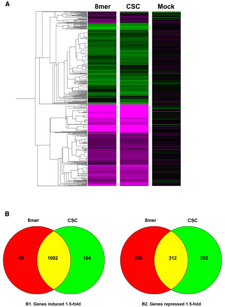
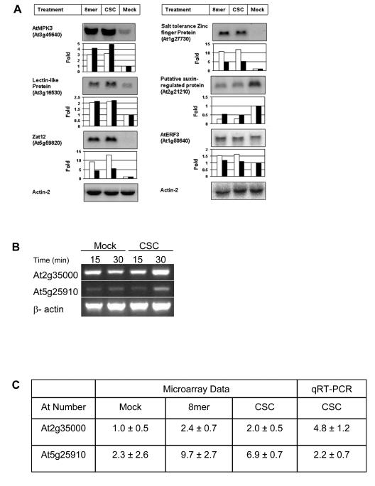
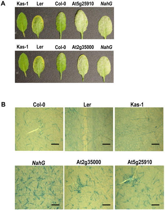
Chitin is a major component of fungal walls and insect exoskeletons. Plants produce chitinases upon pathogen attack and chito-oligomers induce defense responses in plants, though the exact mechanism behind this response is unknown. Using the ATH1 Affymetrix microarrays consisting of about 23,000 genes, we examined the response of Arabidopsis (Arabidopsis thaliana) seedlings to chito-octamers and hydrolyzed chitin after 30 min of treatment. The expression patterns elicited by the chito-octamer and hydrolyzed chitin were similar. Microarray expression profiles for several genes were verified via northern analysis or quantitative reverse transcription-PCR. We characterized T-DNA insertion mutants for nine chito-oligomer responsive genes. Three of the mutants were more susceptible to the fungal pathogen, powdery mildew, than wild type as measured by conidiophore production. These three mutants included mutants of genes for two disease resistance-like proteins and a putative E3 ligase. The isolation of loss-of-function mutants with enhanced disease susceptibility provides direct evidence that the chito-octamer is an important oligosaccharide elicitor of plant defenses. Also, this study demonstrates the value of microarray data for identifying new components of uncharacterized signaling pathways.
Figures
Similar articles
-
Abscisic acid negatively regulates post-penetration resistance of Arabidopsis to the biotrophic powdery mildew fungus.Sci China Life Sci. 2017 Aug;60(8):891-901. doi: 10.1007/s11427-017-9036-2. Epub 2017 Apr 10. Sci China Life Sci. 2017. PMID: 28702742
-
Overexpression of a wheat stearoyl-ACP desaturase (SACPD) gene TaSSI2 in Arabidopsis ssi2 mutant compromise its resistance to powdery mildew.Gene. 2013 Jul 25;524(2):220-7. doi: 10.1016/j.gene.2013.04.019. Epub 2013 Apr 24. Gene. 2013. PMID: 23624392
-
Arabidopsis phospholipase dδ is involved in basal defense and nonhost resistance to powdery mildew fungi.Plant Physiol. 2013 Oct;163(2):896-906. doi: 10.1104/pp.113.223503. Epub 2013 Aug 26. Plant Physiol. 2013. PMID: 23979971 Free PMC article.
-
Suppression of Chitin-Triggered Immunity by Plant Fungal Pathogens: A Case Study of the Cucurbit Powdery Mildew Fungus Podosphaera xanthii.J Fungi (Basel). 2023 Jul 21;9(7):771. doi: 10.3390/jof9070771. J Fungi (Basel). 2023. PMID: 37504759 Free PMC article. Review.
-
Hide-and-Seek: Chitin-Triggered Plant Immunity and Fungal Counterstrategies.Trends Plant Sci. 2020 Aug;25(8):805-816. doi: 10.1016/j.tplants.2020.03.006. Epub 2020 Apr 5. Trends Plant Sci. 2020. PMID: 32673581 Review.
Cited by
-
The Arabidopsis ubiquitin ligases ATL31 and ATL6 control the defense response as well as the carbon/nitrogen response.Plant Mol Biol. 2012 Jun;79(3):217-27. doi: 10.1007/s11103-012-9907-0. Epub 2012 Apr 7. Plant Mol Biol. 2012. PMID: 22481162
-
Verticillium longisporum infection affects the leaf apoplastic proteome, metabolome, and cell wall properties in Arabidopsis thaliana.PLoS One. 2012;7(2):e31435. doi: 10.1371/journal.pone.0031435. Epub 2012 Feb 20. PLoS One. 2012. PMID: 22363647 Free PMC article.
-
AtIQM1, a novel calmodulin-binding protein, is involved in stomatal movement in Arabidopsis.Plant Mol Biol. 2012 Jul;79(4-5):333-46. doi: 10.1007/s11103-012-9915-0. Epub 2012 May 10. Plant Mol Biol. 2012. PMID: 22572939
-
Arabidopsis snc2-1D activates receptor-like protein-mediated immunity transduced through WRKY70.Plant Cell. 2010 Sep;22(9):3153-63. doi: 10.1105/tpc.110.074120. Epub 2010 Sep 14. Plant Cell. 2010. PMID: 20841424 Free PMC article.
-
Endophytic ability of the insecticidal bacterium Brevibacillus laterosporus in Brassica.PLoS One. 2019 May 22;14(5):e0216341. doi: 10.1371/journal.pone.0216341. eCollection 2019. PLoS One. 2019. PMID: 31116753 Free PMC article.
References
-
- Adam L, Somerville SC (1996) Genetic characterization of five powdery mildew disease resistance loci in Arabidopsis thaliana. Plant J 9: 341–356 - PubMed
-
- Adams DJ (2004) Fungal cell wall chitinases and glucanases. Microbiol 150: 2029–2035 - PubMed
-
- Alonso JM, Stepanova AN, Leisse TJ, Kim CJ, Chen H, Shinn P, Stevenson DK, Zimmerman J, Barajas P, Cheuk R, et al (2003) Genome-wide insertional mutagenesis of Arabidopsis thaliana. Science 301: 653–657 - PubMed
-
- Boller T (1995) Chemoperception of microbial signals in plant cells. Annu Rev Plant Physiol 46: 189–214
-
- Dellaporta SL, Wood J, Hicks JB (1983) A plant DNA minipreparation: version II. Plant Mol Biol Rep 1: 19–20
Publication types
MeSH terms
Substances
LinkOut - more resources
Full Text Sources
Other Literature Sources
Molecular Biology Databases
