. 2005 Dec;53(12):1481-9.
doi: 10.1369/jhc.4A6552.2005. Epub 2005 May 27.

Polarized expression of CD74 by gastric epithelial cells

Affiliations

Polarized expression of CD74 by gastric epithelial cells

Carlos A Barrera et al. J Histochem Cytochem. 2005 Dec.

Abstract

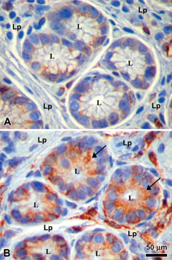
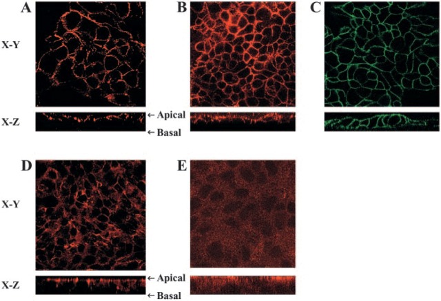
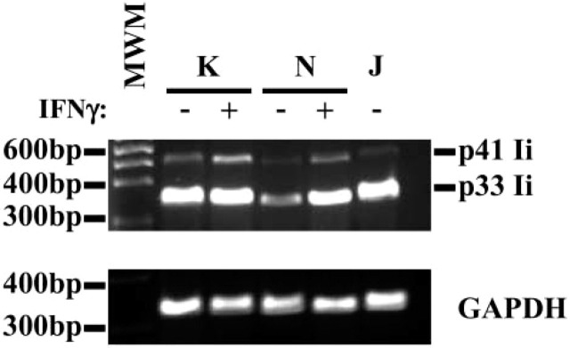
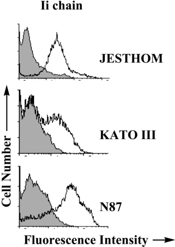
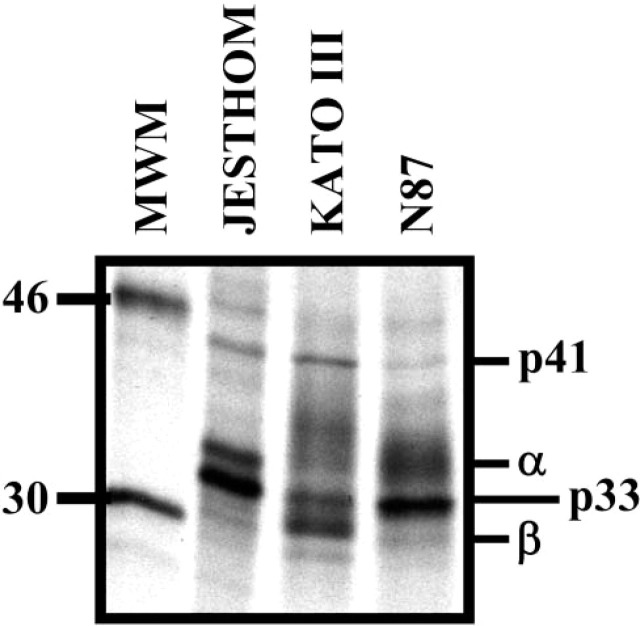
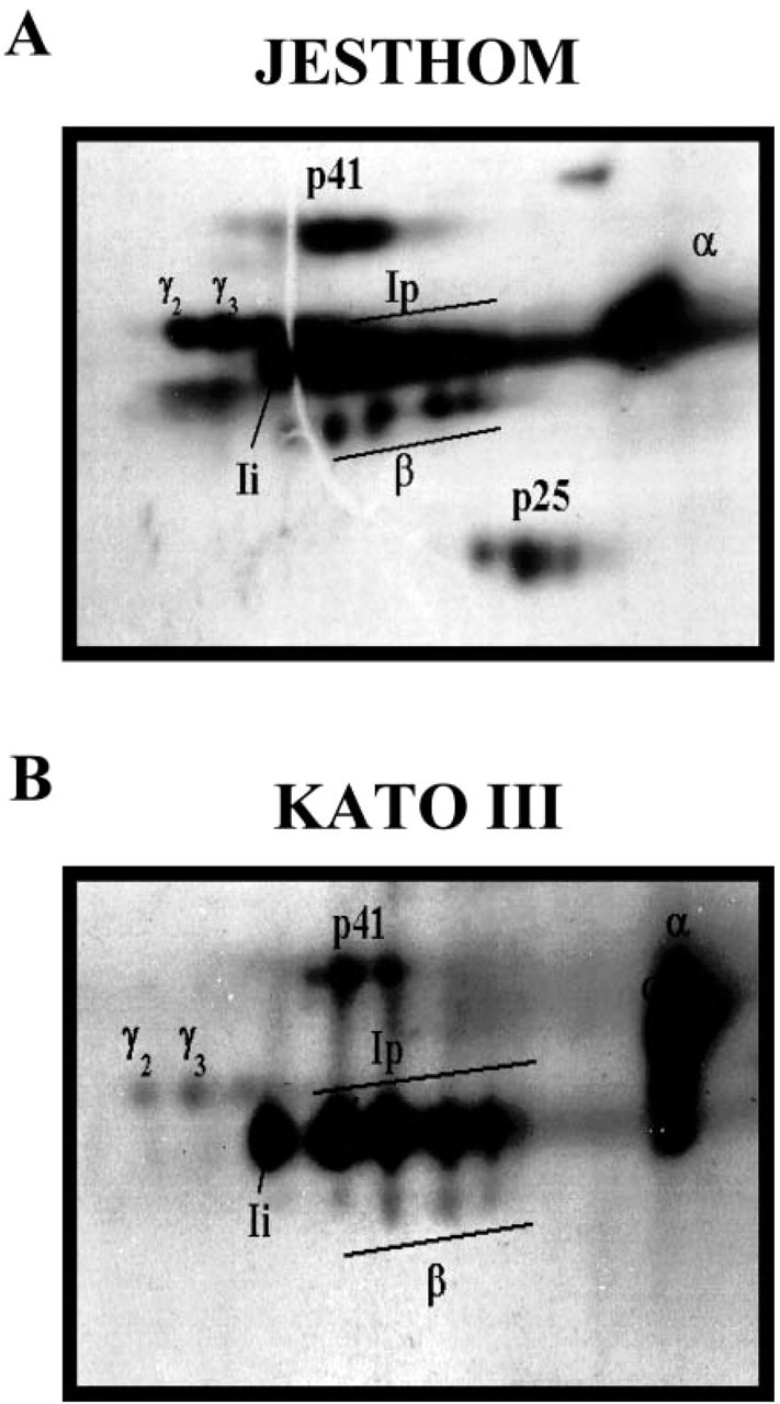
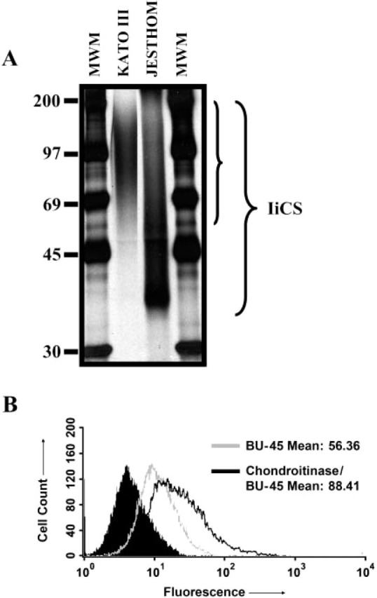
CD74 is known as the major histocompatibility complex (MHC) class II-associated invariant chain (Ii) that regulates the cell biology and functions of MHC class II molecules. Class II MHC and Ii expression was believed to be restricted to classical antigen-presenting cells (APC); however, during inflammation, other cell types, including mucosal epithelial cells, have also been reported to express class II MHC molecules. Given the importance of Ii in the biology of class II MHC, we sought to examine the expression of Ii by gastric epithelial cells (GEC) to determine whether class II MHC molecules in these nonconventional APC cells were under the control of Ii and to further support the role that these cells may play in local immune and inflammatory responses during Helicobacter pylori infection. Thus we examined the expression of Ii on GEC from human biopsy samples and then confirmed this observation using independent methods on several GEC lines. The mRNA for Ii was detected by RT-PCR, and the various protein isoforms were also detected. Interestingly, these cells have a high level expression of surface Ii, which is polarized to the apical surface. These studies are the first to demonstrate the constitutive expression of Ii by human GEC.


Figures

Figure 1
Figure 1
Immunohistochemistry of gastric biopsies. The expression of the invariant chain (Ii) was compared in gastric biopsies from Helicobacter pylori infected (B) vs noninfected donors (A). L = lumen; Lp = lamina propria. Arrow points to intense apical staining on epithelial cells.
Figure 2.
Figure 2.
Surface distribution of invariant chain (Ii) on N87 cells grown on filter inserts. Confocal microscopy images are shown here of the horizontal interfaces (X-Y) and vertical interfaces (X-Z). (A) N87 cells stained with anti-DAF antibody (Alexa 568) as an apical marker, (B) N87 cells stained with anti-Ii antibody (Alexa 568), and (C) N87 cells stained with anti-epithelial antigen with apical and basal staining (FITC). Cells were stained with saturating concentration of antibodies, which reach both apical and baso-lateral sides of the cells. As additional controls, the cells were stained with antibodies to (D) the apical junctional complex protein Zona Occludens-1 and a secondary goat anti-mouse IgG conjugated with Alexa 568 and to (E) the cytoplasmic tail of Ii also with the same secondary antibody conjugated with Alexa 568.
Figure 3.
Figure 3.
Invariant chain (Ii) mRNA detected in gastric epithelial cells by RT-PCR analysis. RNA extracted from KATO III and N87 gastric epithelial cells was reverse transcribed, amplified, and separated by electrophoresis. Specific products were detected in these cells by RT-PCR using the Ii chain primers described in Materials and Methods. As an internal control, B (Jesthom) cells showed the mRNA for those proteins. The amplification of housekeeping gene GAPDH was included as a control for the procedure. This is a representative of three experiments.
Figure 4.
Figure 4.
Invariant chain expression by gastric epithelial cells detected by flow cytometry. Ii chain molecules were expressed in gastric epithelial cells (A) KATO III and (B) N87 before and after IFNγ treatment. For comparison (C), the Jesthom B cell line was used as an internal positive control. The cells were stained with anti-Ii BU45 mAb. The shaded curve represents the staining with the isotype control antibody. The displacement of the fluorescence intensity curve to the right indicated an increase in expression level.
Figure 5.
Figure 5.
Immunoprecipitation of invariant chain isoforms. One-dimensional gel electrophoresis of invariant chain (Ii) proteins from metabolically labeled Jesthom (B cells), KATO III, and N87 (gastric epithelial cells) demonstrates the expression of those isoforms. The Ii isoforms p33 and p41 are presented after immunoprecipitation with anti-BU45 mAb.
Figure 6.
Figure 6.
Microheterogeneity of class II MHC-associated invariant chain. Two-dimensional gel electrophoresis of invariant chain (Ii) proteins from metabolically labeled (A) Jesthom and (B) KATO III demonstrates the similarities in expression of all of the components. The α and β chains of MHC class II and different isoforms of Ii (Ii, Ip, p41, γ2, and γ3) are presented after immunoprecipitation with anti-BU45 mAb.
Figure 7.
Figure 7.
Characterization of invariant chain (Ii)-chondroitin sulfate (CS) in gastric epithelial cells. (A) One-dimensional gel electrophoresis showed expression of Ii-CS in KATO III gastric epithelial cells and Jesthom B-cell line controls. Ii-CS was enriched through a DEAE-sephacel column and then run on SDS/PAGE after immunoprecipitation with anti-Ii VicY-1 antibody. (B) Flow cytometric analysis of N87 cells stained for Ii with BU-45 antibodies after chondroitinase treatment to reveal the relative amount of Ii-CS. The solid peak is the isotype control.

References

    1. Aruffo A, Stamenkovic I, Melnick M, Underhill CB, Seed B. (1990) CD44 is the principal cell surface receptor for hyaluronate. Cell 61: 1303–1313 - PubMed
    1. Bakke O, Dobberstein B. (1990) MHC class II-associated invariant chain contains a sorting signal for endosomal compartments. Cell 63: 707–716 - PubMed
    1. Bamford KB, Fan X, Crowe SE, Leary JF, Gourley WK, Luthra GK, Brooks EG, et al. (1998) Lymphocytes in the human gastric mucosa during Helicobacter pylori have a T helper cell 1 phenotype. Gastroenterology 114: 482–492 - PubMed
    1. Barrera C, Ye G, Espejo R, Gunasena S, Almanza R, Leary J, Crowe S, et al. (2001) Expression of cathepsins B, L, S, and D by gastric epithelial cells implicates them as antigen presenting cells in local immune responses. Hum Immunol 62: 1081–1091 - PubMed
    1. Barrera C, Espejo R, Reyes VE. (2002) Differential glycosylation of MHC class II molecules on gastric epithelial cells: implications in local immune responses. Hum Immunol 63: 384–393 - PubMed

Publication types

MeSH terms