

Effect of triamcinolone acetonide on proliferation of retinal endothelial cells in vitro and in vivo
- PMID: 15923512
- PMCID: PMC1772672
- DOI: 10.1136/bjo.2004.052563
Effect of triamcinolone acetonide on proliferation of retinal endothelial cells in vitro and in vivo
Abstract
Aim: To assess the effect of crystalline triamcinolone acetonide on retinal endothelial cell proliferation in vivo and in vitro.
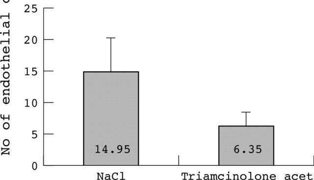
Methods: For in vitro analysis, a sprouting assay was employed. Bovine retinal endothelial cells were stimulated with basic fibroblast growth factor (bFGF) and incubated with different concentrations of triamcinolone acetonide (0.05 mg/ml to 8 mg/ml). For in vivo analysis, a retinopathy of prematurity (ROP) model was used. 16 C57BL/J6 mice were exposed to 75% oxygen from postnatal day 7 to day 12. On day 12, triamcinolone acetonide was intravitreally injected into one eye ("study eye") and isotonic saline into the contralateral eye ("control eye"). On day 17, the mice were sacrificed and the eyes removed for quantitative analysis of preretinal neovascularisation. Four non-exposed mice served as negative control.
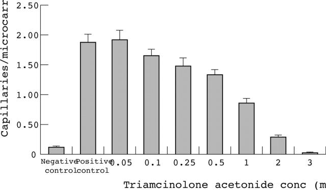
Results: The sprouting assay demonstrated a dose dependent inhibition of bovine retinal endothelial cell proliferation from 0.05 mg triamcinolone acetonide/ml (no inhibition) to 3 mg triamcinolone acetonide/ml (complete inhibition). Dosages of more than 2 mg/ml resulted in cytotoxic changes of endothelial cells. The ROP model demonstrated a significantly lower neovascular cell count of 58% in the study group compared to the control group (6.35 (SD 2.1) cells per histological section versus 14.9 (SD 5.3) cells; p<0.005).
Conclusions: Triamcinolone acetonide inhibits bFGF induced proliferation of retinal endothelial cells in vivo and in vitro. These findings contribute to understanding the mode of action and effects of triamcinolone acetonide on retinal neovascularisation.
Figures
Similar articles
-
Triamcinolone reduces neovascularization, capillary density and IGF-1 receptor phosphorylation in a model of oxygen-induced retinopathy.Invest Ophthalmol Vis Sci. 2006 Nov;47(11):4975-82. doi: 10.1167/iovs.06-0450. Invest Ophthalmol Vis Sci. 2006. PMID: 17065516 Free PMC article.
-
Impact of triamcinolone acetonide on retinal endothelial cells in a retinopathy of prematurity mouse model.Acta Ophthalmol Scand. 2007 Nov;85(7):791-4. doi: 10.1111/j.1600-0420.2007.00945.x. Epub 2007 May 4. Acta Ophthalmol Scand. 2007. PMID: 17488319
-
Structural consequences after intravitreal bevacizumab injection without increasing apoptotic cell death in a retinopathy of prematurity mouse model.Acta Ophthalmol. 2012 Sep;90(6):564-70. doi: 10.1111/j.1755-3768.2010.01963.x. Epub 2010 Aug 4. Acta Ophthalmol. 2012. PMID: 20698831
-
Herbimycin A inhibits angiogenic activity in endothelial cells and reduces neovascularization in a rat model of retinopathy of prematurity.Exp Eye Res. 2004 May;78(5):987-95. doi: 10.1016/j.exer.2003.12.008. Exp Eye Res. 2004. PMID: 15051479
-
Ocular neovascularization: clarifying complex interactions.Am J Pathol. 1998 Sep;153(3):665-70. doi: 10.1016/S0002-9440(10)65607-6. Am J Pathol. 1998. PMID: 9736014 Free PMC article. Review. No abstract available.
Cited by
-
Targeting Neovascularization in Ischemic Retinopathy: Recent Advances.Expert Rev Ophthalmol. 2013 Jun;8(3):267-286. doi: 10.1586/eop.13.17. Expert Rev Ophthalmol. 2013. PMID: 25598837 Free PMC article.
-
Triamcinolone reduces neovascularization, capillary density and IGF-1 receptor phosphorylation in a model of oxygen-induced retinopathy.Invest Ophthalmol Vis Sci. 2006 Nov;47(11):4975-82. doi: 10.1167/iovs.06-0450. Invest Ophthalmol Vis Sci. 2006. PMID: 17065516 Free PMC article.
-
Biomarker identification of immune-related genes in pheochromocytoma and paraganglioma.Transl Androl Urol. 2023 Feb 28;12(2):249-260. doi: 10.21037/tau-22-800. Epub 2023 Feb 27. Transl Androl Urol. 2023. PMID: 36915875 Free PMC article.
-
Aberrant kinetics of bone marrow-derived endothelial progenitor cells in the murine oxygen-induced retinopathy model.Invest Ophthalmol Vis Sci. 2011 Oct 3;52(11):7835-41. doi: 10.1167/iovs.10-5880. Invest Ophthalmol Vis Sci. 2011. PMID: 21896844 Free PMC article.
-
Effect of intravitreal triamcinolone acetonide on retinal apoptosis in experimental retinal neovascularization.Graefes Arch Clin Exp Ophthalmol. 2008 Jul;246(7):1069-70. doi: 10.1007/s00417-008-0814-7. Epub 2008 Apr 17. Graefes Arch Clin Exp Ophthalmol. 2008. PMID: 18418624 No abstract available.
References
-
- Stolk RP, Vingerling JR, de Jong PT, et al. Retinopathy, glucose, and insulin in an elderly population. The Rotterdam Study. Diabetes 1995;44:11–15. - PubMed
-
- Mitchell P, Smith W, Wang JJ, et al. Prevalence of diabetic retinopathy in an older community. The Blue Mountains Eye Study. Ophthalmology 1998;105:406–11. - PubMed
-
- The Diabetic Retinopathy Study Research Group. Photocoagulation treatment of proliferative diabetic retinopathy: The second report of Diabetic Retinopathy Study findings. Ophthalmology 1978;85:82–106. - PubMed
-
- Jonas JB, Hayler JK, Söfker A, et al. Intravitreal injection of crystalline cortisone as adjunctive treatment of proliferative diabetic retinopathy. Am J Ophthalmol 2001;131:468–71. - PubMed
MeSH terms
Substances
LinkOut - more resources
Full Text Sources
Medical