

The use of human serum in supporting the in vitro and in vivo proliferation of human conjunctival epithelial cells
- PMID: 15923513
- PMCID: PMC1772683
- DOI: 10.1136/bjo.2004.055046
The use of human serum in supporting the in vitro and in vivo proliferation of human conjunctival epithelial cells
Abstract
Aim: To evaluate the use of human serum (HS) in supporting the in vitro and in vivo proliferation of human conjunctival epithelial cells, and compare it with fetal bovine serum (FBS) and bovine pituitary extract (BPE).
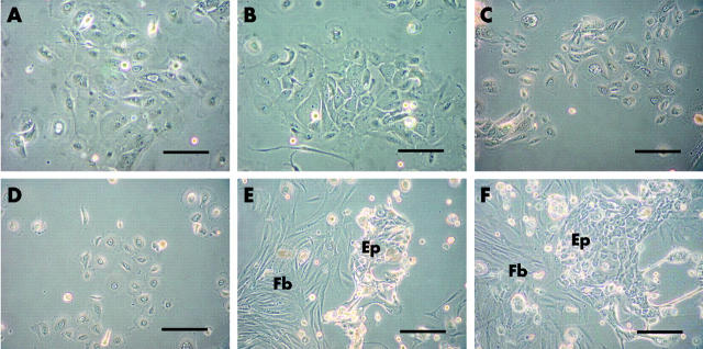
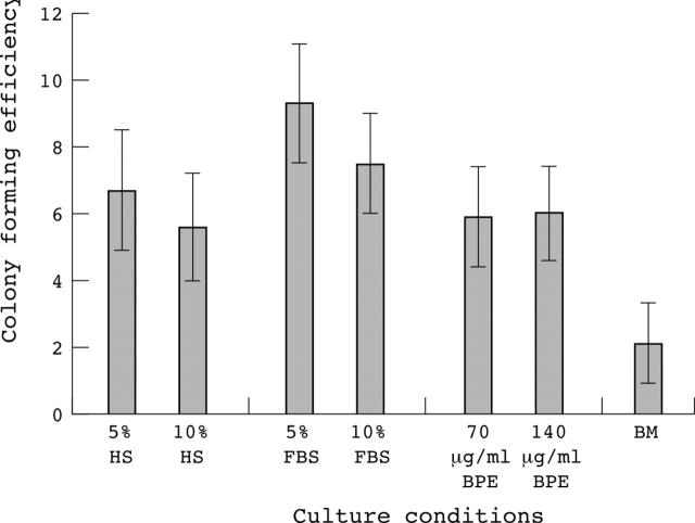
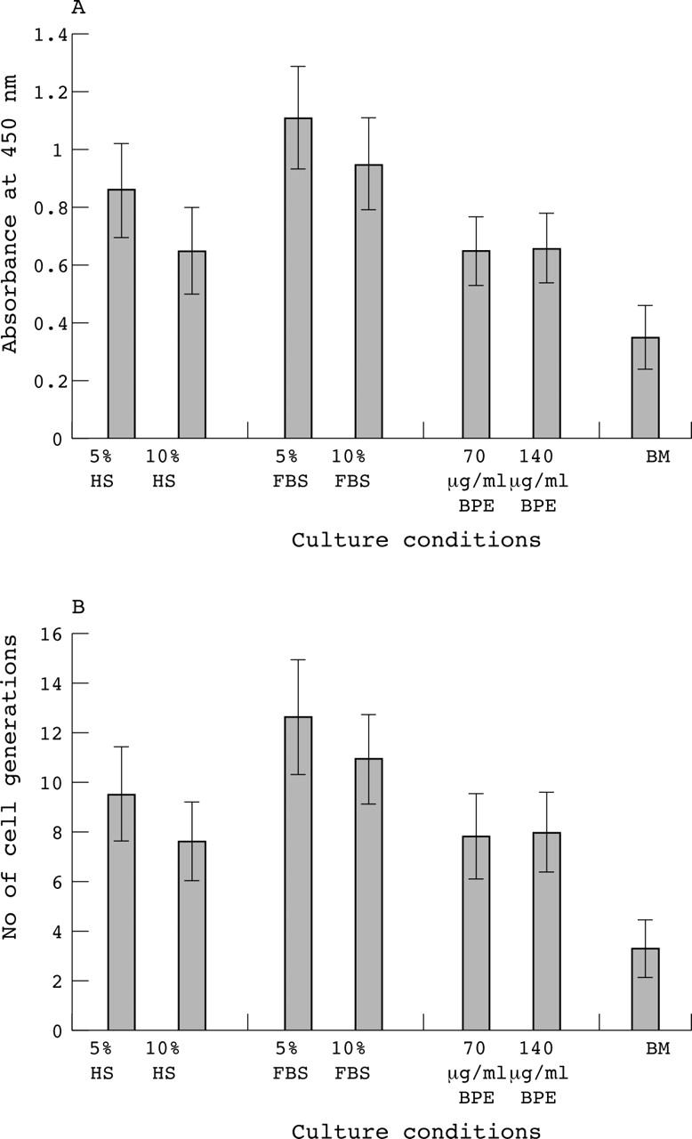
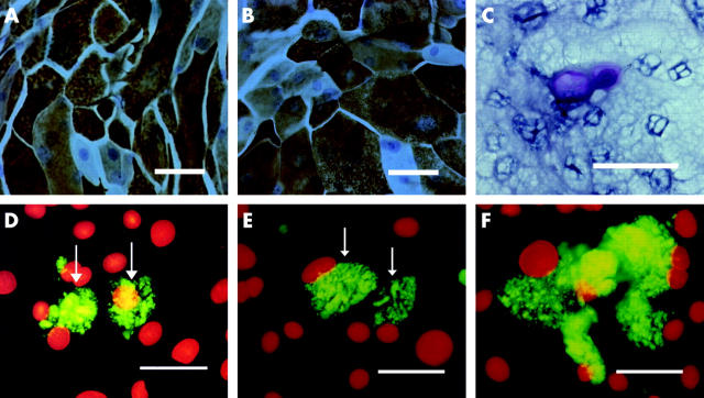
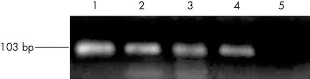
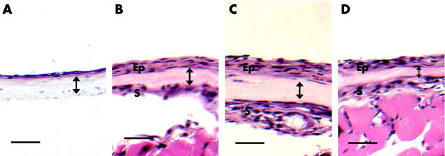
Methods: Conjunctival epithelial cells were cultivated in media supplemented with HS (5%, 10%), FBS (5%, 10%), and BPE (70 microg/ml, 140 microg/ml). The colony forming efficiency (CFE), bromodeoxyuridine (BrdU) ELISA proliferation assay, and cell generations were analysed. Cells were evaluated for keratin (K4, K19, and K3) and MUC5AC expression by immunostaining and RT-PCR. Conjunctival equivalents constructed on amniotic membranes were transplanted onto severe combined immune deficient (SCID) mice for 10 days and analysed histologically.
Results: The proliferation assays of HS supplemented cultures (CFE, 6.7% (SD 1.8%); BrdU absorbance, 0.86 (0.16)) were comparable to FBS supplemented (CFE, 9.3% (1.8%); BrdU absorbance, 1.11 (0.18)) and BPE supplemented cultures (CFE, 5.9 (1.5); BrdU absorbance, 0.65 (0.12)). Goblet cell densities for HS, FBS, and BPE supplemented media were 52 cells/cm(2), 60 cells/cm(2), and 50 cells/cm(2), respectively. HS supplemented cultures formed stratified epithelial sheets in vivo following transplantation.
Conclusions: The proliferative capacity of conjunctival epithelial cells cultivated in HS supplemented cultures was comparable to FBS and BPE supplemented cultures. The elimination of animal material from the culture system is advantageous when cultivating cells for clinical transplantation.
Figures
Similar articles
-
The use of autologous serum in the development of corneal and oral epithelial equivalents in patients with Stevens-Johnson syndrome.Invest Ophthalmol Vis Sci. 2006 Mar;47(3):909-16. doi: 10.1167/iovs.05-1188. Invest Ophthalmol Vis Sci. 2006. PMID: 16505023
-
The development of a serum-free derived bioengineered conjunctival epithelial equivalent using an ultrathin poly(epsilon-caprolactone) membrane substrate.Invest Ophthalmol Vis Sci. 2006 Jan;47(1):105-12. doi: 10.1167/iovs.05-0512. Invest Ophthalmol Vis Sci. 2006. PMID: 16384951
-
Ex vivo expansion of conjunctival and limbal epithelial cells using cord blood serum-supplemented culture medium.Invest Ophthalmol Vis Sci. 2011 Aug 3;52(9):6138-47. doi: 10.1167/iovs.10-6527. Invest Ophthalmol Vis Sci. 2011. PMID: 21474776
-
Goat serum: an alternative to fetal bovine serum in biomedical research.Indian J Exp Biol. 2004 Jan;42(1):26-35. Indian J Exp Biol. 2004. PMID: 15274477 Review.
-
[Progresses of in vitro culture and differentiation in conjunctival epithelial stem cells].Zhonghua Yan Ke Za Zhi. 2011 Jan;47(1):79-82. Zhonghua Yan Ke Za Zhi. 2011. PMID: 21418932 Review. Chinese.
Cited by
-
Epithelial microfilament regulators show regional distribution in mouse conjunctiva.Mol Vis. 2010 Oct 31;16:2215-24. Mol Vis. 2010. PMID: 21151337 Free PMC article.
-
Conjunctival goblet cells: Ocular surface functions, disorders that affect them, and the potential for their regeneration.Ocul Surf. 2020 Jan;18(1):19-26. doi: 10.1016/j.jtos.2019.11.005. Epub 2019 Nov 14. Ocul Surf. 2020. PMID: 31734511 Free PMC article. Review.
-
Cultured human ocular surface epithelium on therapeutic contact lenses.Br J Ophthalmol. 2007 Apr;91(4):459-64. doi: 10.1136/bjo.2006.103895. Epub 2006 Sep 20. Br J Ophthalmol. 2007. PMID: 16987897 Free PMC article.
-
Regeneration of the corneal epithelium with conjunctival epithelial equivalents generated in serum- and feeder-cell-free media.Mol Vis. 2013 Dec 16;19:2542-50. eCollection 2013. Mol Vis. 2013. PMID: 24357922 Free PMC article.
-
Effect of growth factors on the proliferation and gene expression of human meibomian gland epithelial cells.Invest Ophthalmol Vis Sci. 2013 Apr 5;54(4):2541-50. doi: 10.1167/iovs.12-11221. Invest Ophthalmol Vis Sci. 2013. PMID: 23493293 Free PMC article.
References
-
- Pellegrini G, Traverso CE, Franzi AT, et al. Long-term restoration of damaged corneal surfaces with autologous cultivated corneal epithelium. Lancet 1997;349:990–3. - PubMed
-
- Tsai RJ, Li LM, Chen JK. Reconstruction of damaged corneas by transplantation of autologous limbal epithelial cells. N Engl J Med 2000;343:86–93. - PubMed
-
- Koizumi N, Inatomi T, Suzuki T, et al. Cultivated corneal epithelial stem cell transplantation in ocular surface disorders. Ophthalmology 2001;108:1569–74. - PubMed
-
- Shimazaki J, Aiba M, Goto E, et al. Transplantation of human limbal epithelium cultivated on amniotic membrane for the treatment of severe ocular surface disorders. Ophthalmology 2002;109:1285–90. - PubMed
-
- Tsai RJ, Ho YS, Chen JK. The effects of fibroblasts on the growth and differentiation of human bulbar conjunctival epithelial cells in an in vitro conjunctival equivalent. Invest Ophthalmol Vis Sci 1994;35:2865–75. - PubMed
Publication types
MeSH terms
Substances
LinkOut - more resources
Full Text Sources
Other Literature Sources