. 2005 Jun;25(12):4813-25.
doi: 10.1128/MCB.25.12.4813-4825.2005.

Proximity of the U12 snRNA with both the 5' splice site and the branch point during early stages of spliceosome assembly

Affiliations

Proximity of the U12 snRNA with both the 5' splice site and the branch point during early stages of spliceosome assembly

Mikko J Frilander et al. Mol Cell Biol. 2005 Jun.

Abstract

U12 snRNA is required for branch point recognition in the U12-dependent spliceosome. Using site-specific cross-linking, we have captured an unexpected interaction between the 5' end of the U12 snRNA and the -2 position upstream of the 5' splice site of P120 and SCN4a splicing substrates. The U12 snRNA nucleotides that contact the 5' exon are the same ones that form the catalytically important helix Ib with U6atac snRNA in the spliceosome catalytic core. However, the U12/5' exon interaction is transient, occurring prior to the entry of the U4atac/U6atac.U5 tri-snRNP to the spliceosome. This suggests that the helix Ib region of U12 snRNA is positioned near the 5' splice site early during spliceosome assembly and only later interacts with U6atac to form helix Ib. We also provide evidence that U12 snRNA can simultaneously interact with 5' exon sequences near 5' splice site and the branch point sequence, suggesting that the 5' splice site and branch point sequences are separated by <40 to 50 A in the complex A of the U12-dependent spliceosome. Thus, no major rearrangements are subsequently needed to position these sites for the first step of catalysis.


Figures

FIG. 1.
FIG. 1.
(A) Reported snRNA/pre-mRNA interactions near the 5′ss. At early stages of spliceosome assembly, U11 can be cross-linked to the +2 position of the 5′ss consensus sequence in the intron. At late stages of assembly, the +2 position interacts with U6atac whereas the −2 position (in the exon) interacts with U5. Predicted cross-links are shown as lightning bolts, but the exact nucleotides within each snRNA shown have not been identified. (B) Combined psoralen and 4SU cross-linking analysis. The 4SU-containing P120[−2/+2] splicing substrate was incubated in HeLa nuclear extract under splicing conditions for 90 min. 4′-aminomethyl-4,5′,8-trimethyl-psoralen was then added to each reaction, followed by irradiation at 365 nm and Northern analysis using a U12-specific probe as described in Materials and Methods. The conditions for individual reactions are indicated above each panel, and the positions of the cross-linked RNA species are indicated on the left. The control substrate in lane 3 did not contain 4SU. (C) Identification of U12-specific cross-links by RNase H cleavage. The reactions were performed as for panel B except that psoralen was omitted and the incubation time was 60 min. The cross-linked RNA samples were incubated with RNase H plus 100 pmol of DNA oligonucleotides complementary to snRNAs or P120 splicing substrate as indicated above each lane. Subsequently, cross-links were visualized by Northern blotting with a U12-specific probe as described for panel B.
FIG. 2.
FIG. 2.
(A) Localization of the 4SU cross-linked position in the P120 pre-mRNA. P120 splicing substrates containing either two (P120[−2/+2], lanes 1 and 3 to 8) or one 4SU residue (P120[−2], lanes 9 to 14) were cross-linked after incubation at 30°C for 40 min (as in Fig. 1C) and analyzed by Northern blotting using probes specific for U5, U11, or U12 as indicated. The specific 2ome oligonucleotide included in each reaction is indicated above each lane. The identity of each specific cross-link is indicated on the right, and the sizes of molecular weight marker (M, in nucleotides), a 32P-labeled pBR322 MspI digest, are shown on the left. The asterisk denotes a nonspecific cross-reacting band. (B) ATP dependence of the U12 cross-link formation. P120[−2] was incubated in HeLa nuclear extract under normal splicing conditions (lane 2) or in conditions without exogenously added ATP or creatine phosphate (lane 3) and which has also been preincubated at 30°C for 20 min to hydrolyze endogenous ATP. The asterisk denotes a nonspecific cross-reacting band. (C) Protocol for the RNase H assay. (Top) The location and effect of DNA oligonucleotides used in the RNase H assay. In the spliceosome A complex only the intron-specific DNA oligonucleotide is able to bind P120 pre-mRNA and support RNase H hydrolysis, while the U11/U12 di-snRNP binds 5′ss and BPS sequences and protects those sites from DNA oligonucleotide-directed RNase H cleavage as indicated in the figure. (Bottom) Experimental protocol for the RNase H assay. (D) Protection of the U12/5′exon cross-link from the DNA oligonucleotide directed RNase H cleavage. The experimental protocol and the DNA oligonucleotides used have been described for panel C.
FIG. 3.
FIG. 3.
(A) Time course of cross-link formation. The P120[−2/+2] splicing substrate containing a 47-nucleotide 5′ exon was cross-linked as in Fig. 1C at time points indicated above each lane and subsequently analyzed by Northern blotting. The filters were probed for U11, U12, U6atac, or U5 snRNAs, as indicated. The identity of each cross-linked band is indicated on the right. (B) Time course analysis with P120[−2/+2] substrate containing an 8-nt 5′ exon. The reactions were analyzed as in panel A. The strong U11/5′ss signals in lane 1 are the results of saturating concentrations of the substrate used in this experiment (see Materials and Methods). (C) Reversal of the U12-5′ exon interaction. Lanes 1 to 6, time course of U12 cross-link formation under standard reaction conditions. Lanes 7 to 12, a block/release reaction in which a tailed version of the 5′ exon 2ome oligonucleotide (9) was used to arrest the U12-specific cross-link for 45 min (lanes 7 and 8) followed by the addition of a complementary release oligonucleotide (lanes 9 to 11) as indicated above each lane. Lane 12 shows a control reaction without release oligonucleotide. The reactions were analyzed by Northern blotting as in Fig. 1C.
FIG.4.
FIG.4.
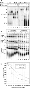
The effect of a 5′ exon oligonucleotide on spliceosomal complex formation. (A) Spliceosome assembly performed in the absence or presence of the 5′ exon 2ome oligonucleotide using unlabeled P120 splicing substrate. The reactions were subsequently separated on native gels and analyzed by Northern blotting with probes indicated above each panel. The migration of spliceosomal complexes A and B and the U11- and U12-specific snRNA complexes (on lanes 1 to 6) is shown on the left. (B and C) Stability of complex A in the presence of the 5′ exon 2ome oligonucleotide. P120 splicing substrate was incubated under splicing conditions for 30 min in the presence of 1 μM 5′ exon 2ome oligonucleotide (lanes 1 to 6) or a mock oligonucleotide of the same length (lanes 7 to 12). The samples were subsequently transferred to ice, and heparin was added to each sample to the final concentration shown above each lane; the samples were then incubated on ice for an additional 2 min. The samples were then loaded onto a 4% native gel. (B) In top and middle panels, unlabeled P120 splicing substrate was used and the gels were analyzed by Northern blotting as in panel A using U11- and U12-specific riboprobes as indicated. Reactions in the bottom panel contained 32P-labeled P120 splicing substrate. The positions of spliceosomal complexes B, A1 (contains both U11 and U12 snRNPs), A2 (contains only U12 snRNP), and the unspecific complex H are indicated. (C) Complex A1 stability as a function of heparin concentration. Complexes A1 and A2 were quantified with a phosphoimager in six independent experiments with the 32P-labeled P120 substrate. The relative stability of A1 was calculated using the formula A1/(A1 + A2).
FIG. 5.
FIG. 5.
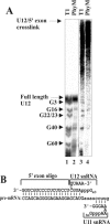
Mapping the 4SU cross-link position on U12 snRNA. (A) 3′-End-labeled un-cross-linked and cross-linked U12 snRNAs (see Materials and Methods) were partially hydrolyzed with RNase T1 or PhyM and analyzed on a 5% polyacrylamide gel. Lanes 1 and 2, partial hydrolysis of the uncross-linked U12 snRNA. Lanes 3 and 4, partial hydrolysis of the cross-linked U12 species. The positions of the detected G residues are indicated on the left. (B) Proposed base-pairing interaction of the U12 snRNA with the 5′ exon sequences of the cross-linked P120 substrate (uppercase, 5′ exon sequence; lowercase, intron sequence). The position of the 5′ exon oligonucleotide and the U11 snRNA base paired with the 5′ss is also indicated.
FIG. 6.
FIG. 6.
The significance of U12/5′exon base pairing. (A) Sequences of the cross-linking substrates used in panels B to D. The total length of the P120 cross-linking substrate is 250 nt, and that of SCN4a substrates is 275 nt. (B) U12/5′ exon cross-link formation using P120[−2] and P120-CG substrates. The cross-linking substrates and conditions or oligonucleotides used are indicated above each lane. Otherwise the reactions were carried out as in Fig. 2A and probed with a U12-specific probe. (C) U12/5′exon cross-link formation using SCN4a[−2] and SCN4a-CG substrates. The reactions were performed in SCN4a splicing conditions (see Materials and Methods), except for lane 7, which was performed as in panel B. Following the cross-linking the reactions were separated on 5% denaturing PAGE gel, and the U12-specific cross-links were detected by Northern blotting using U12-specific probes. The labels are as in Fig. 2A. (D) The same as in panel C, but the reactions were run on 8% denaturing page gel.
FIG. 7.
FIG. 7.
The effect of U12/5′exon binding on splicing activity as measured using a substrate competition assay in which competing 5′ss have been positioned in tandem on a single splicing substrate. (A) 5′ss sequences of the modified substrates used in the experiment. In WT substrate the 5′ss has been duplicated (indicated by bolded letters), and both sites contain a sequence element complementary to the U12 snRNA helix Ib region (underlined). In the following four mutant substrates one of the complementary sequences (either upstream of the proximal or distal site) has been mutated by inversion (substrates 1 and 2) or by substitution (substrates 3 and 4) as indicated in the figure. (B) Quantification of the splicing activities at proximal and distal sites. Equal amounts of each splicing substrate were incubated under splicing conditions for 3 h and processed as described in Materials and Methods. The splicing activities at proximal and distal sites were quantified by measuring the lariat product formation representing the proximal or distal splice site usage using a phosphoimager. The bar chart shows mean values from three to six individual experiments, which were normalized to WT distal activity, which was set at 1.00. Additionally, the proximal splice site values were multiplied by 1.190 to take account the difference in the number of the 32P-labeled U-residues on the lariat products resulting from splicing at proximal versus distal sites (21 versus 25, respectively). Standard deviation is indicated by error bars. The autoradiograph of a denaturing polyacrylamide gel under the bar chart shows a lariat formation from an individual experiment. (C) Relative splice site usage of the different mutant substrates described in panel A. Black circles represent wild-type 5′ exon sequence upstream of the 5′ss and hollow circles represent mutated sites. The preferred 5′ splice sites are indicated on bold. The values were obtained from the same data set as in panel B.
FIG. 8.
FIG. 8.
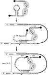
A model for U12-specific interactions with the P120 splicing substrate during the early stages of spliceosome assembly. Following the recognition of the BPS in the spliceosome A complex, the helix Ib region of the U12 snRNA is released from the stem-loop structure and positioned near the 5′ss. Transient base-pairing interaction with the 5′ exon sequences can take place if a suitable complementary sequence is present in the 5′ exon near the 5′ss. The trimethyl cap is depicted as a black circle.

Similar articles

Cited by

References

    1. Abovich, N., and M. Rosbash. 1997. Cross-intron bridging interactions in the yeast commitment complex are conserved in mammals. Cell 89:403-412. - PubMed
    1. Ast, G., and A. M. Weiner. 1996. A U1/U4/U5 snRNP complex induced by a 2′-O-methyl-oligoribonucleotide complementary to U5 snRNA. Science 272:881-884. - PubMed
    1. Brow, D. A. 2002. Allosteric cascade of spliceosome activation. Annu. Rev. Genet. 36:333-360. - PubMed
    1. Burge, C. B., T. Tuschl, and P. A. Sharp. 1999. Splicing of precursors to mRNAs by the spliceosome, p. 525-560. In R. F. Gesteland, T. R. Cech, and J. F. Atkins (ed.), The RNA world, vol. 2. Cold Spring Harbor Laboratory Press, Cold Spring Harbor, N.Y.
    1. Cunningham, S. A., A. J. Else, B. V. Potter, and I. C. Eperon. 1991. Influences of separation and adjacent sequences on the use of alternative 5′ splice sites. J. Mol. Biol. 217:265-281. - PubMed

Publication types

LinkOut - more resources