. 2005 Jun;25(12):4826-40.
doi: 10.1128/MCB.25.12.4826-4840.2005.

Progestins induce transcriptional activation of signal transducer and activator of transcription 3 (Stat3) via a Jak- and Src-dependent mechanism in breast cancer cells

Affiliations

Progestins induce transcriptional activation of signal transducer and activator of transcription 3 (Stat3) via a Jak- and Src-dependent mechanism in breast cancer cells

Cecilia Proietti et al. Mol Cell Biol. 2005 Jun.

Abstract

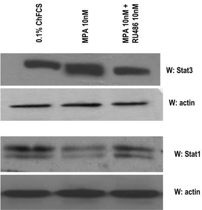
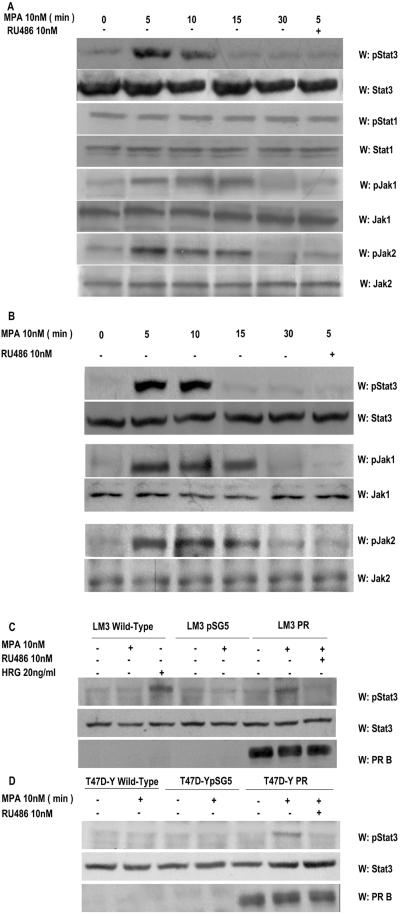
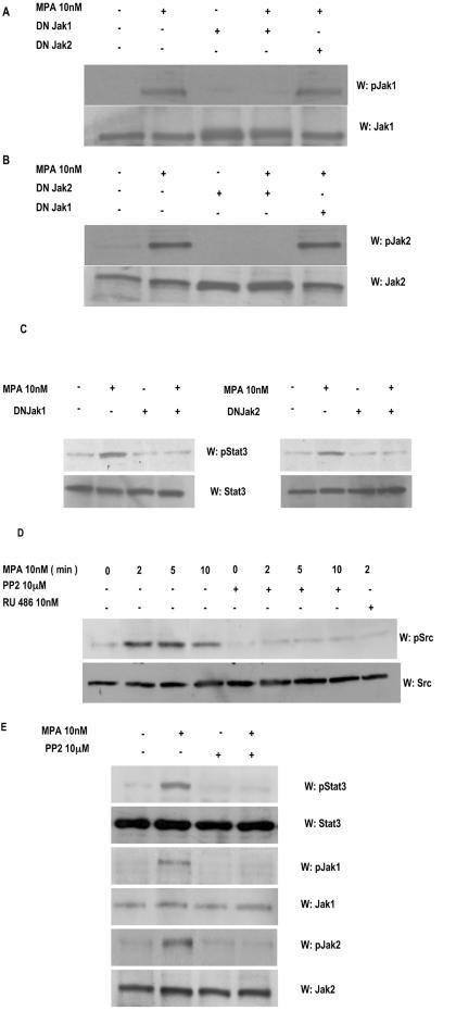
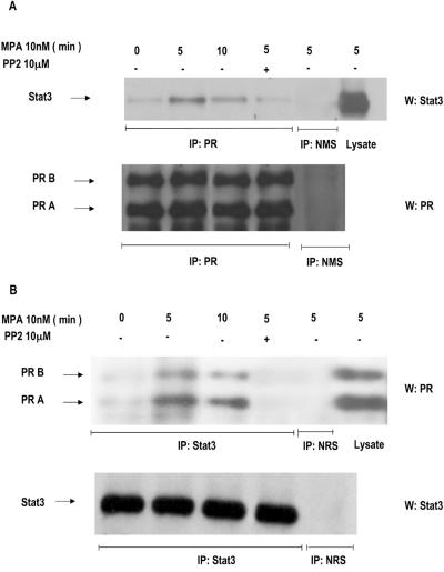
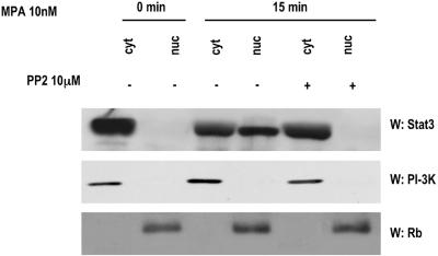
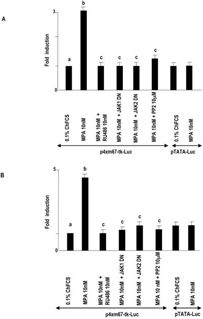
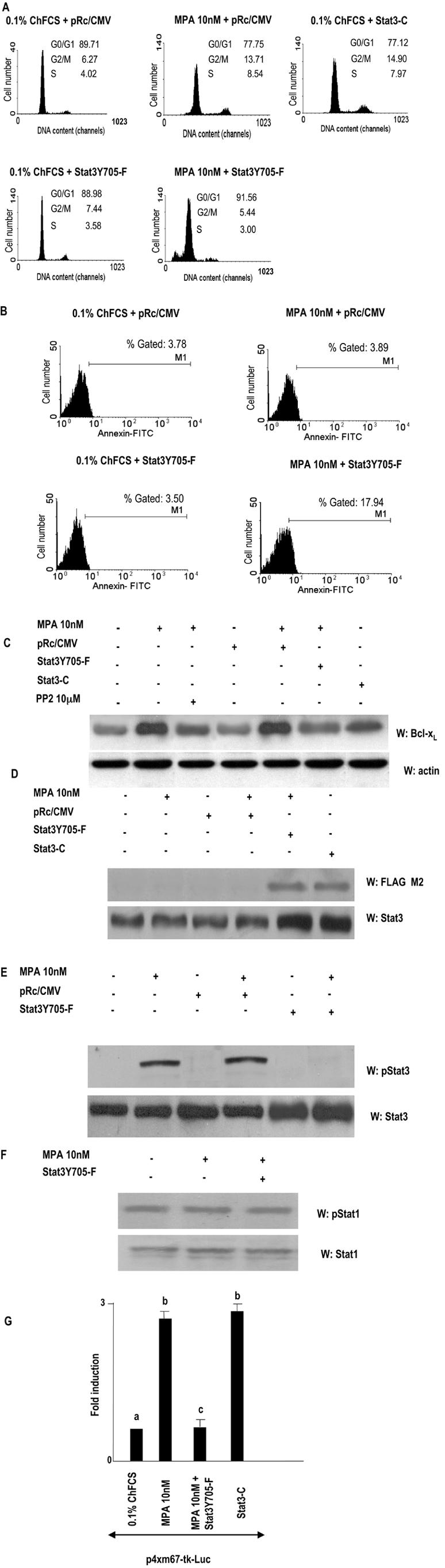
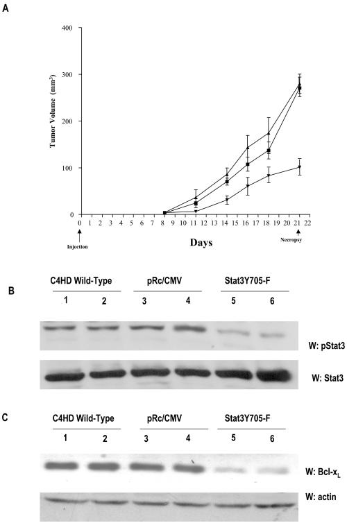
Interactions between steroid hormone receptors and signal transducer and activator of transcription (Stat)-mediated signaling pathways have already been described. In the present study, we explored the capacity of progestins to modulate Stat3 transcriptional activation in an experimental model of hormonal carcinogenesis in which the synthetic progestin medroxyprogesterone acetate (MPA) induced mammary adenocarcinomas in BALB/c mice and in the human breast cancer cell line T47D. We found that C4HD epithelial cells, from the MPA-induced mammary tumor model, expressed Stat3 and that MPA treatment of C4HD cells up-regulated Stat3 protein expression. In addition, MPA induced rapid, nongenomic Stat3, Jak1, and Jak2 tyrosine phosphorylation in C4HD and T47D cells. MPA treatment of C4HD cells also resulted in rapid c-Src tyrosine phosphorylation. These effects were completely abolished by the progestin antagonist RU486. Abrogation of Jak1 and Jak2 activity by transient transfection of C4HD cells with dominant negative (DN) Jak1 or DN Jak2 vectors, or inhibition of Src activity by preincubation of cells with the Src family kinase inhibitor PP2, blocked the capacity of MPA to induce Stat3 phosphorylation. Treatment of C4HD cells with MPA induced Stat3 binding to DNA. In addition, MPA promoted strong Stat3 transcriptional activation in C4HD and T47D cells that was inhibited by RU486 and by blockage of Jak1, Jak2, and Src activities. To investigate the correlation between MPA-induced Stat3 activation and cell growth, C4HD cells were transiently transfected with a DN Stat3 expression vector, Stat3Y705-F, or with a constitutively activated Stat3 mutant, Stat3-C. While expression of Stat3Y705-F mutant had an inhibitory effect on MPA-induced growth of C4HD cells, transfection with the constitutively activated Stat3-C vector resulted in MPA-independent proliferation. Finally, we addressed the effect of targeting Stat3 in in vivo growth of C4HD breast tumors. Blockage of Stat3 activation by transfection of C4HD cells with the DN Stat3Y705-F expression vector significantly inhibited these cells' ability to form tumors in syngeneic mice. Our results have for the first time demonstrated that progestins are able to induce Stat3 transcriptional activation, which is in turn an obligatory requirement for progestin stimulation of both in vitro and in vivo breast cancer growth.


Figures

FIG. 1.
FIG. 1.
MPA up-regulates Stat3 protein expression. Primary cultures of C4HD cells were treated for 48 h in medium with ChFCS supplemented with 10 nM MPA or MPA-10 nM RU486. Fifty micrograms of protein from cell lysates was electrophoresed and immunoblotted for Stat3 and Stat1. A Western blot assay using an antiactin antibody was carried out using identical protein lysates as a control for the specificity of the effect of MPA. This is a representative experiment of a total of four in which the standard error of the mean was within 10%. W, Western blot assay.
FIG. 2.
FIG. 2.
MPA induces tyrosine phosphorylation by Stat3, Jak1, and Jak2. Cultures of C4HD (A) and T47D (B) cells were treated with 10 nM MPA or MPA-10 nM RU486 for the indicated times. Fifty micrograms of protein from cell lysates was electrophoresed, and Western blot assays were performed with antiphosphotyrosine 705 Stat3, antiphosphotyrosine 701 Stat1, antiphosphotyrosine 1022/1023 Jak1, and antiphosphotyrosine 1007/1008 Jak2 antibodies. Membranes were then stripped and hybridized with anti-Stat3, -Stat1, -Jak1, and -Jak2 antibodies. This experiment was repeated six times for C4HD cells and three times for T47D cells with similar results. LM3 (C) or T47D-Y (D) cells were transfected with PRB or with the empty pSG5 plasmidor remained untreated. Cells were then stimulated for 5 min with MPA or pretreated with RU486 before MPA stimulation. LM3 cells were also treated with heregulin for 10 min. Fifty micrograms of protein from cell lysates was electrophoresed, and Western blot assays were performed with antiphosphotyrosine 705 Stat3 (upper parts of panels C and D). Membranes were then stripped and hybridized with anti-Stat3 (middle panes of parts C and D) and anti-PR (lower parts of panels C and D) antibodies. This experiment was repeated three times with similar results. W, Western blot assay.
FIG. 3.
FIG. 3.
Jak1 and Jak2 are involved in MPA-induced Stat3 phosphorylation. C4HD cells were transiently transfected with 2 μg of DN Jak1 or DN Jak2 vector and then treated with MPA for 5 min or left untreated. Fifty micrograms of protein from cell lysates was electrophoresed, and Western blot assays were performed with antiphosphotyrosine Jak1 (A, upper part) or antiphosphotyrosine Jak2 (B, upper part) antibodies. Membranes were then stripped and hybridized with anti-Jak1 (A, lower part) and anti-Jak2 (B, lower part) antibodies. (C) Fifty micrograms of protein from cells transfected with the DN Jak1 (left part) or the DN Jak2 (right part) vector and subsequentlytreated with MPA for 5 min or left untreated was electrophoresed, and Western blot assays were performed with antiphosphotyrosine Stat3 (upper parts). Membranes were then stripped and hybridized with anti-Stat3 (lower parts) antibodies. (D) C4HD cells were treated with MPA for the indicated times or preincubated with the selective Src family kinase inhibitor PP2 or RU486 for 90 min and then treated with MPA. Fifty micrograms of protein from cell lysates was electrophoresed and immunoblotted with an antiphosphotyrosine c-Src antibody (upper part). Membrane was then stripped and hybridized with anti-c-Src antibody (lower part). (E) C4HD cells were preincubated with the selective Src family kinase inhibitor PP2 for 90 min and then treated with MPA for 5 min. Fifty micrograms of protein from cell lysates was electrophoresed and immunoblotted with antiphosphotyrosine Stat3, antiphosphotyrosine Jak1, and antiphosphotyrosine Jak2 antibodies. Membranes were then stripped and hybridized with anti-Stat3, anti-Jak1, and anti-Jak2 antibodies, respectively. These experiments were repeated three times with similar results. W, Western blot assay.
FIG. 4.
FIG. 4.
MPA induces association of Stat3 with PR. (A) C4HD cells were treated with 10 nM MPA for the indicated times or preincubated with PP2 before MPA treatment for 5 min, and PR was immunoprecipitated from 500 μg of protein extracts. As a control, lysates were also immunoprecipitated with normal mouse serum (NMS). Immunocomplexes were subjected to SDS-PAGE and analyzed by Western blotting with an anti-Stat3 antibody (upper part). Twenty micrograms of protein from cell extracts was directly immunoblotted with the Stat3 antibody (last lane, upper part). Identical aliquots of each immunoprecipitate were subjected to immunoblot analysis with anti-PR antibody to verify that nearly equal amounts of immunoprecipitated proteins were loaded (lower part). (B) Protein lysates (500 μg) from cells treated as indicated in panel A were immunoprecipitated with an anti-Stat3 antibody or with normal rabbit serum (NRS). Immunocomplexes were subjected to SDS-PAGE and analyzed by Western blotting with an anti-PR antibody (upper part). Twenty micrograms of protein from cell extracts was directly immunoblotted with the PR antibody (last lane, upper part). Identical aliquots of each immunoprecipitate were subjected to immunoblot analysis with anti-Stat3 antibody to verify that nearly equal amounts of immunoprecipitated proteins were loaded (lower part). This is a representative experiment out of a total of three. W, Western blot assay; IP, immunoprecipitation.
FIG. 5.
FIG. 5.
MPA induces Stat3 nuclear translocation. C4HD cells were treated with 10 nM MPA for the time indicated or were pretreated with PP2 before MPA stimulation. Nuclear (nuc) and cytosolic (cyt) fractions were prepared, and 30 μg of protein from cell extracts was analyzed by Western blot assay for Stat3 expression level. Membranes were then stripped and hybridized with an anti-p85 PI-3K subunit antibody (middle) or an anti-retinoblastoma (Rb) antibody (bottom) in order to control cellular fractionation efficiency. W, Western blot assay.
FIG. 6.
FIG. 6.
MPA induces Stat3 binding to the high-affinity mutant of the SIE from the human c-fos promoter. C4HD cells were treated for 15 min at 37°C with MPA or MPA-RU486 or were left untreated growing in ChFCS. Twenty micrograms of protein from nuclear extracts was incubated for 20 min at room temperature with 1 ng of 32P-labeled double-stranded DNA containing the high-affinity mutant of the SIE from the human c-fos promoter (5′GTGCATTTCCCGTAAATCTTGTCTACA3′) (m67) used as a probe and analyzed by EMSA. The specificity of the Stat3-DNA complexes is shown by competition with 25- and 100-fold mass excesses unlabeled m67 oligonucleotide and by the lack of competition with a 100-fold mass excess of mutant m67 (100xm67 mut). The right panel shows a supershift analysis that was performed by including either anti-Stat3 or anti-Stat1 antibodies. An equivalent amount of preimmune rabbit serum was used as a control in the EMSA reaction mixture (NRS [normal rabbit serum]). This experiment was repeated six times with similar results. wt, wild type.
FIG. 7.
FIG. 7.
MPA induces Stat3 transcriptional activation. C4HD (A) and T47D (B) cells were transiently transfected with 2 μg/well of a luciferase reporter plasmid containing four copies of the m67 high-affinity binding site and with 1 μg/well of a CMV-βgal expression vector as an internal control. In the indicated lanes, C4HD cells were cotransfected with the DN Jak1 and DN Jak2 expression vectors or pretreated with PP2. Cells were also transfected with a pTATA-Luc reporter lacking the m67 insertion. The total amount of transfected DNA was standardized by adding the empty vector. After transfection, cells were treated with MPA and MPA-RU486 at 37°C for 48 h or left untreated growing in ChFCS. C4HD cells were then harvested and lysed. Luciferase and β-galactosidase activities were measured as described in Materials and Methods. Results are presented as n-fold induction of luciferase activity with respect to cells growing in ChFCS. The data shown represent the mean of six independent experiments ± the standard error of the mean. For b versus a and c versus b, P < 0.001.
FIG. 8.
FIG. 8.
Stat3 is involved in MPA-induced proliferation of C4HD cells. (A) C4HD cells were transiently transfected with 2 μg DN Stat3 expression vector, Stat3Y705-F, with 2 μg constitutively activated Stat3 mutant, Stat3-C, or with 2 μg empty pRc/CMV vector, as a control, for 48 h. Cells were treated with MPA for another 48 h or remained untreated and were then stained with PI and analyzed for cell cycle distribution by flow cytometry. The percentages of total cells in the cell cycle phases are indicated. (B) C4HD cells were transiently transfected with 2 μg DN Stat3 expression vector or with 2 μg empty pRc/CMV vector, as a control, for 48 h. Cells were treated as described for panel A, and cell surface Annexin V binding was measured by flow cytometry. (C) Fifty micrograms of protein from lysates of cells transfected with Stat3Y705-F, Stat3-C, and empty pRc/CMV plasmids and from nontransfected cells, treated with MPA for 48 h or left untreated, was electrophoresed, and Western blot assays were performed with an anti Bcl-xL antibody (upper part). Membrane was then stripped and hybridized with an antiactin antibody (lower part). (D) Fifty micrograms of protein from lysates of cells treated as described for panel C and stimulated or not with MPA for 5 min was electrophoresed, and Western blot assays were performed with an anti-FLAG M2 antibody (upper part). Membrane was then stripped and hybridized with an anti-Stat3 antibody (lower part). (E) Fifty micrograms of protein from C4HD cells transfected with 2 μg Stat3Y705-F vector or with empty pRc/CMV plasmid and subsequently left untreated treated or with MPA for 5 min was electrophoresed, and Western blot assays were performed with antiphosphotyrosine Stat3 antibody (upper part). Membranes were then stripped and hybridized with anti-Stat3 antibodies (lower part). (F) Fifty micrograms of protein from C4HD cells transfected with 2 μg Stat3Y705-F vector and then treated with MPA for 5 min or left untreated was electrophoresed, and Western blot assays were performed with antiphosphotyrosine 701 Stat1 antibody (upper part). Membrane was then stripped and hybridized with anti-Stat1 antibody (lower part). Experiments described in panels A to F were repeated three times with similar results. W, Western blot assay. (G) C4HD cells were transiently transfected with 2 μg/well of the m67-Luc reporter plasmid and with 1 μg/well of a CMV-βgal expression vector as an internal control. In the indicated lanes, C4HD cells were cotransfected with either Stat3Y705-F or Stat3-C plasmid. The total amount of transfected DNA was standardized by adding the empty vector. After transfection, cells were treated when indicated with MPA for 48 h and were then harvested and lysed. Luciferase and β-galactosidase activities were measured as described in Materials and Methods. Results are presented as n-fold induction of luciferase activity with respect to cells growing in ChFCS. Data shown represent the mean of two independent experiments ± the standard error of the mean. For b versus a and c versus b, P < 0.001. FITC, fluorescein isothiocyanate.
FIG. 9.
FIG. 9.
In vivo blockage of Stat3 expression. (A) C4HD cells growing in 10 nM MPA were transiently transfected with the DN Stat3Y705-F expression vector (▾) or the empty pRc/CMV vector (▪) or remained untreated (▴), as described in the legend to Fig. 8. After 48 h of transfection, 106 cells from each experimental group were inoculated s.c. into animals treated with a 40-mg MPA depot in the flank opposite the cell inoculum, and tumor width and length were measured three times a week in order to calculate volume as described in Materials and Methods. Each point represents the mean volume ± the standard error of nine independent tumors for each control group and of four tumors that developed in mice injected with Stat3Y705-F-transfected cells. (B) One hundred micrograms of protein from tumor lysates was electrophoresed and immunoblotted with an anti-phospho-Stat3 antibody (upper part). Shown are two representative samples of mice injected with C4HD wild-type cells (lanes 1 and 2), C4HD cells transfected with the empty pRc/CMV vector (lanes 3 and 4), and C4HD cells transfected with the DN Stat3Y705-F expression vector (lanes 5 and 6). Membrane was stripped and hybridized with an anti-Stat3 antibody (lower part). This is a representative experiment out of a total of three. Densitometric analysis of Stat3 phosphorylated bands from the four tumors that developed in mice injected with C4HD cells transfected with the DN Stat3Y705-F vector and from multiple C4HD tumors that developed in mice injected with either wild-type C4HD cells or with C4HD cells transfected with the empty pRc/CMV vector showed a significant decrease in Stat3 tyrosine phosphorylation in tumors from mice injected with cells transfected with the DN Stat3Y705-F vector, with respect to tumors growing in control animals (P < 0.001). W, Western blot assay. (C) One hundred micrograms of protein from tumor lysates was electrophoresed and immunoblotted with an anti-Bcl-xL antibody (upper part). Shown are two representative samples of mice injected with C4HD wild-type cells (lanes 1 and 2), with C4HD cells transfected with the empty pRc/CMV vector (lanes 3 and 4), andwith C4HD cells transfected with the DN Stat3Y705-F expression vector (lanes 5 and 6). Membrane was then stripped and hybridized with an antiactin antibody (lower part), as a control for the specificity of the DN Stat3Y705-F effect. Densitometric analysis of the Bcl-xL band from tumors that developed in mice injected with C4HD cells transfected with the DN Stat3Y705-F, expressed as a percentage of the control values (i.e., tumors growing in control groups), ranged between 25 and 40% for tumors growing in mice injected with DN Stat3Y705-F-transfected cells. There was significant inhibition of Bcl-xL expression in mice injected with DN Stat3Y705-F-transfected cells with respect to mice injected with empty pRc/CMV vector-transfected cells or with wild-type C4HD cells (P < 0.001).

References

    1. Aaronson, D. S., and C. M. Horvath. 2002. A road map for those who don't know JAK-STAT. Science 296:1653-1655. - PubMed
    1. Amin, H. M., T. J. McDonnell, Y. Ma, Q. Lin, Y. Fujio, K. Kunisada, V. Leventaki, P. Das, G. Z. Rassidakis, C. Cutler, L. J. Medeiros, and R. Lai. 2004. Selective inhibition of STAT3 induces apoptosis and G1 cell cycle arrest in ALK-positive anaplastic large cell lymphoma. Oncogene 23:5426-5434. - PubMed
    1. Balana, M. E., L. Labriola, M. Salatino, F. Movsichoff, G. Peters, E. H. Charreau, and P. V. Elizalde. 2001. Activation of ErbB-2 via a hierarchical interaction between ErbB-2 and type I insulin-like growth factor receptor in mammary tumor cells. Oncogene 20:34-47. - PubMed
    1. Balana, M. E., R. Lupu, L. Labriola, E. H. Charreau, and P. V. Elizalde. 1999. Interactions between progestins and heregulin (HRG) signaling pathways: HRG acts as mediator of progestins proliferative effects in mouse mammary adenocarcinomas. Oncogene 18:6370-6379. - PubMed
    1. Ballare, C., M. Uhrig, T. Bechtold, E. Sancho, M. Di Domenico, A. Migliaccio, F. Auricchio, and M. Beato. 2003. Two domains of the progesterone receptor interact with the estrogen receptor and are required for progesterone activation of the c-Src/Erk pathway in mammalian cells. Mol. Cell. Biol. 23:1994-2008. - PMC - PubMed

Publication types

MeSH terms