

MafA is a key regulator of glucose-stimulated insulin secretion
- PMID: 15923615
- PMCID: PMC1140590
- DOI: 10.1128/MCB.25.12.4969-4976.2005
MafA is a key regulator of glucose-stimulated insulin secretion
Abstract
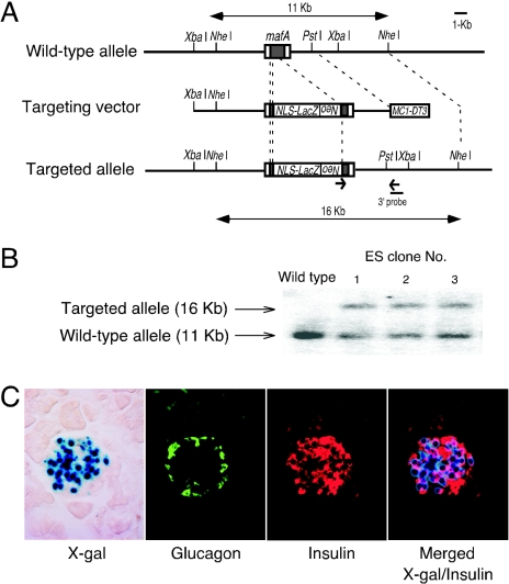
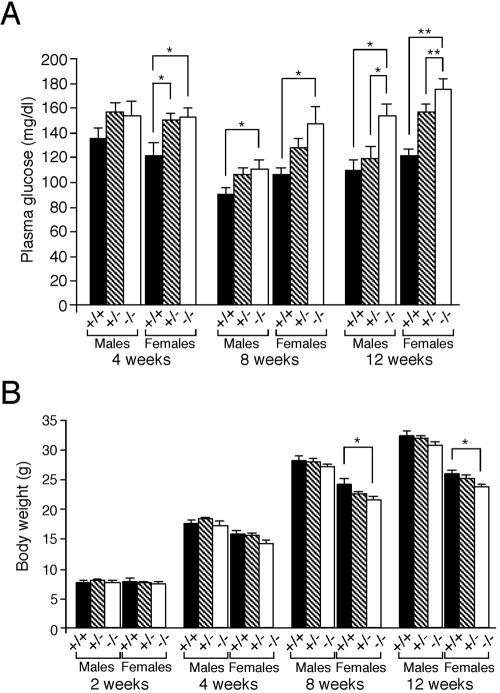
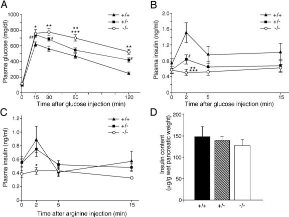
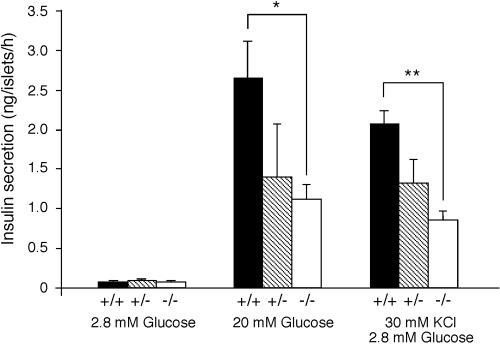
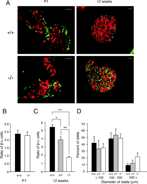
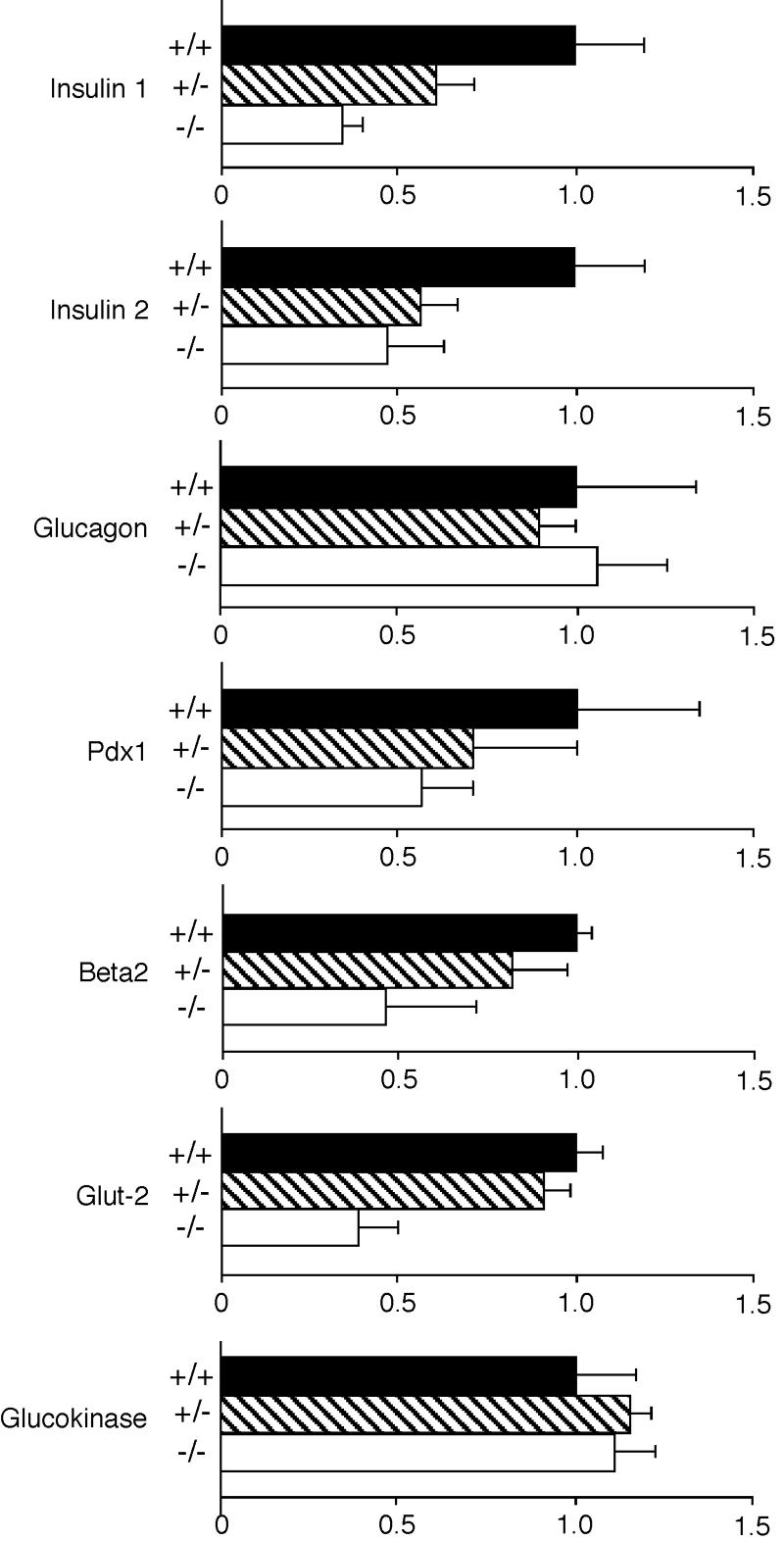
MafA is a transcription factor that binds to the promoter in the insulin gene and has been postulated to regulate insulin transcription in response to serum glucose levels, but there is no current in vivo evidence to support this hypothesis. To analyze the role of MafA in insulin transcription and glucose homeostasis in vivo, we generated MafA-deficient mice. Here we report that MafA mutant mice display intolerance to glucose and develop diabetes mellitus. Detailed analyses revealed that glucose-, arginine-, or KCl-stimulated insulin secretion from pancreatic beta cells is severely impaired, although insulin content per se is not significantly affected. MafA-deficient mice also display age-dependent pancreatic islet abnormalities. Further analysis revealed that insulin 1, insulin 2, Pdx1, Beta2, and Glut-2 transcripts are diminished in MafA-deficient mice. These results show that MafA is a key regulator of glucose-stimulated insulin secretion in vivo.
Figures
Similar articles
-
MafA is a glucose-regulated and pancreatic beta-cell-specific transcriptional activator for the insulin gene.J Biol Chem. 2002 Dec 20;277(51):49903-10. doi: 10.1074/jbc.M206796200. Epub 2002 Oct 3. J Biol Chem. 2002. PMID: 12368292
-
Members of the large Maf transcription family regulate insulin gene transcription in islet beta cells.Mol Cell Biol. 2003 Sep;23(17):6049-62. doi: 10.1128/MCB.23.17.6049-6062.2003. Mol Cell Biol. 2003. PMID: 12917329 Free PMC article.
-
An Inducible Diabetes Mellitus Murine Model Based on MafB Conditional Knockout under MafA-Deficient Condition.Int J Mol Sci. 2020 Aug 5;21(16):5606. doi: 10.3390/ijms21165606. Int J Mol Sci. 2020. PMID: 32764399 Free PMC article.
-
PDX1, Neurogenin-3, and MAFA: critical transcription regulators for beta cell development and regeneration.Stem Cell Res Ther. 2017 Nov 2;8(1):240. doi: 10.1186/s13287-017-0694-z. Stem Cell Res Ther. 2017. PMID: 29096722 Free PMC article. Review.
-
Roles and regulation of transcription factor MafA in islet beta-cells.Endocr J. 2007 Dec;54(5):659-66. doi: 10.1507/endocrj.kr-101. Epub 2007 Aug 30. Endocr J. 2007. PMID: 17785922 Review.
Cited by
-
High-fat diet induced insulin resistance in pregnant rats through pancreatic pax6 signaling pathway.Int J Clin Exp Pathol. 2015 May 1;8(5):5196-202. eCollection 2015. Int J Clin Exp Pathol. 2015. PMID: 26191217 Free PMC article.
-
Role of large MAF transcription factors in the mouse endocrine pancreas.Exp Anim. 2015;64(3):305-12. doi: 10.1538/expanim.15-0001. Epub 2015 Apr 27. Exp Anim. 2015. PMID: 25912440 Free PMC article.
-
GDF15 plays a critical role in insulin secretion in INS-1 cells and human pancreatic islets.Exp Biol Med (Maywood). 2023 Feb;248(4):339-349. doi: 10.1177/15353702221146552. Epub 2023 Feb 5. Exp Biol Med (Maywood). 2023. PMID: 36740767 Free PMC article.
-
Uncovering the role of MAFB in glucagon production and secretion in pancreatic α-cells using a new α-cell-specific Mafb conditional knockout mouse model.Exp Anim. 2020 Apr 24;69(2):178-188. doi: 10.1538/expanim.19-0105. Epub 2019 Dec 2. Exp Anim. 2020. PMID: 31787710 Free PMC article.
-
MafA and MafB regulate genes critical to beta-cells in a unique temporal manner.Diabetes. 2010 Oct;59(10):2530-9. doi: 10.2337/db10-0190. Epub 2010 Jul 13. Diabetes. 2010. PMID: 20627934 Free PMC article.
References
-
- Benkhelifa, S., S. Provot, O. Lecoq, C. Pouponnot, G. Calothy, and M. P. Felder-Schmittbuhl. 1998. mafA, a novel member of the maf proto-oncogene family, displays developmental regulation and mitogenic capacity in avian neuroretina cells. Oncogene 17:247-254. - PubMed
-
- Brissova, M., M. Shiota, W. E. Nicholson, M. Gannon, S. M. Knobel, D. W. Piston, C. V. Wright, and A. C. Powers. 2002. Reduction in pancreatic transcription factor PDX-1 impairs glucose-stimulated insulin secretion. J. Biol. Chem. 277:11225-11232. - PubMed
-
- Furuta, H., Y. Horikawa, N. Iwasaki, M. Hara, L. Sussel, M. M. Le Beau, E. M. Davis, M. Ogata, Y. Iwamoto, M. S. German, and G. I. Bell. 1998. β-Cell transcription factors and diabetes: mutations in the coding region of the BETA2/NeuroD1 (NEUROD1) and Nkx2.2 (NKX2B) genes are not associated with maturity-onset diabetes of the young in Japanese. Diabetes 47:1356-1358. - PubMed
-
- Guillam, M. T., E. Hummler, E. Schaerer, J. I. Yeh, M. J. Birnbaum, F. Beermann, A. Schmidt, N. Deriaz, and B. Thorens. 1997. Early diabetes and abnormal postnatal pancreatic islet development in mice lacking Glut-2. Nat. Genet. 17:327-330. - PubMed
Publication types
MeSH terms
Substances
Grants and funding
LinkOut - more resources
Full Text Sources
Other Literature Sources
Medical
Molecular Biology Databases