. 2005 Jun;25(12):4977-92.
doi: 10.1128/MCB.25.12.4977-4992.2005.

Mechanism of Aurora-B degradation and its dependency on intact KEN and A-boxes: identification of an aneuploidy-promoting property

Affiliations

Mechanism of Aurora-B degradation and its dependency on intact KEN and A-boxes: identification of an aneuploidy-promoting property

Hao G Nguyen et al. Mol Cell Biol. 2005 Jun.

Abstract

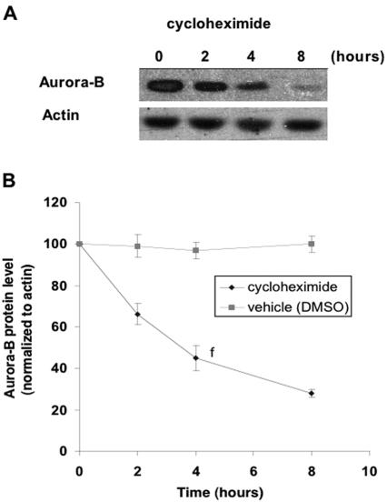
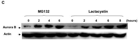
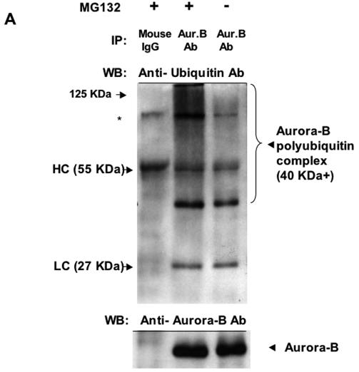
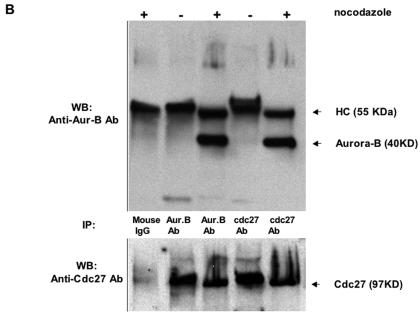
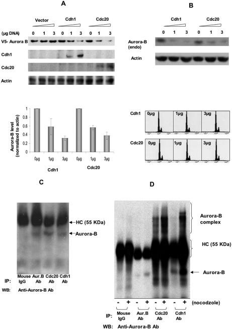
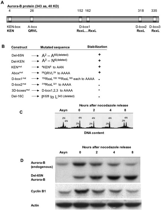
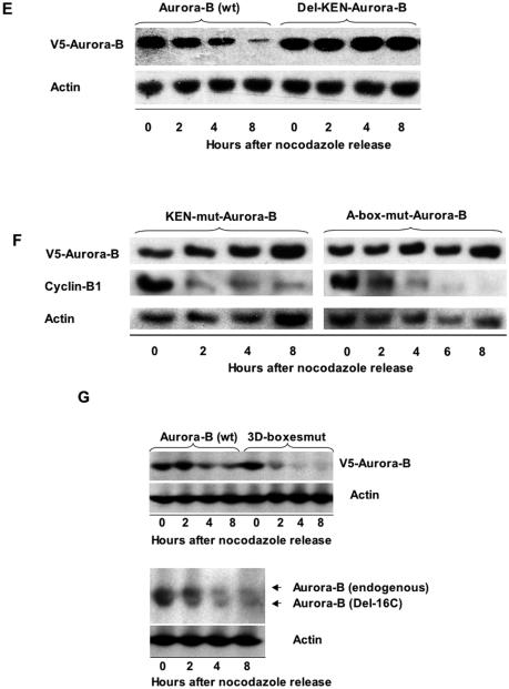
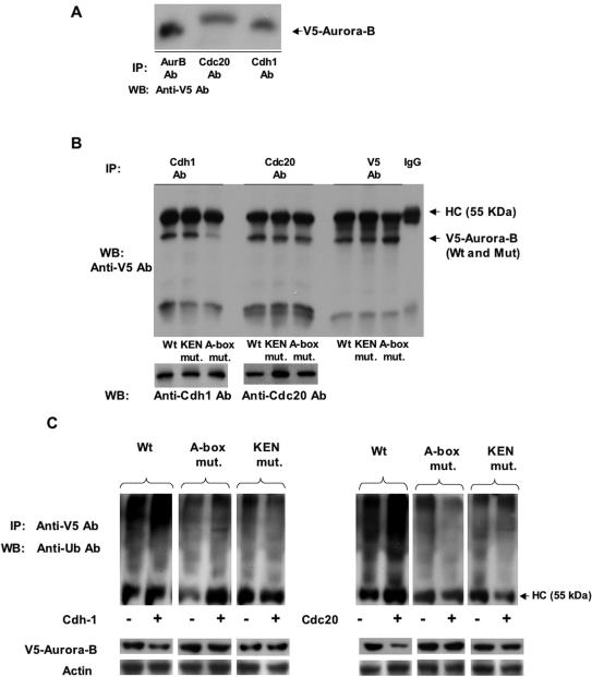
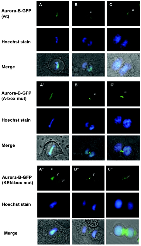
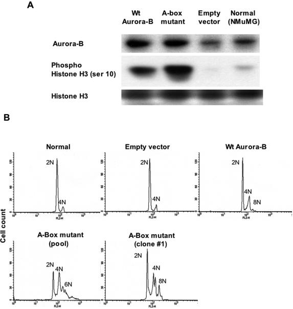
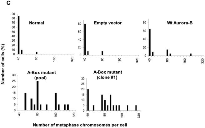
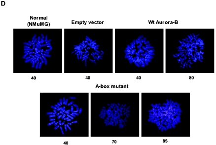
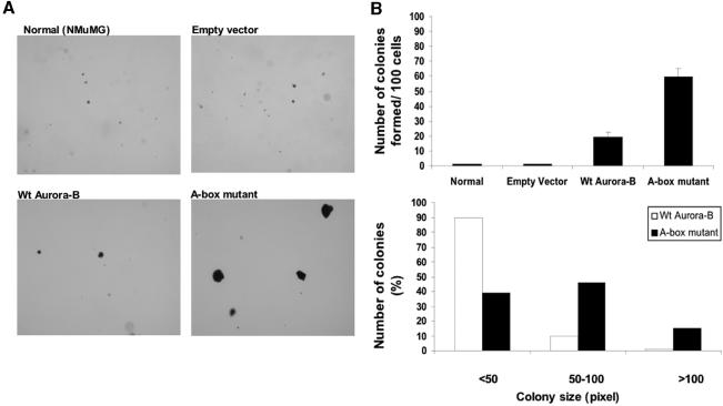
The kinase Aurora-B, a regulator of chromosome segregation and cytokinesis, is highly expressed in a variety of tumors. During the cell cycle, the level of this protein is tightly controlled, and its deregulated abundance is suspected to contribute to aneuploidy. Here, we provide evidence that Aurora-B is a short-lived protein degraded by the proteasome via the anaphase-promoting cyclosome complex (APC/c) pathway. Aurora-B interacts with the APC/c through the Cdc27 subunit, Aurora-B is ubiquitinated, and its level is increased upon treatment with inhibitors of the proteasome. Aurora-B binds in vivo to the degradation-targeting proteins Cdh1 and Cdc20, the overexpression of which accelerates Aurora-B degradation. Using deletions or point mutations of the five putative degradation signals in Aurora-B, we show that degradation of this protein does not depend on its D-boxes (RXXL), but it does require intact KEN boxes and A-boxes (QRVL) located within the first 65 amino acids. Cells transfected with wild-type or A-box-mutated or KEN box-mutated Aurora-B fused to green fluorescent protein display the protein localized to the chromosomes and then to the midzone during mitosis, but the mutated forms are detected at greater intensities. Hence, we identified the degradation pathway for Aurora-B as well as critical regions for its clearance. Intriguingly, overexpression of a stable form of Aurora-B alone induces aneuploidy and anchorage-independent growth.


Figures

FIG. 1.
FIG. 1.
Aurora-B has a short half-life and accumulates in the presence of proteasome inhibitors. A. HeLa cells were treated with 250 μg/ml cycloheximide and collected at the indicated times for Western blot analysis. Proteins (50 μg/lane) were loaded onto an SDS-polyacrylamide gel, transferred to a filter, and incubated with anti-Aurora-B antibody (showing a 40-kDa protein). β-Actin level was also blotted for a loading control (43-kDa protein). B. To quantitatively evaluate the half-life of Aurora-B, its protein level, as determined by Western blotting, was normalized to β-actin level in five independent experiments. Averages shown were calculated using the NIH Image V1.62 software (freeware from the National Institutes of Health). Error bars represent standard deviations. C. HeLa cells were treated for different lengths of time with a proteasome inhibitor, MG132 (20 μM) or lactasystin (10 μM). The cells were lysed and subjected to Western blot analysis with anti-Aurora-B and -β-actin antibodies. Results shown are representative of five independent experiments.
FIG. 1.
FIG. 1.
Aurora-B has a short half-life and accumulates in the presence of proteasome inhibitors. A. HeLa cells were treated with 250 μg/ml cycloheximide and collected at the indicated times for Western blot analysis. Proteins (50 μg/lane) were loaded onto an SDS-polyacrylamide gel, transferred to a filter, and incubated with anti-Aurora-B antibody (showing a 40-kDa protein). β-Actin level was also blotted for a loading control (43-kDa protein). B. To quantitatively evaluate the half-life of Aurora-B, its protein level, as determined by Western blotting, was normalized to β-actin level in five independent experiments. Averages shown were calculated using the NIH Image V1.62 software (freeware from the National Institutes of Health). Error bars represent standard deviations. C. HeLa cells were treated for different lengths of time with a proteasome inhibitor, MG132 (20 μM) or lactasystin (10 μM). The cells were lysed and subjected to Western blot analysis with anti-Aurora-B and -β-actin antibodies. Results shown are representative of five independent experiments.
FIG. 2.
FIG. 2.
Aurora-B is ubiquitinated and capable of binding in vivo to the APC component Cdc27. A. HeLa cells were incubated with (+) or without (−) MG132 (20 μM) for 6 h, lysed, and collected for immunoprecipitation (IP) with anti-Aurora-B antibody (Aur.B Ab) or with mouse IgG as a control. Equal amounts of immunoprotein complex were loaded and resolved by SDS-PAGE. To detect the presence of ubiquitin in the Aurora-B-immunocomplex, antiubiquitin monoclonal antibody was used in the Western blot (WB) analysis shown in the top panel. Polyubiquitinated Aurora-B appears in multiple forms with a molecular mass range greater than 40 kDa. The positions of heavy-chain (HC) and light-chain (LC) mouse IgG and a nonspecific band(which also appears with mouse IgG) (asterisk) are also indicated. The presence of Aurora-B in the immunocomplex was further verified by blotting the same membrane with anti-Aurora-B antibody, as shown in the lower panel. B. To test whether Aurora-B can bind directly to the APC/c, HeLa cells were arrested at M phase with nocodazole (40 ng/ml) for 16 h, lysed, and subjected to IP with anti-Cdc27 or anti-Aurora-B mouse monoclonal antibodies. IP with normal mouse IgG was also performed as a negative control. The same blot was reprobed with anti-Cdc27 to show the efficiency of Cdc27 immunoprecipitation. As expected, the heavy-chain (HC) mouse IgG showed as a 55-kDa protein. The data shown are representative of two experiments. The Aurora-B-Cdc27 immunocomplex was present in synchronized mitotic cells but was scarce in asynchronized cells, as attested by Western blotting and reaction with anti-Aurora B.
FIG. 2.
FIG. 2.
Aurora-B is ubiquitinated and capable of binding in vivo to the APC component Cdc27. A. HeLa cells were incubated with (+) or without (−) MG132 (20 μM) for 6 h, lysed, and collected for immunoprecipitation (IP) with anti-Aurora-B antibody (Aur.B Ab) or with mouse IgG as a control. Equal amounts of immunoprotein complex were loaded and resolved by SDS-PAGE. To detect the presence of ubiquitin in the Aurora-B-immunocomplex, antiubiquitin monoclonal antibody was used in the Western blot (WB) analysis shown in the top panel. Polyubiquitinated Aurora-B appears in multiple forms with a molecular mass range greater than 40 kDa. The positions of heavy-chain (HC) and light-chain (LC) mouse IgG and a nonspecific band(which also appears with mouse IgG) (asterisk) are also indicated. The presence of Aurora-B in the immunocomplex was further verified by blotting the same membrane with anti-Aurora-B antibody, as shown in the lower panel. B. To test whether Aurora-B can bind directly to the APC/c, HeLa cells were arrested at M phase with nocodazole (40 ng/ml) for 16 h, lysed, and subjected to IP with anti-Cdc27 or anti-Aurora-B mouse monoclonal antibodies. IP with normal mouse IgG was also performed as a negative control. The same blot was reprobed with anti-Cdc27 to show the efficiency of Cdc27 immunoprecipitation. As expected, the heavy-chain (HC) mouse IgG showed as a 55-kDa protein. The data shown are representative of two experiments. The Aurora-B-Cdc27 immunocomplex was present in synchronized mitotic cells but was scarce in asynchronized cells, as attested by Western blotting and reaction with anti-Aurora B.
FIG. 3.
FIG. 3.
Aurora-B protein level decreases in cells transiently transfected with the APC/c modulator protein Cdh1 or Cdc20. A. HeLa cells were transiently transfected with different concentrations (1 to 3 μg DNA as shown) of hCdh1 or hCdc20 expression vectors. Mock transfections were also pursued with empty vector. Cells were lysed after 48 h of incubation and evaluated for Aurora-B (40-kDa), Cdh1 (55.2-kDa), and Cdc20 (54.7-kDa) protein levels by Western blot analysis with appropriate antibodies. The Western blot was also probed with anti-β-actin (43 kDa) for a loading control. The Western blots at the top of the figure show the results of a representative experiment involving detection of transfected protein, and the graph shows quantification (as in Fig. 1) of three experiments (averages ± standard deviations). B. (Top panel) Similarly, endogenous (endo) Aurora-B too, followed by Western blot analysis with anti-Aurora-B, was also found to be reduced upon Cdc20 or Cdh1 upregulation. Reaction with anti-β-actin served as a loading control. Data shown are representative of two experiments. The bottom graphs show fluorescence-activated cell sorting analysis with increasing ectopic expression of Cdh1 or Cdc20. C. Asynchronized HeLa cell extracts were subjected to immunoprecipitation (IP) with equal amounts of control mouse IgG, anti-Aurora-B (Aur.B Ab), anti-Cdh1, or anti-Cdc20 antibody and resolved by SDS-PAGE. The immunoprecipitate was subjected to Western blotting (WB) with anti-Aurora-B antibody. D. To show the binding between Aurora-B and the APC/c modulator protein, HeLa cells were synchronized with nocodazole as described for Fig. 2B or nonsynchronized, followed by IP with anti-Aurora-B (Aur.B Ab) or anti-Cdc20 or anti-Cdc20 or as a control with mouse IgG. The immunoprecipitate was subjected to SDS-PAGE and Western blotting with anti-Aurora-B. The positions of Aurora-B as a single band or as a high-molecular-mass complex, as well as the 55-kDa immunoglobulin heavy chains (HC), are shown. Results shown are representative of three experiments.
FIG. 4.
FIG. 4.
Stability of wild-type versus mutated Aurora-B. A. A schematic presentation of Aurora-B (a 343-amino-acid, 40-kDa protein) denoting the five putative degradation signals, including three D-boxes (RXXL) within the C terminus, one KEN box, and an A-box. These degradation sequences are conserved in rat (GenBank accession number BAA23794), mouse (GenBank accession number XP_181344), human (GenBank access number AAH09751), and Xenopus laevis (GenBank accession number AAM76715) Aurora-B proteins. The numbers on the top of the scheme denote the amino acid number at which the consensus sequence starts in relation to the start codon. The conserved amino acids are indicated below the bar in bold print, and the nonconserved ones are denoted by x. B. Summary of different deletions and mutations in Aurora-B and consequences on Aurora-B protein stability. Its stability was examined as illustrated in Fig. 4C to G. C. HeLa cells were arrested at M phase by nocodazole (40 ng/ml) treatment for 16 h, followed by washing off the drug and cell collection at different time points postrelease from synchrony for flow cytometry analysis (A) or for Western blotting (B-D). For flow cytometry analysis, the cells were first stained with PI as detailed in Materials and Methods. Asynchronous (Asyn) cells were not treated with nocodazole. Results shown are from a representative experiment. D. HeLa cells were transfected with Aurora-B construct deleted of the first 65 amino acids (Del-65N Aurora-B). Two days posttransfection, the cells were synchronized and then released from synchrony as in panel A. Cells collected at different time points were subjected to Western blot analysis using an antibody to Aurora-B, which recognizes on the same blot the endogenous and transfected proteins, with the latter being of a lowermolecular weight. Reaction with anti-cyclin B1 served as an additional mitotic marker, while anti-β-actin confirmed equal loading of protein. E and F. HeLa cells were transiently transfected with wild-type (wt) Aurora-B construct or Aurora-B construct deleted of the KEN box (Del-KEN-Aurora-B) or with mutated KEN (to AAN) (KEN-mut-Aurora-B) or with mutated A-box (QRVL to AAAA) (A-box-mut-Aurora-B), as indicated. Each of these constructs is tagged at the 5′ end with V5. Two days posttransfection, cells were synchronized and released from synchrony as in panel A and collected for Western blotting with anti-V5 or anti-cyclin B1 or antiactin (control). Results shown are representative of five experiments. G. The top two blots show the results of a similar assay with HeLa cells transiently transfected with wild-type Aurora-B or 3D-boxesmut (in which the three D-boxes were mutated). The bottom two blots shows HeLa cells transfected with Del-16C mutant. This mutant migrated at a lower molecular weight than endogenous Aurora-B, as the last 16 amino acids at the C terminus were truncated.
FIG. 4.
FIG. 4.
Stability of wild-type versus mutated Aurora-B. A. A schematic presentation of Aurora-B (a 343-amino-acid, 40-kDa protein) denoting the five putative degradation signals, including three D-boxes (RXXL) within the C terminus, one KEN box, and an A-box. These degradation sequences are conserved in rat (GenBank accession number BAA23794), mouse (GenBank accession number XP_181344), human (GenBank access number AAH09751), and Xenopus laevis (GenBank accession number AAM76715) Aurora-B proteins. The numbers on the top of the scheme denote the amino acid number at which the consensus sequence starts in relation to the start codon. The conserved amino acids are indicated below the bar in bold print, and the nonconserved ones are denoted by x. B. Summary of different deletions and mutations in Aurora-B and consequences on Aurora-B protein stability. Its stability was examined as illustrated in Fig. 4C to G. C. HeLa cells were arrested at M phase by nocodazole (40 ng/ml) treatment for 16 h, followed by washing off the drug and cell collection at different time points postrelease from synchrony for flow cytometry analysis (A) or for Western blotting (B-D). For flow cytometry analysis, the cells were first stained with PI as detailed in Materials and Methods. Asynchronous (Asyn) cells were not treated with nocodazole. Results shown are from a representative experiment. D. HeLa cells were transfected with Aurora-B construct deleted of the first 65 amino acids (Del-65N Aurora-B). Two days posttransfection, the cells were synchronized and then released from synchrony as in panel A. Cells collected at different time points were subjected to Western blot analysis using an antibody to Aurora-B, which recognizes on the same blot the endogenous and transfected proteins, with the latter being of a lowermolecular weight. Reaction with anti-cyclin B1 served as an additional mitotic marker, while anti-β-actin confirmed equal loading of protein. E and F. HeLa cells were transiently transfected with wild-type (wt) Aurora-B construct or Aurora-B construct deleted of the KEN box (Del-KEN-Aurora-B) or with mutated KEN (to AAN) (KEN-mut-Aurora-B) or with mutated A-box (QRVL to AAAA) (A-box-mut-Aurora-B), as indicated. Each of these constructs is tagged at the 5′ end with V5. Two days posttransfection, cells were synchronized and released from synchrony as in panel A and collected for Western blotting with anti-V5 or anti-cyclin B1 or antiactin (control). Results shown are representative of five experiments. G. The top two blots show the results of a similar assay with HeLa cells transiently transfected with wild-type Aurora-B or 3D-boxesmut (in which the three D-boxes were mutated). The bottom two blots shows HeLa cells transfected with Del-16C mutant. This mutant migrated at a lower molecular weight than endogenous Aurora-B, as the last 16 amino acids at the C terminus were truncated.
FIG. 5.
FIG. 5.
Mutation of KEN box or A-box inhibits Aurora-B polyubiquitination. A. HeLa cell extracts with ectopic expression of wild-type Aurora-B (V5 tagged) were subjected to immunoprecipitation (IP) with anti-Aurora-B (AurB Ab), anti-Ccd20, or anti-Cdh1 antibody and probed with anti-V5 antibody. WB, Western blotting. B. To show the interaction/lack of interaction of Aurora-B mutant with Cdh1 and Cdc20, an IP assay similar to the IP assay in panel A was performed, using HeLa cell extracts with ectopic expression of wild-type (Wt) and mutated (mut.) Aurora-B. HC, heavy-chain IgG. C. HeLa cells were transiently cotransfected with equal amounts of wild-type V5-Aurora-B, or A-box-mut-Aurora-B or KEN-box-mut-Aurora-B constructs, each together with Cdh-1 or Cdc20 or empty vector, and isolated protein extracts were subjected to immunoprecipitation (IP) with anti-V5 antibody. The immunocomplexes were resolved by SDS-PAGE and Western blotted (WB) with antiubiquitin antibody (Anti-Ub Ab). Samples overexpressing wild-type V5-Aurora-B displayed Aurora-B-polyubiquitin complex (heavy-chain IgG [HC], 55 kDa) as a smear pattern. However, this pattern was significantly reduced in samples containing mutant forms of Aurora-B. To confirm similar expression of Aurora protein (wild type or mutated) derived from the different constructs, protein extracts were also used in Western blot analysis with anti-V5 (bottom panel). Results shown are representative of three experiments.
FIG. 6.
FIG. 6.
A-box Aurora-B or KEN box-mutated Aurora-B accumulates in the midbody zone. HeLa cells were transiently transfected with equal concentrations of wild-type (wt) Aurora-B-GFP construct (panels A to C) or mutant Aurora-B-GFP, containing AAAA instead of QRVL in the A-box (panels A′ to C′). Cells were stained with 1 μM Hoechst stain and visualized using a fluorescence microscope (Olympus IX70, 100× objective) to view GFP (green) or Hoechst stain (blue) or the fluorescence images merged with a phase-contrast microscope. Pictures shown are representative of at least 10 images taken at each phase of the cell cycle, guided by the presence or lack of chromosome condensation viewed viaHoechst staining. Starting at metaphase, both wild-type and A-box-mutated Aurora-B-GFP are localized on chromosomes (A and A′). They are then clearly viewed at the midzone (indicated by arrow) during anaphase (B and B′). In late cytokinesis (C and C′), wild-type Aurora-B-GFP dissociates from the midbody and it reaches undetectable levels. In contrast, mutant Aurora-B-GFP remains at the midzone (B′) and persists there. Consequently, the frequency of cells captured at late anaphase (likely arrested) was much greater (at least threefold) in cultures transfected with the mutated construct. Cells were also transfected with a KEN box-mutated Aurora-B-GFP construct (panels A", B", and C" cells at metaphase, late anaphase, and cytokinesis, respectively). A greater abundance of Aurora-B-GFP in the midbody zone, compared with that of the wild-type construct, was evident; however, a modest level was also localized in the cytosol (indicated by the three arrows in panel A").
FIG. 7.
FIG. 7.
A-box mutated Aurora-B increases mitotic histone H3-Ser10 phosphorylation and contributes to chromosome number instability. A. Shown is a representative Western blot analysis of cellular extract prepared from NMuMG stably transfected with empty vector or with wild-type (Wt) or A-box-mutated Aurora-B (both forms have GFP linked to the N terminus), using anti-Aurora-B. Phosphorylated (Phospho) histone H3 (Ser10) level in the stable transfectants was evaluated by blotting with phosphor-S10-H3-specific antibody. Longer exposure of the blots revealed a ban in the vector-transfected or control samples as well. Total histone H3 levels were also evaluated to verify equal loading of samples. Normalization based on the latter showed that these wild-type or mutated Aurora-B clones (which express the protein at levels similar to those shown here) display comparable levels of histone H3 phosphorylation. B. Fluorescence-activated cell sorting (FACS) analysis showing the ploidy state in NMuMG (Normal), empty vector-, wild-type (Wt)- and mutated Aurora-B-stable transfectants. To show clonal variation, FACS analysis was also performed on a population of cells (clone 1) derived from a single cell via limited dilution. C. The effect of nondegradable Aurora-B on chromosome number instability was also evaluated by chromosome analysis (100 cells analyzed per each sample) and tabulated according to the number of metaphase chromosomes. NMuMG stably expressed with empty vector or with wild-type and A-box mutated Aurora-B were treated with Colcemid (15 ng/ml) for 18 h, chromosomes were prepared by acetic acid-methanol fixation, stained with DAPI, and visualized with a fluorescence microscope with a 1,000× objective as detailed in Materials and Methods. D. Typical metaphase chromosome spreads in NMuMG (Normal),and cells stably expressing empty vector or wild-type (Wt) and A-box-mutated Aurora-B. The numbers of chromosomes (40 for diploid cells) are indicated below the images.
FIG. 7.
FIG. 7.
A-box mutated Aurora-B increases mitotic histone H3-Ser10 phosphorylation and contributes to chromosome number instability. A. Shown is a representative Western blot analysis of cellular extract prepared from NMuMG stably transfected with empty vector or with wild-type (Wt) or A-box-mutated Aurora-B (both forms have GFP linked to the N terminus), using anti-Aurora-B. Phosphorylated (Phospho) histone H3 (Ser10) level in the stable transfectants was evaluated by blotting with phosphor-S10-H3-specific antibody. Longer exposure of the blots revealed a ban in the vector-transfected or control samples as well. Total histone H3 levels were also evaluated to verify equal loading of samples. Normalization based on the latter showed that these wild-type or mutated Aurora-B clones (which express the protein at levels similar to those shown here) display comparable levels of histone H3 phosphorylation. B. Fluorescence-activated cell sorting (FACS) analysis showing the ploidy state in NMuMG (Normal), empty vector-, wild-type (Wt)- and mutated Aurora-B-stable transfectants. To show clonal variation, FACS analysis was also performed on a population of cells (clone 1) derived from a single cell via limited dilution. C. The effect of nondegradable Aurora-B on chromosome number instability was also evaluated by chromosome analysis (100 cells analyzed per each sample) and tabulated according to the number of metaphase chromosomes. NMuMG stably expressed with empty vector or with wild-type and A-box mutated Aurora-B were treated with Colcemid (15 ng/ml) for 18 h, chromosomes were prepared by acetic acid-methanol fixation, stained with DAPI, and visualized with a fluorescence microscope with a 1,000× objective as detailed in Materials and Methods. D. Typical metaphase chromosome spreads in NMuMG (Normal),and cells stably expressing empty vector or wild-type (Wt) and A-box-mutated Aurora-B. The numbers of chromosomes (40 for diploid cells) are indicated below the images.
FIG. 8.
FIG. 8.
Nondegradable Aurora-B mutant promotes anchorage-independent growth in soft agar. A. To study the cellular functions of nondegradable Aurora-B, we used NMuMG (Normal) engineered to stably overexpress wild-type (Wt) or nondegradable Aurora-B mutant. NMuMG were plated in soft agar as described in Materials and Methods. Pictures shown are typical light-phase fields viewed with a 4× objective. B. Colony numbers were determined by scoring the ones with a diameter greater than 35 pixels, representing three cells (using digital image by OpenLab software in conjunction with Olympus IX70 fluorescence microscope) (top panel). The bottom panel displays colony number segregated based on colony size, comparing mammary epithelial cells stably expressing wild-type (Wt) and A-box-mutated Aurora-B. All experiments were repeated three times.

Similar articles

Cited by

References

    1. Adams, R. R., M. Carmena, and W. C. Earnshaw. 2001. Chromosomal passengers and the (aurora) ABCs of mitosis. Trends Cell Biol. 11:49-54. - PubMed
    1. Brandeis, M., and T. Hunt. 1996. The proteolysis of mitotic cyclins in mammalian cells persists from the end of mitosis until the onset of S phase. EMBO J. 15:5280-5289. - PMC - PubMed
    1. Castro, A., Y. Arlot-Bonnemains, S. Vigneron, J. C. Labbe, C. Prigent, and T. Lorca. 2002. APC/Fizzy-related targets Aurora-A kinase for proteolysis. EMBO Rep. 3:457-462. - PMC - PubMed
    1. Castro, A., S. Vigneron, C. Bernis, J. C. Labbe, and T. Lorca. 2003. Xkid is degraded in a D-box, KEN-box, and A-box-independent pathway. Mol. Cell. Biol. 23:4126-4138. - PMC - PubMed
    1. Castro, A., S. Vigneron, C. Bernis, J. C. Labbe, C. Prigent, and T. Lorca. 2002. The D-Box-activating domain (DAD) is a new proteolysis signal that stimulates the silent D-Box sequence of Aurora-A. EMBO Rep. 3:1209-1214. - PMC - PubMed

Publication types

MeSH terms

Substances

LinkOut - more resources