. 2005 Jun;25(12):5040-51.
doi: 10.1128/MCB.25.12.5040-5051.2005.

DNA topoisomerase I is a cofactor for c-Jun in the regulation of epidermal growth factor receptor expression and cancer cell proliferation

Affiliations

DNA topoisomerase I is a cofactor for c-Jun in the regulation of epidermal growth factor receptor expression and cancer cell proliferation

Antoine Mialon et al. Mol Cell Biol. 2005 Jun.

Abstract

DNA topoisomerase I (Topo I) is a molecular target for the anticancer agent topotecan in the treatment of small cell lung cancer and ovarian carcinomas. However, the molecular mechanisms by which topotecan treatment inhibits cancer cell proliferation are unclear. We describe here the identification of Topo I as a novel endogenous interaction partner for transcription factor c-Jun. Reciprocal coimmunoprecipitation analysis showed that Topo I and c-Jun interact in transformed human cells in a manner that is dependent on JNK activity. c-Jun target gene epidermal growth factor receptor (EGFR) was identified as a novel gene whose expression was specifically inhibited by topotecan. Moreover, Topo I overexpression supported c-Jun-mediated reporter gene activation and both genetic and chemical inhibition of c-Jun converted cells resistant to topotecan-elicited EGFR downregulation. Topotecan-elicited suppression of proliferation was rescued by exogenously expressed EGFR. Furthermore, we demonstrate the cooperation of the JNK-c-Jun pathway, Topo I, and EGFR in the positive regulation of HT-1080 cell proliferation. Together, these results have identified transcriptional coactivator Topo I as a first endogenous cofactor for c-Jun in the regulation of cell proliferation. In addition, the results of the present study strongly suggest that inhibition of EGFR expression is a novel mechanism by which topotecan inhibits cell proliferation in cancer therapy.


Figures
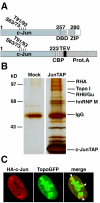
FIG. 1.
FIG. 1.
Identification of novel c-Jun-interacting protein complex. (A) Structure of c-Jun and c-Jun1-223TAP fusion proteins. DBD, DNA-binding domain; ZIP, leucine zipper; CBP, calmodulin binding peptide; TEV, tobacco etch virus protease cleavage site. (B) HEK293 cells were transiently transfected with an mammalian expression construct coding for Jun1-223TAP fusion protein or left untransfected (Mock). At 24 h after transfection, nuclear proteins from both cell cultures were subjected to tandem affinity purification, and proteins in the final eluates were analyzed by sodium dodecyl sulfate-polyacrylamide gel electrophoresis and silver staining. The indicated proteins from Jun1-23TAP eluates were identified thereafter by tandem mass spectrometric analysis. (C) c-Jun and Topo I colocalize in the perinucleolar region. HT-1080 cells were transiently transfected with HA-c-Jun and TopoGFP expression constructs. After 24 h cells were fixed and stained for HA epitope, and the colocalization of proteins was then analyzed by confocal microscopy. The white arrowheads in the merged panel indicate sites for colocalization between c-Jun and Topo I.
FIG. 2.
FIG. 2.
Topo I is a novel endogenous interaction partner for c-Jun. (A and B) HT-1080 cell nuclear lysates were subjected to immunoprecipitation (IP) with either c-Jun goat antibody and preimmune goat sera (PI) (A) or with c-Jun rabbit antibody and GST antibody (B). After washing, immunoprecipitates and 5% of the nuclear proteins (NP 5%) used as a starting material for the assay were analyzed by Western blotting with the indicated antibodies. (C) Reciprocal immunoprecipitation analysis of c-Jun-Topo I interaction. HT-1080 cells were transiently transfected with GFP or TopoGFP expression construct. At 24 h after transfection, nuclear proteins of each samples were subjected to immunoprecipitation analysis by GFP antibody. Immunoprecipitates and 5% of the nuclear proteins (NP 5%) used as a starting material were analyzed for endogenous c-Jun and for TopoGFP by Western blotting. (D) Topo I preferentially interacts with phosphorylated c-Jun. HT-1080 cells were pretreated with JNK inhibitor SP600125 for 2 h, and the interaction between endogenous c-Jun and Topo I was studied by coimmunoprecipitation with c-Jun rabbit antibody. Immunoprecipitates were analyzed for endogenous total and phosphorylated c-Jun and for Topo I by Western blotting with the indicated antibodies. (E) HT-1080 cells were transiently transfected with HAc-Junwt, with HAc-JunAla, or with empty expression plasmid (Mock). At 24 h after the transfections, cellular lysates were subjected to coimmunoprecipitation analyses with HA antibody. Immunoprecipitates (IP) and 5% of the nuclear proteins (NP 5%) used as a starting material were analyzed for endogenous Topo I and for HA-tagged c-Jun proteins by Western blotting. (A to E) All panels show representative examples of two or three experiments with similar results. (F) HT-1080 cells were transiently transfected with 5xGal luciferase reporter plasmid, JunGal, or DBDGal expression constructs, together with the indicated amounts of pEGFP or TopoGFP constructs. The luciferase activity in cell lysates was measured 24 h after transfection. Transfection efficiency was monitored by cotransfecting the cell with Ubi-Renilla luciferase construct. Shown are mean values + the SD of three experiments done with four parallel samples.
FIG. 3.
FIG. 3.
Topo I activity is selectively required for EGFR expression (A and B) HT-1080 cells were transiently transfected with dominant-negative c-Jun expression construct (A) or with c-Jun specific siRNA construct (B). Expression levels of His-tagged recombinant c-Jun (His), endogenous c-Jun, EGFR, and β-actin proteins were studied 24 h (A) or 72 h (B) after transfection by Western blotting. (C) Concentration-dependent inhibition of EGFR expression by topotecan. HT-1080 cells were treated with indicated concentrations of topotecan for 24 h, and the expression levels of EGFR and β-actin were studied by Western blotting. The relative expression levels of EGFR are indicated below the actin panel. Quantitation has been done by MCID imaging analysis software and correlated to actin expression from the same sample. (D) HT-1080 cells were treated with topotecan (1 μM) for 12 h, and the amounts of total and phosphorylated EGFR were determined by Western blotting with EGFR and p-EGFR antibodies. (E and F) HT-1080 cells were treated with topotecan (1 μM) for 12 h, and the expression levels of the indicated proteins were determined by Western blotting. (A to F) All panels show representative examples of two or three experiments with similar results.
FIG. 4.
FIG. 4.
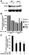
Inhibition of EGFR expression by topotecan is dependent on c-Jun. (A) MEFs lacking c-Jun (Jun−/−) and their wild-type counterparts (WT) were treated with topotecan (1 μM) for 12 h, and the expression levels of EGFR and actin were studied by Western blotting. (B) Quantification of the EGFR protein expression levels in Jun−/− and WT cells in response to topotecan treatment. Quantitation has been done by MCID imaging analysis software and correlated to actin expression from the same sample. Shown are actual values for three separate experiments and mean values + the SD of these experiments taken together. (C) Inhibition of JNK activity converts HT-1080 cells resistant to topotecan. HT-1080 cells were pretreated for 2 h with SP600125 and treated thereafter with topotecan. After 12 h the expression levels of EGFR and actin were determined by Western blotting. Quantitation of EGFR expression was done by MCID imaging analysis software and correlated to actin expression from the same sample. Mean values + the SD of two separate experiments done in duplicate are shown.
FIG. 5.
FIG. 5.
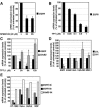
Topo I promotes EGFR gene expression at the mRNA level in both transformed and nontransformed cells. (A) JNK pathway stimulates EGFR mRNA expression. HT-1080 cells were treated with chemical JNK inhibitor SP600125 for 3 and 6 h, and the EGFR mRNA expression levels were determined by real-time PCR analysis. (B) Low micromolar concentrations of topotecan inhibits EGFR mRNA expression in HT-1080 cells. HT-1080 cells were treated with indicated concentrations of topotecan for 6 h, and EGFR mRNA expression levels were determined by real-time PCR analysis. (C) HT-1080 cells were treated with topotecan (1 μM) for the indicated periods of time, and the expression levels of EGFR and ErbB2 mRNAs were determined by real-time PCR analysis. (D) Topo I is selectively required for EGFR mRNA expression. HT-1080 cells were treated with topotecan (1 μM) for 6 h, and the expression levels of indicated mRNAs were determined by real-time PCR analysis. In panels A to D, the data are presented as mean values + the SD of two to three independent experiments. (E) Inhibition of EGFR gene expression by topotecan is not specific for HT-1080 cells. The indicated cancer cell lines and normal human skin fibroblast cultures (NSF) were incubated with 1 μM topotecan for 3 or 6 h, and the expression levels of EGFR and ErbB2 mRNAs were determined by real-time PCR analysis. In all panels, each sample was measured in duplicate, and the expression of each transcript is presented as the percentage of indicated mRNA expression relative to control β-actin mRNA expression.
FIG. 6.
FIG. 6.
JNK-c-Jun pathway and Topo I cooperate on positive regulation of HT-1080 cell proliferation. (A) Analysis of JNK phosphorylation status in HT-1080 cells. HT-1080 cells were left untreated or were pretreated with 1 μM topotecan for 30 min before treatment with UVC for 1 h as indicated. The JNK1 (p46), JNK2 (p54), and c-Jun phosphorylation status was then determined by Western blotting with phospho-specific antibodies. (B and C) c-Jun positively regulates HT-1080 cell proliferation. HT-1080 cells were transiently transfected with dominant-negative c-Jun expression construct (B) or with c-Jun specific siRNA construct (C). The expression levels of His-tagged recombinant c-Jun (His), EGFR, β-actin, and PCNA as an indicator of proliferation were studied 24 h (B) or 72 h (C) after transfection by Western blotting. (D) The JNK-c-Jun pathway and Topo I cooperatively regulate HT-1080 cell proliferation. HT-1080 cells were treated with the indicated concentrations of JNK inhibitor SP600125 or topotecan alone or in combination. After 24 h the rate of proliferation was determined by using a BrdU incorporation assay. Mean values + the SD of three independent experiments are shown each with four parallel samples and datum points.
FIG. 7.
FIG. 7.
Inhibition of EGFR expression is a novel mechanism by which topotecan inhibits cancer cell proliferation. (A) EGFR activity supports HT-1080 cell proliferation. HT-1080 cells were treated with topotecan (1 μM), AG1478 (20 μM), or thymidine (2 mM) for 12 h, and EGFR phosphorylation and expression of cyclin E, PCNA, MEK1,2, ERK1,2, and actin were determined by Western blotting. A representative figure of two experiments with similar results is presented. (B) HT-1080 cells were treated with AG1478 (20 μM), topotecan (0.05 μM), or Herceptin (10 μg/ml) alone or in combinations for 24 h, and proliferation was thereafter determined by BrdU incorporation assay. Mean values + the SD of two to three independent experiments are presented, each with four parallel samples or datum points. (C) Overexpression of EGFR desensitizes HT-1080 cells to topotecan-elicited proliferation inhibition. HT-1080 cells were transiently transfected with empty expression vector (pcDNA) or with EGFR expression vector (pcDNAEGFR) or left untransfected. At 24 h after transfection cells were treated with topotecan, and the rate of proliferation was determined 24 h later by BrdU assay. The mean + the SD of relative rate of proliferation compared to untreated cultures from three independent experiments is presented, each with four parallel samples or datum points. (D) Analysis of EGFR, cyclin E, and actin expression levels from the cells transfected parallel with BrdU assay shown in panel C. (F) Proposed model for regulation of cancer cell proliferation through cooperation of Topo I and c-Jun on positive regulation of EGFR gene expression.

Similar articles

Cited by

References

    1. Basehoar, A. D., S. J. Zanton, and F. B. Pugh. 2004. Identification and distinct regulation of yeast TATA box-containing genes. Cell 116:699-709. - PubMed
    1. Blaney, S. M., D. E. Cole, F. M. Balis, K. Godwin, and D. G. Poplack. 1993. Plasma and cerebrospinal fluid pharmacokinetic study of topotecan in nonhuman primates. Cancer Res. 53:725-727. - PubMed
    1. Bost, F., R. McKay, M. Bost, O. Potapova, N. M. Dean, and D. Mercola. 1999. The Jun kinase 2 isoform is preferentially required for epidermal growth factor-induced transformation of human A549 lung carcinoma cells. Mol. Cell. Biol. 19:1938-1949. - PMC - PubMed
    1. Brummelkamp, T. R., R. Bernards, and R. Agami. 2002. A system for stable expression of short interfering RNAs in mammalian cells. Science 296:550-553. - PubMed
    1. Daoud, S. S., P. J. Munson, W. Reinhold, L. Young, V. V. Prabhu, Q. Yu, J. LaRose, K. W. Kohn, J. N. Weinstein, and Y. Pommier. 2003. Impact of p53 knockout and topotecan treatment on gene expression profiles in human colon carcinoma cells: a pharmacogenomic study. Cancer Res. 63:2782-2793. - PubMed

Publication types

MeSH terms