. 2005 Jun;25(12):5073-83.
doi: 10.1128/MCB.25.12.5073-5083.2005.

Connexin 40, a target of transcription factor Tbx5, patterns wrist, digits, and sternum

Affiliations

Connexin 40, a target of transcription factor Tbx5, patterns wrist, digits, and sternum

Anne Pizard et al. Mol Cell Biol. 2005 Jun.

Abstract

Haploinsufficiency of T-box transcription factor 5 (TBX5) causes human Holt-Oram syndrome (HOS), a developmental disorder characterized by skeletal and heart malformations. Mice carrying a Tbx5 null allele (Tbx5(+/Delta)) have malformations in digits, wrists, and sternum joints, regions where Tbx5 is expressed. We demonstrate that mice deficient in connexin 40 (Cx40), a Tbx5-regulated gap junction component, shared axial and appendicular skeletal malformations with Tbx5(+/Delta) mice. Although no role in skeleton patterning has been described for gap junctions, we demonstrate here that Cx40 is involved in formation of specific joints, as well as bone shape. Even a 50% reduction in either Tbx5 or Cx40 produces bone abnormalities, demonstrating their crucial control over skeletal development. Further, we demonstrate that Tbx5 exerts in part its key regulatory role in bone growth and maturation by controlling via Cx40 the expression of Sox9 (a transcription factor essential for chondrogenesis and skeleton growth). Our study strongly suggests that Cx40 deficiency accounts for many skeletal malformations in HOS and that Tbx5 regulation of Cx40 plays a critical role in the exquisite developmental patterning of the forelimbs and sternum.


Figures
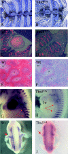
FIG. 1.
FIG. 1.
Skeletal expression of Tbx5 and Cx40 in wild-type (Wt) and Tbx5+/Δ mice. (A to D) In situ hybridization with antisense 35S-labeled Tbx5 (A and B; black) and Cx40 (C and D; red) riboprobes to the sternum and ribs from E15.5 wild-type (A and C) and Tbx5+/Δ (B and D) embryos. Note Tbx5 (A and B) and Cx40 (C and D) expression in the perichondrium surrounding sternal bands and where ribs contact the sternum (asterisk). Cx40 was also expressed in the rib perichondrium. (E and F) In situ hybridization of 35S-labeled Tbx5 and Cx40 probes to wild-type wrist sections demonstrated colocalization of Cx40 and Tbx5 expression (dIV, digit IV; H, hamate; L, lunate). Hybridization with 35S-labeled sense probes revealed no signal in wild-type and Tbx5+/Δ mice (data not shown). (G to J) Whole-mount in situ hybrid-ization to E12.5 wild-type (G and I) and Tbx5+/Δ (H and J) embryos using digoxigenin-labeled antisense Cx40 riboprobe. Note Cx40 is expressed in the wild-type sternal band (G, arrows), where Tbx5 is also detected (not shown), but its expression is decreased in the sternal band of Tbx5+/Δ embryos (H, arrows). Similarly, Cx40 is expressed in the developing scapula and forelimb region (I, arrow) of wild-type embryos but its expression is diminished in Tbx5+/Δ embryos (J, arrow).
FIG. 2.
FIG. 2.
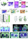
Carpal bone and digit abnormalities in Tbx5+/Δ and Cx40 mutant mice. (A to D) Skeletal preparations of carpal bones from adult mice of the indicated genotypes stained with Alcian Blue and Alizarin Red, which delineate cartilage and bone, respectively. (A) Schematic representation of carpal bones from a wild-type (Wt) mouse identifying six major carpal bones: H, hamate; C, capitate; Tm, trapezium; Td, trapezoid; S, scaphoid; L, lateral cuneiform. (B) Ensemble of carpal bone patterns observed in Tbx5+/Δ wrists. Each schematic representation identifies a carpal bone fusion, indicated in grey. Tm-S fusions occurred most frequently. One animal had no Tm (arrow). (C) Carpal bone fusions observed in Cx40+/− and Cx40−/− wrists. Schematic representations of fused bones (green) demonstrate three fusions involving the trapezium (Tm): Tm-Td, Tm-C, and Tm-Td-C (U shaped). (D) Distribution of carpal fusions observed in wrists of mutant mice. The histograms represent the combined frequency of fusions observed in both left and right wrists because no differences between frequencies of left and right wrists were observed. Note that fusions involve the Tm in 95% of Tbx5+/Δ (grey) and 100% of Cx40 mutant (green) mice. Fusions in Cx40 mutant mice often involved the trapezoid (64 to 94%) and rarely involved the capitate (12 to 50%) bones. Fusions in Tbx5+/Δ mice always (100%) involved the scaphoid and rarely (11%) the lunate bones. In compound Tbx5+/Δ Cx40+/− mice, the Tm fused to both proximal and distal carpal bones (not shown). (E to G) Altered phalanges and metacarpal bones in mutant and wild-type digits. (E) Schematic representation of metacarpal bones and phalanges elongated in Tbx5+/Δ (grey striped) or Cx40 (green) mutant mice compared to the wild type (clear). m, metacarpal bones; pp, proximal phalanges; ip, intermediate phalanges; dp, distal phalanges. (F) Metacarpal bone (m) and proximal phalanges of digit I from adult Tbx5+/Δ mice are markedly elongated compared to age-matched wild-type littermate. (G) Alcian Blue- and Alizarin Red-stained bone preparations from paws of mutant and wild-type mice. The metacarpal bone and proximal phalange of digit one exhibit a premature ossification center in newborn Tbx5+/Δ mice (denoted by arrow) compared to wild-type and Cx40−/− mice (flanking). (H to J) Sox 9 expression in wild-type, Tbx5+/Δ, and Cx40−/− digits at E13.5 as assessed by in situ hybridization. Note more proximal and higher expression of Sox9 (darker) in the phalangeal region of Tbx5Δ/+ (I) and Cx40 (J) mutant mice compared to their wild-type littermate (H).
FIG. 3.
FIG. 3.
Sternal defects in Tbx5+/Δ, Cx40-deficient, and compound mutant mice. (A) Schematic representation (left) of normal sternal bones (M, manubrium; S1 to -4, sternebra 1 to 4; X, xiphoid). Skeletal preparations of wild-type (Wt) and Tbx5+/Δ specimens (E15.5) demonstrate that the Tbx5+/Δ sternum is shortened, consistent with abnormalities in chondrogenesis. (B) Newborn Cx40+/− mice exhibit comparable sternal bone abnormalities to Tbx5+/Δ mice. Note reduced sternal length results from fusion of the third and fourth sternebrae (asterisk). (C) The S3-S4 junction in newborn wild-type mice is well delineated and contains two components. Tbx5+/Δ S3-S4 junctions show incomplete fusion (red arrow) of two hemisternebrae and disappearance of the S3-S4 junction due to ossification. Cx40+/− and Cx40−/− mice also have abnormal S3-S4 junctions with complete or asymmetric loss of the S4 ossification center (yellow head arrow). (D) Protuberance of the xiphoid process is evident in the lateral view of the rib cages of Tbx5+/Δ and Cx40−/− mice. (E) Additional ossification center detected at the rostral part of the manubrium in newborns may account for the two-piece bone. (F) Abnormal manubrium in adult Cx40+/−, Tbx5+/Δ, and Tbx5+/Δ Cx40+/− mice. Note the presence of an additional joint resulting in a two-piece manubrium only in mutant mice (shown here for Tbx5+/Δ).
FIG. 4.
FIG. 4.
Skeletal phenotypes specific to Tbx5+/Δ mutant mice. (A to D) In situ hybridization of collagen X probe in the epiphyseal growth plate of the humerus identified an expanded hypertrophic chondrocyte zone in Tbx5+/Δ (C and D) compared to wild-type (A and B) mice or Cx40-deficient mice (not shown). 35S-labeled collagen X probe was hybridized to sections, counterstained with toluidine blue, and imaged under bright-field (A and C) and dark-field (B and D) illumination. (E to F) The two-piece scaphoid due to a novel joint (arrow) found in 17% of Tbx5+/Δ mice occurred independently of fusion. (G) Severe sternebral fusion (S2-S3-S4) in Tbx5+/Δ. (H to J) Degrees of bifurcation observed in Tbx5+/Δ (I and J) but not Cx40 mutant mice compared to the wild type (H). (K) Western blot analyses of TGF signaling in wild-type, Tbx5+/Δ, and Cx40−/− mice. Expression of TGF-β2 was similar in Tbx5+/Δ, Cx40, and wild-type mice. While Smad2 and Smad4 protein levels were also comparable, phosphorylated Smad2 was selectively reduced in Tbx5+/Δ mutant mice. Phosphorylated Smad1 (P-Smad1), -5, and-8 remained unchanged among the genotypes.
FIG. 5.
FIG. 5.
Skeletal phenotypes specific to Cx40 mutant mice. (A to D) Abnormal ribs were found in Cx40 mutant mice but not in wild-type mice (A). Variation of phenotype in Cx40+/− mice ranged from bifurcation of the first rib cartilage (B) to C5 elongation (C, arrow) to formation of an additional 1′ rib (D). (E and F) Lower-limb malformations in Cx40 mutant mice. Fusion of the lateral cuneiform (lc) and navicular (na) anklebones in Cx40−/− (F) and Cx40+/− (not shown) mice compared to the wild type (Wt) (E). There is also delayed ossification (red) in multiple anklebones of Cx40 mutant mice.
FIG. 6.
FIG. 6.
Model of Tbx5 and Cx40 regulation. Cx40 and other genes, regulated by Tbx5, produce normal patterning of specific bones. In many instances, Tbx5 activates Cx40 expression and hence determines the proper patterning and shape of sternal and carpal bones and forelimb digit length. Different transcription factors (potentially Tbx4) regulate Cx40 expression in hind limbs and other bones. Tbx5 haploinsufficiency directly or indirectly (hatched arrow) misregulates downstream genes, causing abnormal skeleton formation. Tbx5 haploinsufficiency also modulates other gene products independent of Cx40, such as receptor-activated Smad2 (phosphorylated Smad2 [P-Smad2]) levels.

References

    1. Agarwal, P., J. N. Wylie, J. Galceran, O. Arkhitko, C. Li, C. Deng, R. Grosschedl, and B. G. Bruneau. 2003. Tbx5 is essential for forelimb bud initiation following patterning of the limb field in the mouse embryo. Development 130:623-633. - PubMed
    1. Akiyama, H., M. C. Chaboissier, J. F. Martin, A. Schedl, and B. de Crombrugghe. 2002. The transcription factor Sox9 has essential roles in successive steps of the chondrocyte differentiation pathway and is required for expression of Sox5 and Sox6. Genes Dev. 16:2813-2828. - PMC - PubMed
    1. Ausubel, F., R. Brent, R. E. Kingston, D. D. Moore, J. G. Seidman, J. A. Smith, and K. Struhl. (ed.). 2004. Current protocols in molecular biology. John Wiley & Sons, Inc., New York, N.Y.
    1. Baitner, A. C., S. G. Maurer, M. B. Gruen, and P. E. Di Cesare. 2000. The genetic basis of the osteochondrodysplasias. J. Pediatr. Orthop. 20:594-605. - PubMed
    1. Bamshad, M., R. C. Lin, D. J. Law, W. C. Watkins, P. A. Krakowiak, M. E. Moore, P. Franceschini, R. Lala, L. B. Holmes, T. C. Gebuhr, B. G. Bruneau, A. Schinzel, J. G. Seidman, C. E. Seidman, and L. B. Jorde. 1997. Mutations in human TBX3 alter limb, apocrine and genital development in ulnar-mammary syndrome. Nat. Genet. 16:311-315. [Erratum, 19(1): 102, 1998.] - PubMed

Publication types

MeSH terms