. 2005 Jun;25(12):5084-94.
doi: 10.1128/MCB.25.12.5084-5094.2005.

Oct-3/4 maintains the proliferative embryonic stem cell state via specific binding to a variant octamer sequence in the regulatory region of the UTF1 locus

Affiliations

Oct-3/4 maintains the proliferative embryonic stem cell state via specific binding to a variant octamer sequence in the regulatory region of the UTF1 locus

Masazumi Nishimoto et al. Mol Cell Biol. 2005 Jun.

Abstract

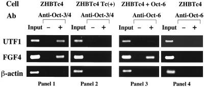
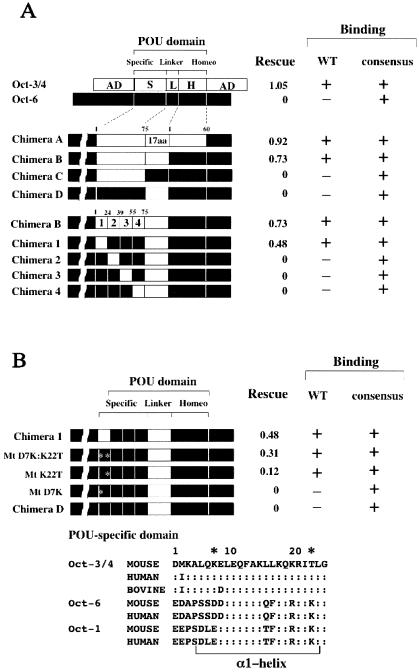
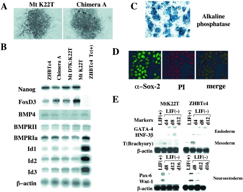
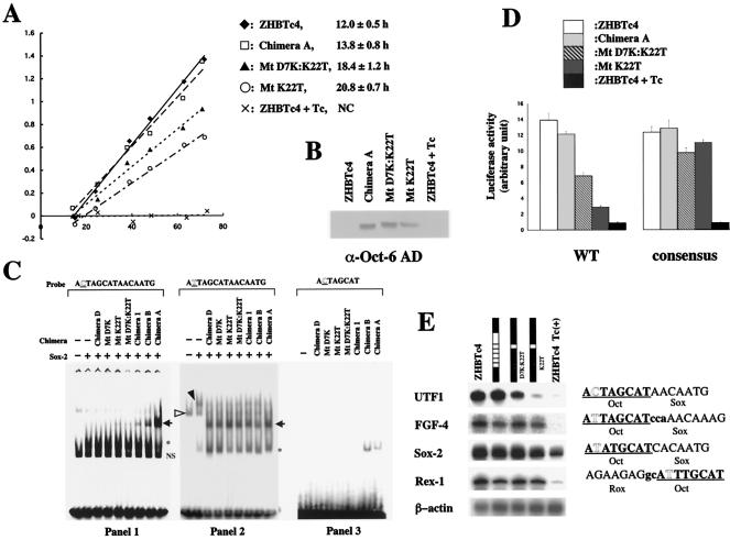
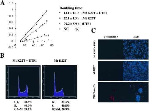
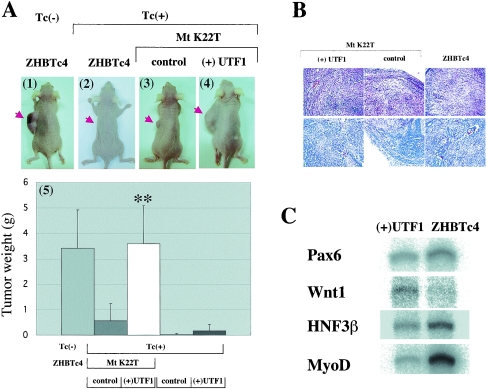
The POU transcription factor Oct-3/4 has been shown to be critical for maintaining embryonic stem (ES) cell character. However, the molecular mechanisms underlying its function remain elusive. We have previously shown that among the POU transcription factor family of proteins, Oct-3/4 alone is able to bind to the regulatory region of the UTF1 gene bearing a variant octamer sequence together with Sox-2. Here, we demonstrate using Oct-3/4-Oct-6 chimeras that there is a precise correlation between the ability of proteins to form a complex on the UTF1 enhancer with Sox-2 and the ability to maintain the stem cell state in ES cells. Different chimeric proteins show differential abilities to form a Sox-2-containing complex on the UTF1 regulatory region, with a decrease in efficiency of the complex formation accompanied by a decrease in the level of UTF1 expression and the rate of cell proliferation. Overexpression of UTF1 in these slow-growing cells was able to restore their proliferation rate to wild-type levels. Moreover, UTF1 was also observed to have an effect on teratoma formation. These results suggest a molecular pathway by which Oct-3/4 induces rapid proliferation and tumorigenic properties of ES cells through activation of the UTF1 gene.


Figures

FIG. 1.
FIG. 1.
The UTF1 enhancer specifically recruited Oct-3/4 in vivo. ZHBTc4 ES cells were maintained in the undifferentiated state, except in the experiments represented in the second panel, in which cells were induced to differentiate by Tc treatment. An Oct-6 expression vector was stably integrated in cells to boost Oct-6 expression to a level comparable to that of Oct-3/4 for the experiments shown in the third panel. Cross-linked chromatin from ES cells was immunoprecipitated with anti-Oct-3/4 or anti-Oct-6 antibodies or corresponding preimmune-sera (−). Precipitated DNA was purified and subjected to PCR to amplify the indicated regulatory regions. Genomic DNA was also prepared from the supernatant of a reaction in which no antibody was added and then diluted 300 times with H2O and used as a control for PCRs.
FIG. 2.
FIG. 2.
Correlation between the ability of Oct-3/4-Oct-6 chimeric proteins to form a complex with Sox-2 on the UTF1 enhancer sequence carrying a variant octamer motif and to maintain ES cell state. (A) Functional characterization of Oct-3/4-Oct-6 chimeric proteins. The open and solid boxes represent Oct-3/4 and Oct-6 regions, respectively. Amino acids in the POU-specific domain and homeodomain are numbered separately (for details, see reference 13). Amino acid sequences of Oct-3/4 and Oct-6 linker portions are shown in the same reference (13). The POU-specific domain can be divided into four helices, abbreviated as 1, 2, 3, and 4. The potential of the chimeric proteins to maintain the self-renewal of ES cells was analyzed by the rescue experiment, as described in Materials and Methods. Rescue indices were determined by arbitrarily setting the values (number of colonies) obtained with ZHBTc4 ES cells cultured in the absence of Tc as 1, and relative values obtained from expression of chimeric proteins were calculated. The DNA binding ability of chimeric proteins was analyzed with EMSA using as probes the wild-type UTF1 enhancer element (WT) or an enhancer in which the variant octamer sequence was converted to the canonical octamer sequence (consensus). Since the reactions were performed in the presence of Sox-2, formation of ternary complexes (Chimera/Sox-2/DNA) was monitored. (B) Identification of a critical amino acid in the POU-specific domain that is required for Oct-3/4-specific activity. The biochemical and biological properties of the depicted Oct-3/4-Oct-6 chimeric mutant proteins were characterized as in panel A. The amino acid sequences of α-helix 1 of the POU-specific domains of three different octamer factors from various species are shown at the bottom. Double dots represent amino acids in Octamer factors that are identical to those in corresponding positions of mouse Oct-3/4.
FIG. 3.
FIG. 3.
The ES cell state of cells rescued with the chimeric protein MtK22T. (A) Morphology of ZHBTc4 ES cells in which Mtk22T or chimera A was expressed in the place of wild-type Oct-3/4. Rescue experiments were performed as for Fig. 2 with the indicated chimeric protein expression vectors. The obtained colonies were observed under a microscope. (B) Examination in rescued cells of the expression levels of marker genes known to play an important role in maintaining pluripotency. RNA was prepared from the parental ES cells (ZHBTc4) cultured in the absence or presence of Tc. RNA was also prepared from ZHBTc4 ES cells whose stem cell state was maintained due to the expression of either chimera A, MtD7K:K22T, or MtK22T. Subsequently, expression levels of genes shown in the panel were analyzed by RNase protection, as described in Materials and Methods. (C) The confirmation of ES-cell status of the rescued cells with alkaline phosphatase staining. ES cells expressing MtK22T were cultured on a feeder layer and subjected to alkaline phosphatase staining procedures, as described in Materials and Methods. In these experiments, feeder layer cells served as a negative control. (D) The ES cells rescued with MtK22T possessed Sox-2 in their nuclei. ES cells bearing MtK22T were analyzed by immunostaining with anti-Sox-2 antibody and counterstained with 4′,6′-diamidino-2-phenylindole to confirm the nuclear localization of the Sox-2 protein. (E) Potential of rescued cells to differentiate into all three germ layers. Rescued cells bearing MtK22T or parental ZHBTc4 ES cells were differentiated in vitro by inducing embryoid body formation, as described in Materials and Methods. The expression of differentiation marker genes was analyzed by RNase protection, as with panel B.
FIG. 4.
FIG. 4.
MtK22T fails to support rapid proliferation due to its diminished ability to sustain elevated UTF1 expression in ES cells. (A) ES cells rescued with MtK22T showed slower proliferation than the parental ES cells. Cells (1 × 10⁴) of the parental ES cell line (ZHBTc4) or those bearing either chimera A, MtD7K:K22T, or MtK22T were transferred to tissue culture dishes. Subsequently, cells were trypsinized at the indicated times, and the total number of cells was counted. ZHBTc4 ES cells induced to differentiate with Tc for 96 h were also used for the analysis. NC, not calculable. (B) Western blot analysis of chimeric proteins expressed in the rescued ES cells. Whole-cell extracts were prepared from the indicated ES cells and used for Western blot analysis using an anti-Oct-6 antibody. (C) The biologically active chimeric proteins display different levels of complex formation on the UTF1 regulatory region bearing a variant octamer sequence. The expression vectors for the indicated chimeric proteins, Sox-2, or empty vector were introduced into HeLa cells, and whole-cell extracts were prepared from these cells. For the experiments shown in panels 1 and 2, these extracts were mixed and EMSA was performed as described in Materials and Methods, while extract containing Sox-2 was not used in panel 3. Arrow indicates the Oct-3/4-Sox-2-UTF1 regulatory element ternary complex. NS indicates nonspecific band, while an asterisk indicates the bands representing binding of either Sox-2 or chimeric protein to the probes. (All of the chimeric proteins can bind to probe bearing the variant octamer through the AT-rich Sox-2 binding site. For details, see reference 26). The open and solid triangles indicate Oct-1 and Oct-1/Sox-2 complex bound to the probe, respectively. (D) MtK22T exhibits much less potent activity than wild-type Oct-3/4 in activating the UTF1 regulatory enhancer. The two types of tk-Luc reporter plasmids (tk-Luc with wild-type UTF1 regulatory region or with the UTF1 enhancer bearing the consensus octamer sequence) were independently introduced into three different types of ES cells bearing either chimera A, Mt D7K:K22T, or Mt K22T. These reporters were also introduced in parental ZHBTc4 ES cells cultured in the absence or presence of Tc. After 48 h posttransfection, whole-cell extracts were prepared and luciferase activities were measured. The activity of these ES cells in which tk-Luc reporter plasmid with no regulatory enhancer had been introduced was arbitrarily set to 1. The data shown were obtained from four independent experiments with comparable results. (E) The UTF1 gene shows differential expression levels depending on the type of chimeric protein expressed. The expression levels of UTF1 as well as other Oct-3/4 target genes in parental ZHBTc4 ES cells cultured in the absence or presence of Tc or carrying one of the depicted chimeric proteins was analyzed by RNase protection assay. Enhancer core sequences of the genes are shown on the right, and putative octamer binding sites are underlined.
FIG. 5.
FIG. 5.
UTF1 supports rapid cell growth of ES cells. (A) The rapid proliferation phenotype characteristic of ES cells was restored to the slow-growing rescued cells by overexpression of UTF1. The UTF1 expression vector (CAG/UTF1/IRES/HIS) or empty vector (CAG/IRES/HIS) was stably integrated into ES cells whose stem cell state was maintained by MtK22T with the aid of histidinol, as described in Materials and Methods. UTF1 and (−) represent differentiated ZHBTc4 cells in which UTF1 and empty vector, respectively, had been stably integrated. After stable selection, these cells were subjected to Tc treatment for 96 h before their cell growth rate was characterized. The cell proliferation rate of these ES cells was measured as with Fig. 4A. NC, not calculable. (B) Examination of the effect of UTF1 on the cell cycle control with FACS analysis. The UTF1-overexpressing and control MtK22T-rescued cells in A were used for FACS analysis. Data shown at the bottom of panels represent averages from five independent experiments. (C) The UTF1-overexpressing and control MtK22T-rescued cells were cultured in the absence of zeocin and blasticidin S, and the numbers of cytokeratin 7-positive cells were compared by immunocytochemistry. The differentiation-induced ZHBTc4 ES cells were also used as a positive control.
FIG. 6.
FIG. 6.
(A) The UTF1 shows a prominent effect on teratoma formation. Panels 1 and 2: injection of ZHBTc4 ES cells into nude mice which had been supplied with Tc-free (panel 1) or Tc-containing (panel 2) drinking water. Panels 3 and 4: tumors in Tc-treated mice derived from injection of MtK22T-rescued ES cells in which empty (panel 3) and UTF1 expression (panel 4) vectors were stably integrated. Panel 5: summary of teratoma formation by the indicated cells. Data shown in the panel were obtained from eight independent injections. The (+)UTF1 and control represent stable integration of UTF1 and empty vectors into the indicated cells, respectively. Double asterisk, P < 0.01 when compared to MtK22T-rescued cells in which the empty vector had been integrated. (B) Histological analysis of teratomas. Teratomas resulting from ES cells containing MtK22T alone, Mt K22T plus UTF1, and the parental ZHBTc4 ES cells were subjected to histological analysis with hematoxylin-eosin staining procedures. (C) Analysis of differentiation marker gene expression in teratomas. RNA was prepared from teratomas derived from UTF1-overexpressing MtK22T-rescued cells and parental ZHBTc4 ES cells, and expression levels of the indicated differentiation marker genes were analyzed by RNase protection.

Similar articles

Cited by

References

    1. Antonchuk, J., G. Sauvageau, and R. K. Humphries. 2002. HOXB4-induced expansion of adult hematopoietic stem cells ex vivo. Cell 109:39-45. - PubMed
    1. Aurora, R., and W. Herr. 1992. Segments of the POU domain influence one another's DNA-binding specificity. Mol. Cell. Biol. 12:455-467. - PMC - PubMed
    1. Avilion, A. A., S. K. Nicolis, L. H. Pevny, L. Perez, N. Vivian, and R. Lovell-Badge. 2003. Multipotent cell lineages in early mouse development depend on Sox-2 function. Genes Dev. 17:126-140. - PMC - PubMed
    1. Ben-Shushan, E., J. R. Thompson, L. J. Gudas, and Y. Bergman. 1998. Rex-1, a gene encoding a transcription factor expressed in the early embryo, is regulated via Oct-3/4 and Oct-6 binding to an octamer site and a novel protein, Rox-1, binding to an adjacent site. Mol. Cell. Biol. 18:1866-1878. - PMC - PubMed
    1. Boiani, M., S. Eckardt, N. A. Leu, H. R. Scholer, and K. J. McLaughlin. 2003. Pluripotency deficit in clones overcome by clone-clone aggregation: epigenetic complementation? EMBO J. 22:5304-5312. - PMC - PubMed

Publication types

MeSH terms