. 2005 Jun;25(12):5119-33.
doi: 10.1128/MCB.25.12.5119-5133.2005.

The MEK1 scaffolding protein MP1 regulates cell spreading by integrating PAK1 and Rho signals

Affiliations

The MEK1 scaffolding protein MP1 regulates cell spreading by integrating PAK1 and Rho signals

Ashok Pullikuth et al. Mol Cell Biol. 2005 Jun.

Abstract

How the extracellular signal-regulated kinase (ERK) cascade regulates diverse cellular functions, including cell proliferation, survival, and motility, in a context-dependent manner remains poorly understood. Compelling evidence indicates that scaffolding molecules function in yeast to channel specific signals through common components to appropriate targets. Although a number of putative ERK scaffolding proteins have been identified in mammalian systems, none has been linked to a specific biological response. Here we show that the putative scaffold protein MEK partner 1 (MP1) and its partner p14 regulate PAK1-dependent ERK activation during adhesion and cell spreading but are not required for ERK activation by platelet-derived growth factor. MP1 associates with active but not inactive PAK1 and controls PAK1 phosphorylation of MEK1. Our data further show that MP1, p14, and MEK1 serve to inhibit Rho/Rho kinase functions necessary for the turnover of adhesion structures and cell spreading and reveal a signal-channeling function for a MEK1/ERK scaffold in orchestrating cytoskeletal rearrangements important for cell motility.


Figures

FIG. 1.
FIG. 1.
MP1/p14 regulates PAK1-dependent MEK1 phosphorylation and activation. (A) Depletion of MP1/p14 inhibits fibronectin-stimulated PAK1 phosphorylation (pS298 MEK1) and activation of MEK1 (pS218/222 MEK1). (B) Selective inhibition of MEK activation by depletion of MP1/p14. REF52 cells transfected with MP1 siRNA or control RNA were stimulated with EGF or PDGF, and MEK activation was assessed as described for panel A. (C) FAK autophosphorylation proceeds normally in cells depleted of MP1. Identical results were seen in cells transfected with either an independent MP1 siRNA or p14 siRNA (data not shown). (D) Fibronectin-stimulated tyrosine phosphorylation events are mostly independent of MP1/p14. Lysates prepared from control or MP1 siRNA-transfected cells were blotted with anti-phosphotyrosine antisera. The arrowhead indicates tyrosine-phosphorylated ERK (see the text).
FIG. 2.
FIG. 2.
MP1 associates with PAK1. (A) MP1 associates with the PAK1 C terminus. COS cells were transiently transfected with V5-tagged PAK1 constructs (WT, wild type; NT, amino acids 1 to 248; CT, amino acids 248 to 545) with or without FLAG-tagged MP1 and Myc-tagged p14. FLAG immunoprecipitates were formed and blotted with V5, p14, or MP1 antisera (top three panels). Expression of V5 PAK1 constructs was confirmed by blotting lysates with V5 antisera (bottom panel). The intense band present in all immunoprecipitates is immunoglobulin G heavy chain. (B) MP1 preferentially associates with activated PAK1. COS cells were transiently transfected with the V5-tagged PAK1 constructs indicated with or without FLAG-tagged MP1 and Myc-p14. FLAG immunoprecipitates were formed and blotted with V5, p14, or MP1 antisera (top three panels). The expression of V5 PAK1 constructs was confirmed by blotting lysates with V5 antisera (bottom panel). TE, T423E; KR, K299R; LF, L107F; LF/KR, L107F K399R. (C) The PAK1 C terminus (residues 232 to 544) binds directly to MP1/p14. GST fusion proteins were incubated with recombinant MP1/p14 complex before capture with glutathione Sepharose. MP1/p14 pulled down by the fusion proteins was assessed by blotting (top panels) and the presence of fusion protein verified by Coomassie blue staining (bottom panel). Blotting of input (1%) established that MP1/p14 was present in each reaction. The upper band seen in the p14 blots is residual MP1 signal remaining after stripping.
FIG. 3.
FIG. 3.
Mutations in the MEK1 proline-rich sequence selectively inhibit MEK1-MP1 binding. (A) Residues present in the MEK1 PRS but not conserved in MEK2 and Xenopus MEK proline-rich sequences (which do not bind MP1 [; data not shown]) were mutated to the corresponding MEK2 residue. (B) Yeast two-hybrid assays identify MEK1 mutants selectively defective for binding MP1. Mutants were analyzed for their ability to bind MP1 and Raf-1 in yeast two-hybrid assays. Growth on media lacking histidine and induction of β-galactosidase activity indicates two-hybrid interaction; growth on media lacking uracil and containing 5-fluoroorotic acid (5-FDA) indicates no bait-prey interaction. The expression of all MEK1 mutants, MP1, and Raf-1 was verified by Western blotting (data not shown). (C) MEK1 L274S and MEK1 Q278P are unable to bind MP1. CCL39 cells were cotransfected with HA-tagged MEK mutants and FLAG-tagged MP1. FLAG immunoprecipitates were formed and blotted with HA antiserum (top panel) or MP1 antiserum (middle panel). HA blotting of lysates confirmed expression of all MEK constructs (bottom panel). (D) MEK1 mutants unable to bind MP1 are competent to bind Raf. CCL39 cells were transiently transfected with HA-tagged MEK1 constructs. HA immunoprecipitates were blotted for endogenous Raf-1 (top) or HA-MEK (middle). Lysates were blotted with Raf-1 antiserum (bottom). (E) MEK1 mutants unable to bind MP1 are phosphorylated by PAK1 during adhesion to fibronectin. REF52 cells were transiently transfected with HA-tagged MEK1 constructs, suspended in serum-free medium, and allowed to adhere to fibronectin-coated plates. HA immunoprecipitates were blotted with anti-phospho S298 MEK1 antiserum (top) or HA antiserum (bottom).
FIG. 4.
FIG. 4.
MP1/p14 regulates cell spreading on fibronectin. (A) REF52 cells transiently transfected with control, MP1, or p14 siRNA were replated on fibronectin-coated petri dishes as described previously (73). Cultures were fixed at the indicated time points and random fields scored for spread cells (left panel) and adherent cells (right panel). Data are representative of three experiments. (B) Typical morphology of cells transfected with siRNA following replating on fibronectin for 10 min. Control transfected cells spread rapidly (arrows), whereas MP1 or p14 siRNA-transfected cells remain round and refractile with multiple membrane blebs (arrowheads). Immunoblots confirmed depletion of MP1 and p14 (not shown).
FIG. 5.
FIG. 5.
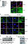
MP1 and MEK regulate Rho-ROCK signaling in adherent cells. (A) Depletion of MP1 and p14 has no effect on Rho-GTP levels in continuously adherent cells. Rho-GTP loading was measured as described previously (62). (B) Depletion of MP1 and p14 stimulates accumulation of Y27632-resistant focal adhesions and stress fibers in continuously adherent cells. Vinculin and F-actin were detected with antivinculin antisera and phalloidin, respectively. Focal adhesions in control RNA-transfected cells are primarily localized at the cell periphery (arrows); in contrast, abundant adhesions form tracks under the cell body in cells depleted of MP1 (arrowheads). Pretreatment with Y27632 (10 μM, 30 min) disrupted stress fibers and adhesions in control transfected cells but not in cells depleted of MP1. (C) Pharmacological inhibition of MEK signaling leads to accumulation of abundant, elongated focal adhesions under the cell body. REF52 cells were treated with or without UO126, fixed, and stained for vinculin and F-actin. Control cells exhibit mostly peripheral focal adhesions (arrows); in contrast, abundant, elongated adhesions form under the cell body in the presence of UO126 (arrowheads). Scale bars, 25 μm. (D) Depletion of MP1/p14 or inhibition of MEK (25 μM UO126; 30 min) stimulates Rho kinase activity. ROCKII was immunoprecipitated and assayed as described in Materials and Methods. Incubation of immunoprecipitates with Y27632 confirmed substrate phosphorylation by Rho kinase. (E) Depletion of MP1/p14 stimulates phosphorylation of cofilin. Continuously adherent cells transfected with control or MP1 siRNA were incubated with or without Y27632 (10 μM; 30 min) prior to lysis. Phosphorylation of endogenous cofilin was assessed by Western blotting.
FIG. 6.
FIG. 6.
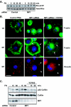
MP1 regulates Rho-GTP loading and function during acute spreading on fibronectin. (A) MP1 controls Rho-GTP loading. REF52 cells transfected with control or MP1 siRNA were suspended and then allowed to adhere to fibronectin-coated plates for the indicated times. Rho-GTP loading was measured as described previously (62). (B) Rho-dependent focal adhesion and stress fiber formation are controlled by MP1. REF52 cells transfected and suspended as described for panel A were treated with or without the Rho kinase inhibitor Y27632 (10 μM) for 30 min prior to replating on fibronectin for the indicated times. Cells depleted of MP1/p14 exhibit dense circumferential stress fibers (arrows) and large vinculin-containing adhesions (arrowheads). Typical stress fiber (arrows) and vinculin staining (arrowheads) are partially restored by pretreatment with the Rho kinase inhibitor. Scale bar, 25 μm. (C) Depletion of MP1/p14 stimulates ROCK-dependent phosphorylation of cofilin in acutely adhering cells. Cells transfected with control or MP1 siRNA were incubated with or without Y27632 (10 μM; 30 min) prior to replating on fibronectin. Phosphorylation of endogenous cofilin was assessed by Western blotting.
FIG. 7.
FIG. 7.
MP1-regulated ERK activation suppresses ROCK function to allow cell spreading. (A) Inhibition of ROCK rescues the spreading defect of cells depleted of MP1/p14. Cells were allowed to attach and spread on fibronectin for 30 min with or without prior treatment with Y27632. A structurally distinct ROCK inhibitor, H1152, was used to confirm that elevated ROCK function underlies the spreading defect. Lysates prepared from REF cells transfected with control or MP1 siRNA were blotted with ROCKI and II antisera (inset); ROCK expression is unaffected by depletion of MP1/p14. (B) Inhibition of ROCK rescues the spreading defect in cells depleted of MP1. Cultures were fixed at 30 min and random fields scored for spread cells. Data are representative of two experiments. (C) Rho kinase is downstream of MP1/p14-regulated MEK1 phosphorylation and ERK activation. Lysates were blotted for MEK1 phosphorylation and ERK activation following replating on fibronectin (30 min) in the presence or absence of Y27632. Scale bar, 25 μm.

Similar articles

Cited by

References

    1. Alahari, S. K., P. J. Reddig, and R. L. Juliano. 2004. The integrin-binding protein Nischarin regulates cell migration by inhibiting PAK. EMBO J. 23:2777-2788. - PMC - PubMed
    1. Arber, S., F. A. Barbayannis, H. Hanser, C. Schneider, C. A. Stanyon, O. Bernard, and P. Caroni. 1998. Regulation of actin dynamics through phosphorylation of cofilin by LIM-kinase. Nature 393:805-809. - PubMed
    1. Arthur, W. T., and K. Burridge. 2001. RhoA inactivation by p190RhoGAP regulates cell spreading and migration by promoting membrane protrusion and polarity. Mol. Biol. Cell 12:2711-2720. - PMC - PubMed
    1. Bobak, D., J. Moorman, A. Guanzon, L. Gilmer, and C. Hahn. 1997. Inactivation of the small GTPase Rho disrupts cellular attachment and induces adhesion-dependent and adhesion-independent apoptosis. Oncogene 15:2179-2189. - PubMed
    1. Bowman, A. B., R. S. Patel-King, S. E. Benashski, J. M. McCaffery, L. S. Goldstein, and S. M. King. 1999. Drosophila roadblock and Chlamydomonas LC7: a conserved family of dynein-associated proteins involved in axonal transport, flagellar motility, and mitosis. J. Cell Biol. 146:165-180. - PMC - PubMed

Publication types

MeSH terms