

Transcriptional regulation of the SCL locus: identification of an enhancer that targets the primitive erythroid lineage in vivo
- PMID: 15923636
- PMCID: PMC1140604
- DOI: 10.1128/MCB.25.12.5215-5225.2005
Transcriptional regulation of the SCL locus: identification of an enhancer that targets the primitive erythroid lineage in vivo
Abstract
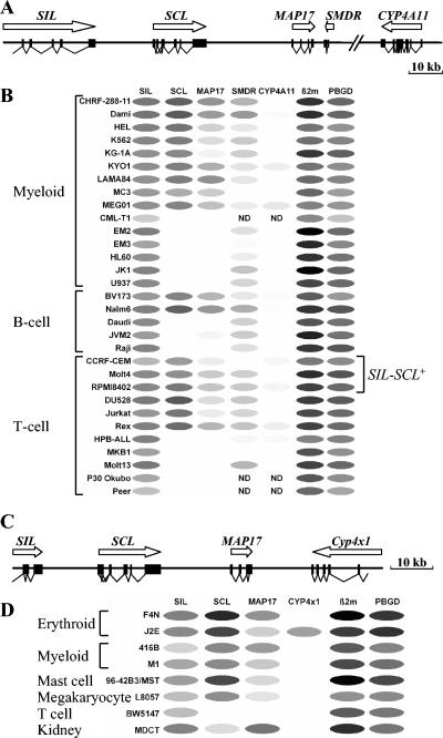
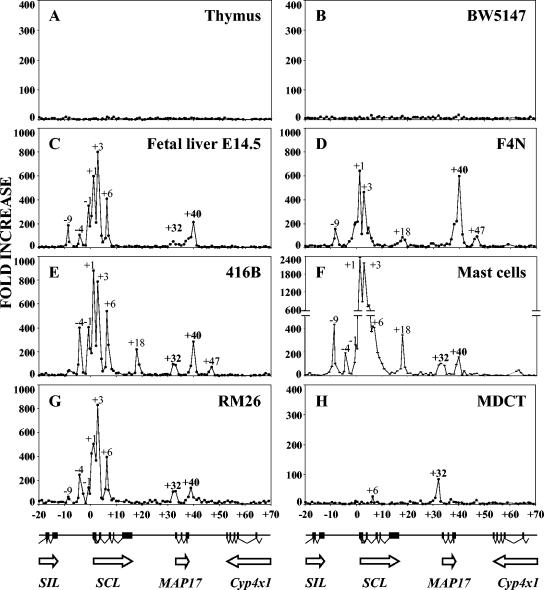
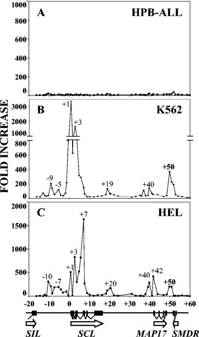
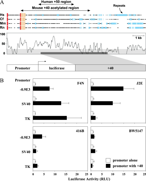
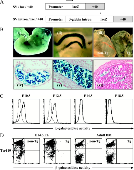
The stem cell leukemia (SCL) gene, also known as TAL-1, encodes a basic helix-loop-helix protein that is essential for the formation of all hematopoietic lineages, including primitive erythropoiesis. Appropriate transcriptional regulation is essential for the biological functions of SCL, and we have previously identified five distinct enhancers which target different subdomains of the normal SCL expression pattern. However, it is not known whether these SCL enhancers also regulate neighboring genes within the SCL locus, and the erythroid expression of SCL remains unexplained. Here, we have quantitated transcripts from SCL and neighboring genes in multiple hematopoietic cell types. Our results show striking coexpression of SCL and its immediate downstream neighbor, MAP17, suggesting that they share regulatory elements. A systematic survey of histone H3 and H4 acetylation throughout the SCL locus in different hematopoietic cell types identified several peaks of histone acetylation between SIL and MAP17, all of which corresponded to previously characterized SCL enhancers or to the MAP17 promoter. Downstream of MAP17 (and 40 kb downstream of SCL exon 1a), an additional peak of acetylation was identified in hematopoietic cells and was found to correlate with expression of SCL but not other neighboring genes. This +40 region is conserved in human-dog-mouse-rat sequence comparisons, functions as an erythroid cell-restricted enhancer in vitro, and directs beta-galactosidase expression to primitive, but not definitive, erythroblasts in transgenic mice. The SCL +40 enhancer provides a powerful tool for studying the molecular and cellular biology of the primitive erythroid lineage.
Figures
Similar articles
-
The scl +18/19 stem cell enhancer is not required for hematopoiesis: identification of a 5' bifunctional hematopoietic-endothelial enhancer bound by Fli-1 and Elf-1.Mol Cell Biol. 2004 Mar;24(5):1870-83. doi: 10.1128/MCB.24.5.1870-1883.2004. Mol Cell Biol. 2004. PMID: 14966269 Free PMC article.
-
Impaired in vitro erythropoiesis following deletion of the Scl (Tal1) +40 enhancer is largely compensated for in vivo despite a significant reduction in expression.Mol Cell Biol. 2013 Mar;33(6):1254-66. doi: 10.1128/MCB.01525-12. Epub 2013 Jan 14. Mol Cell Biol. 2013. PMID: 23319051 Free PMC article.
-
The SCL +40 enhancer targets the midbrain together with primitive and definitive hematopoiesis and is regulated by SCL and GATA proteins.Mol Cell Biol. 2007 Oct;27(20):7206-19. doi: 10.1128/MCB.00931-07. Epub 2007 Aug 20. Mol Cell Biol. 2007. PMID: 17709394 Free PMC article.
-
The Hematopoietic Stem and Progenitor Cell Cistrome: GATA Factor-Dependent cis-Regulatory Mechanisms.Curr Top Dev Biol. 2016;118:45-76. doi: 10.1016/bs.ctdb.2016.01.002. Epub 2016 Feb 26. Curr Top Dev Biol. 2016. PMID: 27137654 Free PMC article. Review.
-
The SCL/TAL1 gene: roles in normal and malignant haematopoiesis.Bioessays. 1997 Jul;19(7):607-13. doi: 10.1002/bies.950190711. Bioessays. 1997. PMID: 9230693 Review.
Cited by
-
Real-time PCR mapping of DNaseI-hypersensitive sites using a novel ligation-mediated amplification technique.Nucleic Acids Res. 2007;35(8):e56. doi: 10.1093/nar/gkm108. Epub 2007 Mar 27. Nucleic Acids Res. 2007. PMID: 17389645 Free PMC article.
-
The modern primitives: applying new technological approaches to explore the biology of the earliest red blood cells.ISRN Hematol. 2013 Oct 3;2013:568928. doi: 10.1155/2013/568928. ISRN Hematol. 2013. PMID: 24222861 Free PMC article. Review.
-
Shared transcription factors contribute to distinct cell fates.Transcription. 2014;5(5):e978173. doi: 10.4161/21541264.2014.978173. Epub 2015 Jan 6. Transcription. 2014. PMID: 25425188 Free PMC article.
-
Dr. Jekyll and Mr. Hyde: MAP17's up-regulation, a crosspoint in cancer and inflammatory diseases.Mol Cancer. 2018 Apr 12;17(1):80. doi: 10.1186/s12943-018-0828-7. Mol Cancer. 2018. PMID: 29650022 Free PMC article. Review.
-
Modeling dynamic functional relationship networks and application to ex vivo human erythroid differentiation.Bioinformatics. 2014 Dec 1;30(23):3325-33. doi: 10.1093/bioinformatics/btu542. Epub 2014 Aug 12. Bioinformatics. 2014. PMID: 25115705 Free PMC article.
References
-
- Acuto, O., R. E. Hussey, K. A. Fitzgerald, J. P. Protentis, S. C. Meuer, S. F. Schlossman, and E. L. Reinherz. 1983. The human T cell receptor: appearance in ontogeny and biochemical relationship of alpha and beta subunits on IL-2 dependent clones and T cell tumors. Cell 34:717-726. - PubMed
-
- Baker, J. R., S. Satarug, R. J. Edwards, M. R. Moore, D. J. Williams, and P. E. Reilly. 2003. Potential for early involvement of CYP isoforms in aspects of human cadmium toxicity. Toxicol. Lett. 137:85-93. - PubMed
-
- Begley, C. G., and A. R. Green. 1999. The SCL gene: from case report to critical hematopoietic regulator. Blood 93:2760-2770. - PubMed
-
- Bockamp, E. O., F. McLaughlin, A. M. Murrell, B. Gottgens, L. Robb, C. G. Begley, and A. R. Green. 1995. Lineage-restricted regulation of the murine SCL/TAL-1 promoter. Blood 86:1502-1514. - PubMed
Publication types
MeSH terms
Substances
Grants and funding
LinkOut - more resources
Full Text Sources
Medical
Research Materials
Miscellaneous