. 2005 Jun;25(12):5242-52.
doi: 10.1128/MCB.25.12.5242-5252.2005.

Most F508del-CFTR is targeted to degradation at an early folding checkpoint and independently of calnexin

Affiliations

Most F508del-CFTR is targeted to degradation at an early folding checkpoint and independently of calnexin

Carlos M Farinha et al. Mol Cell Biol. 2005 Jun.

Abstract

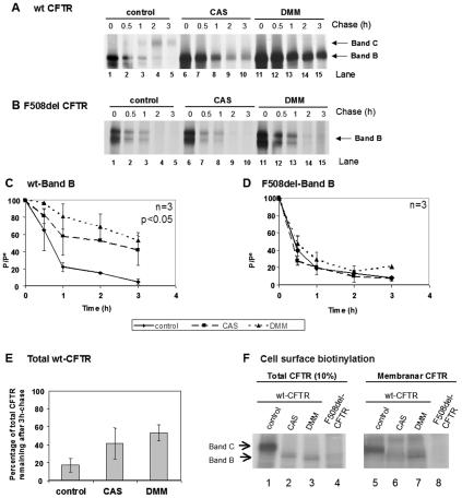
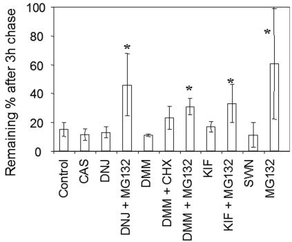
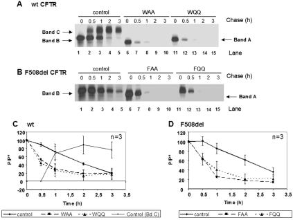
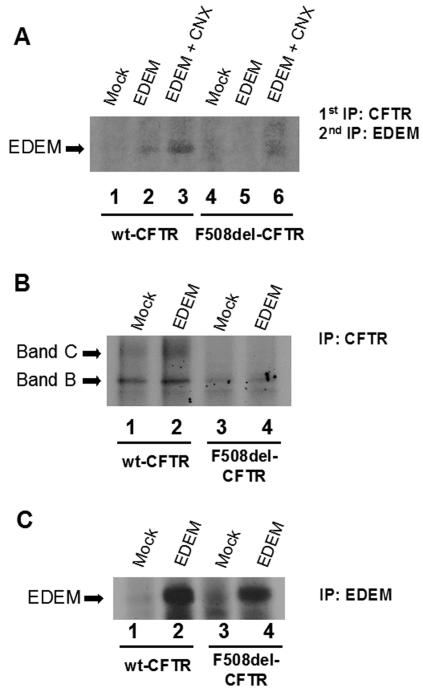
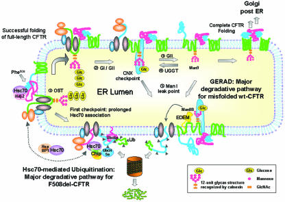
Biosynthesis and folding of multidomain transmembrane proteins is a complex process. Structural fidelity is monitored by endoplasmic reticulum (ER) quality control involving the molecular chaperone calnexin. Retained misfolded proteins undergo ER-associated degradation (ERAD) through the ubiquitin-proteasome pathway. Our data show that the major degradation pathway of the cystic fibrosis transmembrane conductance regulator (CFTR) with F508del (the most frequent mutation found in patients with the genetic disease cystic fibrosis) from the ER is independent of calnexin. Moreover, our results demonstrate that inhibition of mannose-processing enzymes, unlike most substrate glycoproteins, does not stabilize F508del-CFTR, although wild-type (wt) CFTR is drastically stabilized under the same conditions. Together, our data support a novel model by which wt and F508del-CFTR undergo ERAD from two distinct checkpoints, the mutant being disposed of independently of N-glycosidic residues and calnexin, probably by the Hsc70/Hsp70 machinery, and wt CFTR undergoing glycan-mediated ERAD.


Figures

FIG. 1.
FIG. 1.
Detection of calnexin levels by Western blotting after transient transfection. CHO cells stably expressing either wt CFTR (A) or F508del-CFTR (B) were transiently transfected with human calnexin cDNA (see Materials and Methods). Lanes 1, nontransfected cells; lanes 2, mock-transfected cells (with the empty vector); lanes 3, cells analyzed 24 h posttransfection with calnexin cDNA. Blots were probed with an Ab specific for human calnexin, as indicated on the left.
FIG. 2.
FIG. 2.
Turnover and processing of wt and F508del-CFTR under calnexin overexpression. CHO cells stably expressing (A) wt or (B) F508del-CFTR were transiently transfected with the calnexin cDNA construct (lanes 7 to 12) or with the same amount of empty vector as a control (lanes 1 to 6). Twenty-four hours posttransfection, the cells were pulse-labeled for 30 min with [35S]methionine and chased for 0 h (lanes 1 and 7), 0.5 h (lanes 2 and 8), 1 h (lanes 3 and 9), 1.5 h (lanes 4 and 10), 2 h (lanes 5 and 11), and 3 h (lanes 6 and 12). The cells were then lysed and immunoprecipitated with an anti-CFTR Ab (see Materials and Methods). Following electrophoretic separation and fluorography, immature (band B) and mature (band C) forms of CFTR were quantified (see Materials and Methods). Turnover of the core-glycosylated form (band B) of wt CFTR (C) and F508del-CFTR (D) is shown as the ratio between P, the amount of band B at time t, and P0, the amount of band B at the start of the chase (i.e., at the end of pulse). The efficiency of conversion of the core-glycosylated form (band B) into the fully glycosylated form of wt CFTR (band C) was also estimated for wt CFTR (E) and was determined as the ratio between the amount of band C at time t and the amount of band B at the start of the chase (P0). The number of experiments is indicated at the right upper corner of panels C, D, and E. Statistically significant differences are indicated (P < 0.05). (F) (Top) Calnexin cDNA was used to transfect cells stably expressing wt or F508del-CFTR. After being labeled with [35S]methionine, the cells were lysed and CFTR immunoprecipitated with an anti-CFTR Ab. After elution, a second IP was performed (see Materials and Methods) using a mixture (1:1) of human and hamster anticalnexin Abs. Lanes 1 and 3, cells transfected with empty vector (as a control); lanes 2 and 4, cells transfected with calnexin cDNA. (Bottom) Results of direct IP of calnexin in lysates.
FIG. 3.
FIG. 3.
Immunodetection of calnexin levels after transfection with RNAi duplexes specific for this chaperone. (A) CHO cells stably expressing wt CFTR (lanes 1 to 4) or F508del-CFTR (lanes 5 to 8) were transfected with calnexin RNAi duplexes (see Materials and Methods) and analyzed 24 h afterwards. The cells were lysed, and 30 μg of total protein was loaded onto an SDS-PAGE gel. Western blotting was performed using with an Ab recognizing endogenous (hamster) calnexin. Lanes 1 and 5, nontransfected (NT) cells; lanes 2 to 4 and 6 to 8, cells transfected with different amounts of RNAi duplexes (lanes 2 and 6, 20 pmol; lanes 3 and 7, 60 pmol; and lanes 4 and 8, 120 pmol). The arrow indicates detection of calnexin. (B) Blots were scanned, and densitometry was performed for quantification. The results are shown as a plot of the percentage of calnexin present relative to nontransfected cells.
FIG. 4.
FIG. 4.
Turnover and processing of wt and F508del-CFTR under calnexin down-regulation by RNAi. CHO cells stably expressing (A) wt or (B) F508del-CFTR were transfected with 60 pmol of RNAi primers specific for calnexin (lanes 6 to 10) or green fluorescent protein RNAi primers as a negative control (lanes 1 to 5). Twenty-four hours posttransfection, the cells were pulse-labeled and chased as before (Fig. 2) for 0 h (lanes 1 and 6), 0.5 h (lanes 2 and 7), 1 h (lanes 3 and 8), 2 h (lanes 4 and 9), and 3 h (lanes 5 and 10). The cells were then lysed, immunoprecipitated with an anti-CFTR Ab, and analyzed as before (see the legend to Fig. 2) to determine the turnover of immature wt CFTR (C) and F508del-CFTR (D) and the processing efficiency of wt CFTR (E). The number of experiments and statistically significant differences are indicated as in Fig. 2.
FIG. 5.
FIG. 5.
Turnover of wt and F508del-CFTR under CAS and DMM. CHO cells stably expressing (A) wt CFTR or (B) F508del-CFTR were treated with 1 mM CAS (lanes 6 to 10) or 1 mM DMM (lanes 11 to 15) or untreated (lanes 1 to 5). After a 90-min treatment, the cells were pulse-labeled and chased for 0 h (lanes 1, 6, and 11), 0.5 h (lanes 2, 7, and 12), 1 h (lanes 3, 8, and 13), 2 h (lanes 4, 9, and 14), and 3 h (lanes 5, 10, and 15). The cells were then lysed, immunoprecipitated with an anti-CFTR Ab, and analyzed as before (see the legend to Fig. 2) to determine the turnover of immature wt CFTR (C) and F508del-CFTR (D). (E) Percentage of total CFTR (band B plus band C) remaining at the end of the chase period in untreated cells compared with total band B in cells treated with either CAS or DMM. The number of experiments and statistically significant differences are indicated as in Fig. 2. (F) Cell surface biotinylation followed by CFTR IP was performed to determine whether protein produced under CAS or DMM reaches the cell surface. CHO cells expressing wt or F508del-CFTR were incubated for 72 h with CAS or DMM. Cell surface proteins were biotinylated, and CFTR IP was carried out. Samples were divided into two equal portions, and biotin-labeled proteins were captured with streptavidin beads in one of the fractions. Total and biotinylated (membrane) CFTRs were then in vitro phosphorylated using [γ-32P]ATP and cAMP-dependent protein kinase. Ten percent of total immunoprecipitated CFTR (lanes 1 to 4) and all immunoprecipitated membrane CFTR (lanes 5 to 8) were subjected to SDS-PAGE and fluorography. Band B of wt CFTR produced under CAS or DMM could be clearly detected in the membrane fraction.
FIG. 6.
FIG. 6.
Degradation of F508del-CFTR under various inhibitors. CHO cells stably expressing F508del-CFTR were treated with 1 mM CAS, 1 mM DMM, 100 μM KIF, 100 μM SWN, 1 mM DNJ, 25 μM of proteasome inhibitor MG132, or 20 μM of the translation elongation inhibitor CHX, alone or simultaneously. After 90 min of treatment, the cells were pulse-labeled and chased as before (Fig. 2), but at a single time point (3 h). The cells were then lysed, immunoprecipitated with an anti-CFTR Ab, and analyzed as before (see the legend to Fig. 2) to determine the percentage of F508del-CFTR remaining after chase. The obtained data are shown graphically. The asterisk indicates statistically significant differences relative to the control (first bar).
FIG. 7.
FIG. 7.
Turnover of nonglycosylated mutants of wt CFTR (WQQ and WAA) and F508del-CFTR (FQQ and FAA). BHK cells stably expressing wt CFTR (A, lanes 1 to 5), N894A-N900A (WAA) (A, lanes 6 to 10), N894Q-N900Q (WQQ) (A, lanes 11 to 15), F508del-CFTR (B, lanes 1 to 5), F508del-N894A-N900A (FAA) (B, lanes 6 to 10), or F508del-N894Q-N900Q CFTR (FQQ) (B, lanes 11 to 15) were pulse-labeled and chased as before (Fig. 2) for 0 h (lanes 1, 6, and 11), 0.5 h (lanes 2, 7, and 12), 1 h (lanes 3, 8, and 13), 2 h (lanes 4, 9, and 14), and 3 h (lanes 5, 10, and 15). The cells were then lysed, immunoprecipitated with an anti-CFTR Ab, and analyzed as before (see the legend to Fig. 2) to determine the turnover of wt, WAA, and WQQ (C) and F508del-, FAA, and FQQ (D) CFTRs. The number of experiments and statistically significant differences are indicated as in Fig. 2.
FIG. 8.
FIG. 8.
Presence of EDEM in CFTR complexes. (A) EDEM cDNA was used alone or with calnexin (CNX) cDNAs to transfect cells stably expressing wt or F508del-CFTR. After being labeled with [35S]methionine, the cells were lysed and CFTR IP was performed with an anti-CFTR Ab. After elution, a second IP was performed using a specific anti-EDEM Ab. Lanes 1 and 4, cells transfected with empty vector (as a control); lanes 2 and 5, cells transfected with EDEM cDNA; lanes 3 and 6, cells cotransfected with EDEM and calnexin cDNAs. As controls, direct IPs of either CFTR (B) or EDEM (C) were also performed after transient transfection with EDEM cDNA.
FIG. 9.
FIG. 9.
Proposed model for the major degradation pathways of F508del- and wt CFTR. (i) Synthesis of CFTR occurs with its concomitant insertion in the ER membrane and attachment of an Hsc70/Hdj-2 (or Hsp70/Hdj-1) pair to nascent cytosolic domains, as described previously (12, 30). Other authors have described increased levels of Hsc70/Hdj-2 complexes with F508del-CFTR relative to wt CFTR and expression of NBD1 as the earliest point at which Hsc70/Hdj-2 could bind the nascent CFTR polypeptidic chain (30). The same study reported that complex formation between Hdj-2 and nascent wt CFTR was greatly reduced after expression of the R domain, suggesting NBD1-R domain interaction as a critical point in CFTR folding. The cell thus seems to use this Hsc70/Hsp70 control as the first checkpoint to assess CFTR conformation, and we propose that it is the major mechanism to discard F508del-CFTR. Prolonged retention of unfolded F508del-CFTR by Hsc70 at this point enables CHIP to interact with Hsc70/Hsp70 (probably by displacing Hdj-2) and causes the mutant to be degraded through the Hsc70-CHIP-UbcH5a pathway (31, 57). The E2 Ubc6 may also contribute to F508del-CFTR ERAD (24). Contrary to what happens with F508del-CFTR, wt CFTR, for which NBD1-R intramolecular interaction and folding is achieved, proceeds in the folding pathway through interaction of its N-glycosyl residues (ii) with calnexin (iii). Wt CFTR acquires its native conformation through successive rounds of release-deglucosylation (iv) and rebinding-reglucosylation (v) to calnexin, which also constitutes the second ERQC checkpoint. Upon successful folding, CFTR exits the ER, proceeding through the secretory pathway (vi). However, prolonged presence in the calnexin cycle may cause misfolded CFTR to become a substrate of mannosidase I (vii). This enzyme trims mannose residues from the protein glycan moiety, possibly generating the Man8B glycan intermediate that is recognized by EDEM, which targets the client protein to ERAD (viii). We call this ER-degradative pathway GERAD, to indicate its dependence on the glycan moiety. According to this model, F508del-CFTR follows a major degradative pathway from the first (Hsc70-dependent) ERQC checkpoint, whereas misfolded wt CFTR undergoes proteolytic GERAD at the second (calnexin-dependent) one (see the text for a description).

References

    1. Alberti, S., K. Bohse, V. Arndt, A. Schmitz, and J. Hohfeld. 2004. The cochaperone HspBP1 inhibits the CHIP ubiquitin ligase and stimulates the maturation of the cystic fibrosis transmembrane conductance regulator. Mol. Biol. Cell 15:4003-4010. - PMC - PubMed
    1. Ayalon-Soffer, M., M. Shenkman, and G. Z. Lederkremer. 1999. Differential role of mannose and glucose trimming in the ER degradation of asialoglycoprotein receptor subunits. J. Cell Sci. 112:3309-3318. - PubMed
    1. Bergeron, J. J., M. B. Brenner, D. Y. Thomas, and D. B. Williams. 1994. Calnexin: a membrane-bound chaperone of the endoplasmic reticulum. Trends Biochem. Sci. 19:124-128. - PubMed
    1. Cabral, C. M., Y. Liu, K. W. Moremen, and R. N. Sifers. 2002. Organizational diversity among distinct glycoprotein endoplasmic reticulum-associated degradation programs. Mol. Biol. Cell 13:2639-2650. - PMC - PubMed
    1. Cabral, C. M., Y. Liu, and R. N. Sifers. 2001. Dissecting glycoprotein quality control in the secretory pathway. Trends Biochem. Sci. 26:619-624. - PubMed

Publication types

MeSH terms

LinkOut - more resources