. 2005 Jun;25(12):5253-69.
doi: 10.1128/MCB.25.12.5253-5269.2005.

Rab3D regulates a novel vesicular trafficking pathway that is required for osteoclastic bone resorption

Affiliations

Rab3D regulates a novel vesicular trafficking pathway that is required for osteoclastic bone resorption

Nathan J Pavlos et al. Mol Cell Biol. 2005 Jun.

Abstract

Rab3 proteins are a subfamily of GTPases, known to mediate membrane transport in eukaryotic cells and play a role in exocytosis. Our data indicate that Rab3D is the major Rab3 species expressed in osteoclasts. To investigate the role of Rab3D in osteoclast physiology we examined the skeletal architecture of Rab3D-deficient mice and found an osteosclerotic phenotype. Although basal osteoclast number in null animals is normal the total eroded surface is significantly reduced, suggesting that the resorptive defect is due to attenuated osteoclast activity. Consistent with this hypothesis, ultrastructural analysis reveals that Rab3D(-/-) osteoclasts exhibit irregular ruffled borders. Furthermore, while overexpression of wild-type, constitutively active, or prenylation-deficient Rab3D has no significant effects, overexpression of GTP-binding-deficient Rab3D impairs bone resorption in vitro. Finally, subcellular localization studies reveal that, unlike wild-type or constitutively active Rab3D, which associate with a nonendosomal/lysosomal subset of post-trans-Golgi network (TGN) vesicles, inactive Rab3D localizes to the TGN and inhibits biogenesis of Rab3D-bearing vesicles. Collectively, our data suggest that Rab3D modulates a post-TGN trafficking step that is required for osteoclastic bone resorption.


Figures

FIG. 1.
FIG. 1.
Expression of Rab3 isoforms in mouse osteoclasts. (A) Schematic representation of the RT-PCR strategy using degenerative Rab3 oligonucleotides coding for regions D1 and D2. Conserved structural domains between Rab and Ras proteins are designated G1-G5. ED denotes effector-binding loop. The hatched region represents the membrane-binding motif (CXC). (B) Detection of Rab3 GTPases in BMM-derived OCs by degenerative RT-PCR and Southern blot analysis. Sizes (in base pairs) were determined using DNA molecular weight (MW) standards. Brain served as a positive control for Rab3 mRNA expression. Southern blotting was perform under high stringency conditions using an α-P32-labeled mouse Rab3 probe as outlined in the Materials and Methods. (C) mRNA expression and regulation of Rab3 isoforms in RANKL-differentiated RAW 264.7 cells. RAW 264.7 cells were cultured in either the presence or absence of RANKL (100 ng/ml) for 0 to 5 days before being harvested for mRNA extraction. cDNA was synthesized using 2 μg of purified mRNA and then subjected to PCR amplification using gene specific primers to mouse Rab3A, Rab3B, Rab3C, Rab3D, TRAP, Cath K, CTR and the internal control 36B4. (D) Immunodetection of Rab3 proteins in OCs and bone. OC and tissue homogenates were extracted with Triton X-114 to enrich for Rab3 proteins that carry C-terminal geranylgeranyl modification. Proteins (OC, 100 μg; bone, 100 μg) were analyzed by SDS-PAGE and immunoblotting with antibodies directed against Rab3A (K-15), Rab3D and Rab3/A/B/C (42.1). Pancreas (50 μg) and brain (10 μg) serve as positive controls. (E) Cellular localization of Rab3D gene transcripts in primary mouse OCs by FISH. Rab3D mRNA transcripts are evidenced by the green (FITC) hybridization signal. In situ hybridization following RNase treatment show only background activity and served as a control. (F) Immunolocalization of Rab3D in nonpolarized and highly polarized OCs. Isolated mature OCs were seeded onto either glass or dentin slices and cultured overnight before being fixed and immunostained with primary polyclonal rabbit anti-Rab3D antibodies and secondary anti-rabbit antibody-Alexa 488 nm (green). Nuclei and filamentous actin were visualized with Hoechst staining (blue) and rhodamine-labeled phalloidin (red), respectively. Bar, 10 μm.
FIG. 2.
FIG. 2.
Osteopetrosis in Rab3D−/− mice (A) Representative longitudinal sections of distal tibae from 12-week-old, sex-matched wild-type (WT) and Rab3D−/− mice stained with hematoxylin and eosin. More dense and irregular shaped trabecular bone was observed in the metaphyseal region of Rab3D−/− mice but not WT controls. (B) Histomorphometric analysis of tibial bone architecture. Trabecular bone volume (BV/TV), trabecular number (Tb.N), trabecular separation (Tb.Sp), trabecular thickness (Tb.Th), and eroded surface (ES). Data are expressed as means ± SEM. (*, P < 0.025; **, P < 0.001). Results from five mice of each genotype are shown and were measured blindly. (C) Histologic sections of tibial metaphysis of WT and Rab3D−/− littermates stained for TRAP activity (red reaction product) to visualize OCs in vivo (×200). (D) Normal ex vivo OC formation in Rab3D−/− mice. BMMs from either WT or Rab3D−/− 6-week-old female mice were cultured for 7 days in the presence of M-CSF (20 ng/ml) and RANKL (100 ng/ml) before being fixed and stained for TRAP. Note multinucleated cells derived form Rab3D−/− BMMs are indistinguishable from those derived from that of the WT. Bar, 10 μm. (E) Quantitation of TRAP positive multinuclear cells (>3 nuclei) following a 7-day BMM/RANKL/M-CSF culture. Bars represent means ± standard errors of the mean (n = 6).
FIG. 3.
FIG. 3.
(A) Disturbed ruffled border formation in OCs of Rab3D−/− mice. Representative electron micrographs of OCs resident in metaphyseal bone of two individual WT and Rab3D−/− mice. Note whereas the ruffled borders of WT OCs exhibit characteristic villus-like projections, those of Rab3D−/− are irregular and blunted. Bar, 1 μm. (B) Normal actin organization in Rab3D−/− OCs. BMMs from WT and Rab3D−/− mice were cultured on glass coverslips for 7 days in the presence of M-CSF (20 ng/ml) and RANKL (100 ng/ml). Multinucleated cells were then fixed, permeablized, and stained with rhodamine-phalloidin (red) to visual F-actin (red) and Hoechst to identify nuclei (blue). Bar, 10 μm.
FIG. 4.
FIG. 4.
Expression and biochemical characterization of stably expressed EYFP-Rab3D fusion chimeras in RAW 264.7 cells. (A) Western blot analysis of lysates of RAW 264.7 cells transfected with pEYFP and pEYFP-Rab3D, -Rab3DQ81L, -Rab3DN135I and -Rab3DΔCXC. Stably transfected RAW 264.7 cells were lysed in standard sample buffer, and 50 μg of total cell lysate was loaded for SDS-PAGE and subsequently transferred onto a nitrocellulose membrane. Incubation was performed using a polyclonal anti-Rab3D antibody, and bands were visualized by the ECL detection system. The arrowhead denotes endogenous Rab3D protein. (B) GTP-binding blot of Rab3D WT and mutant proteins. Nitrocellulose membranes were probed with either radiolabeled [α-32P]GTP for 1 h or a polyclonal anti-Rab3D antibody and subjected to autoradiography and chemilluminescent development. (C) Geranylgeranylation of EYFP-tagged Rab3D fusions. RAW 264.7 cells expressing EYFP, EYFP-Rab3D, or EYFP-Rab3D mutants were lysed in Triton X-114 to examine posttranslational processing. The lysates, normalized for protein content, were warmed to 33°C, and the detergent (Dt) and aqueous (Aq) phases were separated by centrifugation as described in Materials and Methods. Equal proportions of each fraction were resolved by SDS-PAGE and immunoblotting using an anti-EGFP (A.v. peptide) serum. EYFP is hydrophilic and partitions almost exclusively into the aqueous phase, whereas geranylgeranylation of the CXC motifs of EYFP-Rab3D, EYFP-Rab3DQ81L, and EYFP-Rab3DN135I imparts sufficient hydrophobicity to partition the proteins into the detergent phase. Truncation of the CXC motif for EYFP-Rab3DΔCXC largely results in partitioning to the aqueous phase. Prenylation is complete, since all of the expressed EYFP-Rab3D and its GTP/GDP-mutants shifted to the detergent phase. All results shown are representative of at least three independent experiments.
FIG. 5.
FIG. 5.
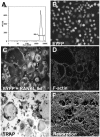
EYFP-expressing RAW 264.7 cells differentiate into functionally authentic osteoclasts. (A) FACS analysis of postsorted stable expressing EYFP-RAW 264.7 cells. (B through F) EYFP RAW 264.7 cells were stimulated with RANKL to induce differentiation. After 5 days, fully differentiated OCs were either examined by confocal microscopy to compare EYFP expression levels with their mononuclear precursors (B and C) or stained with rhodamine-conjugated phalloidin to visualize F-actin rings (D). (E) TRAP activity of EYFP-expressing OCs. (F) Resorptive pits formed after 9 days of culture on dentin slices.
FIG. 6.
FIG. 6.
Overexpression of dominant-negative Rab3DN135I impairs osteoclastic bone resorption in vitro. (A) RAW 264.7 cells stably expressing EYFP-Rab3D and mutant proteins were cultured on dentin slices in the presence of RANKL (100 ng/ml) to analyze resorption activity. After 9 days, TRAP-positive cells were removed and resorptive lacunae were assessed by scanning electron microscopy. Whereas EYFP-Rab3D, constitutively active EYFP-Rab3DQ81L and EYFP-Rab3DΔCXC expressing OCs produce numerous well-defined resorptive lacunae, those formed by EYFP-Rab3DN135I OCs are shallow and poorly demarcated. (B) Resorption parameters of OCs expressing EYFP-Rab3D fusion proteins. Pit number, diameter and percentage resorbed area per dentin slice were examined by scanning electron microscopy. Indices were calculated using image analysis software (NIH). *, P < 0.05; **, P < 0.001 compared with EYFP in all panels; error bars represent standard deviation of means (n = 6).
FIG. 7.
FIG. 7.
Overexpression of EYFP-Rab3D and its mutants does not affect osteoclast formation or F-actin organization. (A) Stably transfected RAW 264.7 cells were cultured on plastic or dentin surfaces in the presence of 100 ng/ml RANKL for 5 and 9 days, respectively. OC-like cells were fixed and then stained for either TRAP alone (left panels) or double stained for TRAP and rhodamine-phalloidin (F-actin; middle and right panels) and visualized by bright field and confocal microscopy. Quantitation of the number of TRAP-positive multinucleate cells (>3 nuclei) per cm2 (B) and number of F-actin rings per dentin slice (C). Error bars represent standard errors of the mean (n = 6).
FIG. 8.
FIG. 8.
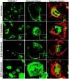
Subcellular distribution of EYFP-Rab3D fusion proteins in RAW 264.7 cells and osteoclast-like cells. RAW 264.7 cells stably expressing EYFP-Rab3D, EYFP-Rab3DQ81L, EYFP-Rab3DN135I and EYFP-Rab3DΔCXC were grown on either glass coverslips or dentin slices in either the absence or the presence of RANKL (100 ng/ml) for 5 to 9 days. Cells were then fixed with 4% paraformaldehyde and processed for confocal microscopy. Actively resorbing OCs were visualized by the presence of F-actin rings using rhodamine-conjugated phalloidin. Insets represent magnifications of hatched regions. Each image is representative of a 0.4-μm section.
FIG. 9.
FIG. 9.
Rab3D modulates the homotypic fusion of granules in living cells. RAW 264.7 cells expressing either EYFP-Rab3D WT or mutant fusion proteins were cultured on Coverglass chamber slides for 24 h before being analyzed by time-lapse confocal microscopy on a heated stage (37°C). Images were recorded at 5 s intervals for 10 min periods. Left panel illustrates merged phase contrast and EYFP images for each cell. Insets represent enlarged selected images from a time-lapse series. Arrows denote EYFP-Rab3D-bearing vesicles/granules undergoing fusion events. Note little dynamics is observed in EYFP-Rab3DN135I expressing cells (see videos S1 through S3 in the supplemental material).
FIG. 10.
FIG. 10.
Rab3D is associated with a nonendosomal/lysosomal post-TGN vesicular compartment. Stably transfected RAW 264.7 cells expressing EYFP-Rab3D fusions were labeled with organelle-specific markers. To visualize the TGN, cells were incubated with anti-TGN38 and Alexa Fluor 546-goat anti-rabbit secondary antibodies. For early and recycling endosomes, cells were loaded with either Alexa Fluor 546-transferrin (30 min) or the fluid phase marker β-rhodamine-dextran (10,000 molecular weight) (4 h) prior to fixation. Acidic compartments were identified with LysoTracker Red. Whereas EYFP-Rab3D partially colabeled with the TGN (yellow), very little overlap can be observed with other endocytic compartment markers.
FIG. 11.
FIG. 11.
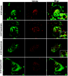
Colocalization of Rab3D with the TGN in osteoclasts. Stably transfected RAW.264.7 cells overexpressing either EYFP-Rab3D or its mutants were cultured on glass coverslips in the presence of RANKL (100 ng/ml). After 5 days, cells were fixed with 4% paraformaldehyde, permeabilized, and labeled with a rabbit polyclonal rat anti-TGN38 (primary) antibody and Alexa Fluor 546-congated goat anti-rabbit (secondary) antibodies as described in Materials and Methods. Cells were then mounted in low fade mounting medium and analyzed by confocal microscopy. Colocalization depicted by yellow color in merged images. Insets represent magnification of hatched regions.
FIG. 12.
FIG. 12.
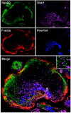
Intracellular localization of Rab3D and TRAP in osteoclasts. RAW 264.7 cell-derived OCs cultured on glass coverslips were fixed with 4% paraformaldehyde before being stained for TRAP activity (magenta) using a fluorescence-based protocol (see Materials and Methods). OCs were subsequently double immunostained for Rab3D (green), and rhodamine-phalloidin (F-actin, red) before being stained with Hoechst to visualize cell nuclei (blue). Each panel shows a projection image of scanned x-y sections through the cell. Merge represents an overlap of all four color channels. Inset represents magnification of hatched region. White color indicates colocalization. Bar, 10 μm.

Similar articles

Cited by

References

    1. Abu-Amer, Y., S. L. Teitelbaum, J. C. Chappel, P. Schlesinger, and F. P. Ross. 1999. Expression and regulation of RAB3 proteins in osteoclasts and their precursors. J. Bone Miner. Res. 14:1855-1860. - PubMed
    1. Alto, N. M., J. Soderling, and J. D. Scott. 2002. Rab32 is an A-kinase anchoring protein and participates in mitochondrial dynamics. J. Cell Biol. 158:659-668. - PMC - PubMed
    1. Altschul, S. F., T. L. Madden, A. A. Schaffer, J. Zhang, Z. Zhang, W. Miller, and D. J. Lipman. 1997. Gapped BLAST and PSI-BLAST: a new generation of protein database search programs. Nucleic Acids Res. 25:3389-3402. - PMC - PubMed
    1. Baldini, G., T. Hohl, H. Y. Lin, and H. F. Lodish. 1992. Cloning of a Rab3 isotype predominantly expressed in adipocytes. Proc. Natl. Acad. Sci. USA 89:5049-5052. - PMC - PubMed
    1. Baldini, G., G. Wang, M. Weber, M. Zweyer, R. Bareggi, J. W. Witkin, and A. M. Martelli. 1998. Expression of Rab3D N135I inhibits regulated secretion of ACTH in AtT-20 cells. J. Cell Biol. 140:305-313. - PMC - PubMed

Publication types

MeSH terms

LinkOut - more resources