. 2005 Jun;25(12):5282-91.
doi: 10.1128/MCB.25.12.5282-5291.2005.

G1/S cell cycle arrest provides anoikis resistance through Erk-mediated Bim suppression

Affiliations

G1/S cell cycle arrest provides anoikis resistance through Erk-mediated Bim suppression

Nicole L Collins et al. Mol Cell Biol. 2005 Jun.

Abstract

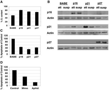
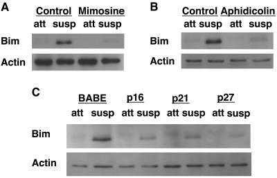
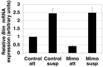
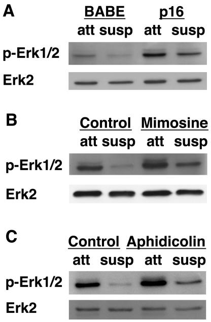
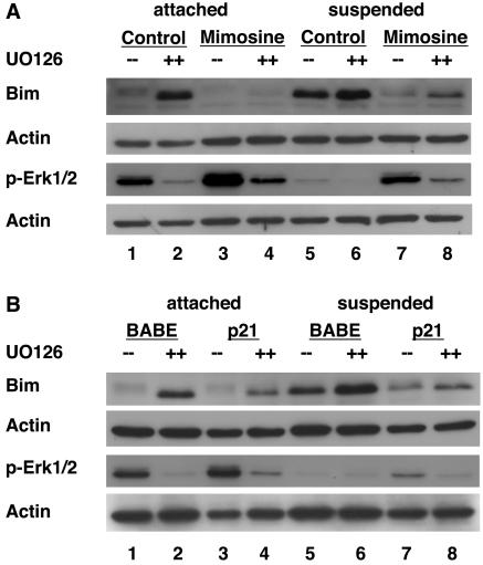
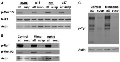
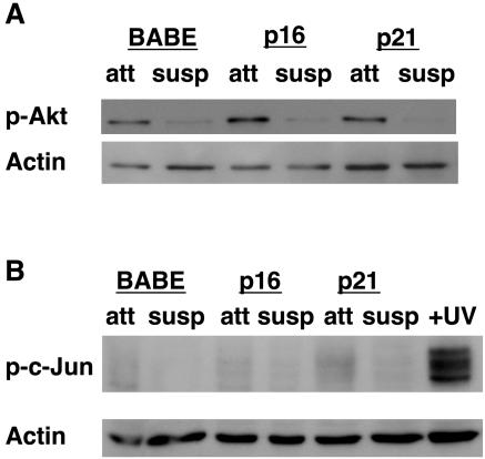
Proper attachment to the extracellular matrix is essential for cell survival. Detachment from the extracellular matrix results in an apoptotic process termed anoikis. Anoikis induction in MCF-10A mammary epithelial cells is due not only to loss of survival signals following integrin disengagement, but also to consequent downregulation of epidermal growth factor (EGFR) and loss of EGFR-induced survival signals. Here we demonstrate that G(1)/S arrest by overexpression of the cyclin-dependent kinase inhibitors p16(INK4a), p21(Cip1), or p27(Kip1) or by treatment with mimosine or aphidicolin confers anoikis resistance in MCF-10A cells. G(1)/S arrest-mediated anoikis resistance involves suppression of the BH3-only protein Bim. Furthermore, in G(1)/S-arrested cells, Erk phosphorylation is maintained in suspension and is necessary for Bim suppression. Following G(1)/S arrest, known proteins upstream of Erk, including Raf and Mek, are not activated. However, retained Erk activation under conditions in which Raf and Mek activation is lost is observed, suggesting that G(1)/S arrest acts at the level of Erk dephosphorylation. Thus, anoikis resistance by G(1)/S arrest is mediated by a mechanism involving Bim suppression through maintenance of Erk activation. These results provide a novel link between cell cycle arrest and survival, and this mechanism could contribute to the survival of nonreplicating, dormant tumor cells that avert apoptosis during early stages of metastasis.


Figures

FIG. 1.
FIG. 1.
G1/S arrest protects from anoikis. (A) Expression of p16INK4a, p21Cip1, or p27Kip1 induces G1 arrest. MCF-10A cells were infected with retroviruses encoding control vector (BABE), p16INK4a, p21Cip1, or p27Kip1. Immediately following puromycin selection for stably infected lines, PI staining and FACS analysis were performed to measure DNA content. Percent G1 content for each condition is shown. These results represent the averages ± SEM from at least three separate experiments. (B) Confirmation of p16INK4a, p21Cip1, or p27Kip1 overexpression under attached and suspended conditions. Following puromycin selection, cells described for panel A were plated on either tissue culture plastic or Poly-HEMA-coated plates in full growth medium for 24 h. Attached (att) and suspended (susp) lysates were harvested and immunoblotted with antisera against p16INK4a, p21Cip1, or p27Kip1 to examine overexpression or with antiserum against actin as a loading control. (C) Expression of p16INK4a, p21Cip1, or p27Kip1 provides anoikis resistance. Cells expressing p16INK4a, p21Cip1, p27Kip1, or control vector (BABE) were plated in Poly-HEMA-coated plates in growth medium containing 0.5% methocellulose for 48 h. Apoptosis was measured by use of a colorimetric DNA fragmentation ELISA. Apoptosis of each sample was normalized compared to that of control vectors. The values shown represent averages of normalized values ± SEM from at least three separate experiments as described in Materials and Methods. (D) G1/S arrest by mimosine or aphidicolin treatment confers anoikis resistance. MCF-10A cells were treated with vehicle control (Control), mimosine (Mimo), or aphidicolin (Aphid) as described in Materials and Methods. Following treatment, cells were plated on Poly-HEMA-coated plates in growth medium containing 0.5% methocellulose with the appropriate drug or vehicle control, and DNA fragmentation ELISAs were performed as described for panel C.
FIG. 2.
FIG. 2.
G1/S arrest inhibits Bim expression following detachment. (A) Mimosine treatment inhibits detachment-induced Bim upregulation. MCF-10A cells were treated with vehicle (Control) or mimosine as described in Materials and Methods. Following vehicle control or mimosine treatment for 24 h, cells were plated on either tissue culture plastic or Poly-HEMA-coated plates in full growth medium for 24 h. Attached (att) and suspended (susp) lysates were harvested and immunoblotted with anti-Bim antiserum to examine Bim expression. (B) Aphidicolin treatment inhibits detachment-induced Bim upregulation. Cells were treated with vehicle (Control) or aphidicolin as described in Materials and Methods. Bim expression was determined 24 h following detachment as described for panel A. (C) CKI expression inhibits detachment-induced Bim upregulation. MCF-10A cells transduced with vectors encoding p16INK4a, p21Cip1, p27Kip1, or empty control vector (BABE) were plated as described for panels A and B. Bim expression was determined following detachment as described for panel A.
FIG. 3.
FIG. 3.
p16INK4a-mediated Bim suppression is dependent on G1/S arrest. (A) CDK4 R24C expression precludes p16INK4a-mediated growth arrest. MCF-10A cells transduced with empty LXSN vector or LXSN encoding CDK4 R24C (denoted R24C) were superinfected with empty BABE vector or BABE encoding p16INK4a. Following double selection with G418 and puromycin, subconfluent, attached cells were harvested for FACS analysis. The percentage of G1 content for each cell type is displayed as a measure of G1 arrest. These results represent averages ± SEM from at least three separate experiments. (B) CDK4 R24C expression allows detachment-induced Bim upregulation in p16INK4a-expressing cells. Following selection, cells described for panel A were plated under attached (att) or suspended (susp) conditions for 24 h, at which point lysates were harvested. The lysates were immunoblotted with antiserum to Bim, p16INK4a, CDK4, or actin (the last as a loading control) under attached and suspended conditions following the coexpression of CDK4 R24C and p16INK4a. These images are of two immunoblots of the same cell lysates. The first blot was probed with antiserum against Bim and then reprobed for actin as a loading control. The second blot was probed with antiserum against p16INK4a and then reprobed for both CDK4 and actin (as a loading control). (C) CDK4 R24C specifically prevents p16INK4a-mediated growth arrest and has no effect on growth arrest mediated by p21Cip1. As described for panel A, MCF-10A cells were transduced with empty LXSN vector or LXSN encoding CDK4 R24C but were superinfected with empty BABE vector or BABE encoding p21Cip1. Following double selection, subconfluent, attached cells were harvested and FACS analysis was performed as described for panel A. (D) CDK4 R24C expression has no effect on detachment-induced Bim upregulation in p21Cip1-expressing cells. Following selection, cells described for panel C were plated under attached or suspended conditions for 24 h and lysates were harvested as described for panel B. Lysates were immunoblotted with antiserum to Bim, CDK4, p21Cip1, or actin. These images are of two separate immunoblots. The first blot was probed with antiserum against Bim and then reprobed for CDK4 and actin (as a loading control). The second blot was probed with antiserum against p21Cip1 and then reprobed for actin as a loading control.
FIG. 4.
FIG. 4.
G1/S arrest does not inhibit detachment-induced Bim mRNA expression. Control and mimosine-treated MCF-10A cells were plated at 400,000 cells/well in either tissue culture-treated (attached) or Poly-HEMA-coated (suspended) six-well plates for 24 h. RNA was harvested, and double-stranded cDNA using oligo(dT) primers was synthesized from 5 μg total RNA. Real-time quantitative PCR was performed on equal amounts of total RNA as described previously (47). The relative Bim mRNA expression for each condition was plotted. Error bars represent the SEM of nine quantitative PCRs from three independent experiments.
FIG. 5.
FIG. 5.
G1/S arrest increases Erk phosphorylation under attached and suspended conditions. (A) Expression of p16INK4a enhances Erk phosphorylation in attached cells and following detachment. MCF-10A cells transduced with control vector (BABE) or BABE encoding p16INK4a were placed under attached or suspended conditions following selection. After 24 h in suspension, cell lysates were harvested and immunoblotted with antiserum against total Erk2 or against phosphorylated Erk1/2 (p-Erk1/2). (B) Mimosine treatment enhances Erk phosphorylation under attached and suspended conditions. MCF-10A cells were treated as described for Fig. 2A, except the lysates were immunoblotted with the Erk antibodies described for Fig. 5A. (C) Aphidicolin treatment enhances Erk phosphorylation in attached and suspended cells. MCF-10A cells were treated as described for Fig. 2B, except the lysates were immunoblotted with the Erk antibodies described for panel A.
FIG. 6.
FIG. 6.
Increased Erk phosphorylation is necessary for G1/S-mediated Bim suppression. (A) Increased Erk activity is necessary for mimosine-mediated Bim suppression. MCF-10A cells were treated with DMSO (control) or 200 μM mimosine as described in Materials and Methods. Following 24 h of DMSO or mimosine treatment, cells were then treated with either DMSO (−−) or the MEK inhibitor UO126 (++) and incubated for another 24 h. Cells were then either left attached or placed in suspension in the presence of all drugs or vehicle for 24 h. At this point, cell lysates were prepared and immunoblotted with antiserum to Bim, actin (as a loading control), or phosphorylated Erk (p-Erk1/2). These images are of two immunoblots of the same cell lysates with corresponding actin loading controls beneath each. (B) Increased Erk phosphorylation is required for p21Cip1-mediated Bim suppression. MCF-10A cells expressing either control vector (BABE) or BABE encoding p21Cip1 were treated with DMSO or UO126, placed under attached or suspended conditions, and cell lysates were prepared. These lysates were immunoblotted with antiserum to Bim, phosphorylated Erk1/2 (p-Erk1/2), or actin (loading control). These images are of two immunoblots of the same cell lysates with corresponding actin loading controls beneath each.
FIG. 7.
FIG. 7.
G1/S arrest does not activate the Ras/MAPK pathway upstream of Erk. (A) CKI expression does not enhance Mek phosphorylation. Attached (att) and suspended (susp) lysates harvested from MCF-10A cells treated similarly to those in Fig. 1B were immunoblotted with antisera against phosphorylated Mek1/2 (p-Mek 1/2), Mek1, and actin (as a loading control). (B) G1/S arrest by mimosine or aphidicolin treatment does not enhance Raf or Mek phosphorylation. Attached (att) and suspended (susp) lysates from cells treated with vehicle control, mimosine (Mimo), or aphidicolin (Aphid) as described for Fig. 2A and B were immunoblotted with antisera against phosphorylated Raf (p-Raf), phosphorylated Mek1/2 (p-Mek1/2), and actin (as a loading control). (C) Overall tyrosine phosphorylation is not enhanced following mimosine treatment. Lysates treated similarly to those in Fig. 2A were immunoblotted with antisera against phosphorylated tyrosine (p-Tyr) and actin (as a loading control).
FIG. 8.
FIG. 8.
The PI3K/Akt and JNK/MAPK pathways do not contribute to G1/S arrest-mediated anoikis resistance. (A) Akt phosphorylation does not change following G1 arrest. MCF-10A cells overexpressing p16INK4a, p21Cip1, or control vector (BABE) were plated under attached (att) or suspended (susp) conditions for 24 h. Lysates were harvested as described for Fig. 1B and immunoblotted with antiserum against phosphorylated Akt (p-Akt) or actin (as a loading control). (B) c-Jun is not phosphorylated following detachment or G1 arrest in MCF-10A cells. Lysates from attached (att) or suspended (susp) cells treated as described for panel A, along with lysates from UV-irradiated MCF-10A cells (as a positive control), were harvested as described for Fig. 1B. Lysates were immunoblotted with antiserum against phosphorylated c-Jun (p-c-Jun) as a measure of the activity of the JNK/MAPK pathway. The immunoblot was then reprobed for actin as a loading control.
FIG. 9.
FIG. 9.
G1/S arrest inhibits Bim upregulation and maintains Erk phosphorylation following growth factor withdrawal. MCF-10A cells transduced with control vector (BABE) or BABE encoding p21Cip1 were cultured with (++) or without (−−) EGF for 24 h and analyzed for Bim expression and Erk phosphorylation (p-Erk1/2) as described for Fig. 6.

References

    1. Allan, L. A., N. Morrice, S. Brady, G. Magee, S. Pathak, and P. R. Clarke. 2003. Inhibition of caspase-9 through phosphorylation at Thr 125 by ERK MAPK. Nat. Cell Biol. 5:647-654. - PubMed
    1. Alpan, R. S., and A. B. Pardee. 1996. p21WAF1/CIP1/SDI1 is elevated through a p53-independent pathway by mimosine. Cell Growth Differ. 7:893-901. - PubMed
    1. Asada, M., T. Yamada, H. Ichijo, D. Delia, K. Miyazono, K. Fukumuro, and S. Mizutani. 1999. Apoptosis inhibitory activity of cytoplasmic p21(Cip1/WAF1) in monocytic differentiation. EMBO J. 18:1223-1234. - PMC - PubMed
    1. Biswas, S. C., and L. A. Greene. 2002. Nerve growth factor (NGF) down-regulates the Bcl-2 homology 3 (BH3) domain-only protein Bim and suppresses its proapoptotic activity by phosphorylation. J. Biol. Chem. 277:49511-49516. - PubMed
    1. Blagosklonny, M. V. 2002. Are p27 and p21 cytoplasmic oncoproteins? Cell Cycle 1:391-393. - PubMed

Publication types

MeSH terms