. 2005 Jun 1;118(Pt 11):2425-33.
doi: 10.1242/jcs.02371.

Localization of all seven messenger RNAs for the actin-polymerization nucleator Arp2/3 complex in the protrusions of fibroblasts

Affiliations

Localization of all seven messenger RNAs for the actin-polymerization nucleator Arp2/3 complex in the protrusions of fibroblasts

Lisa A Mingle et al. J Cell Sci. .

Abstract

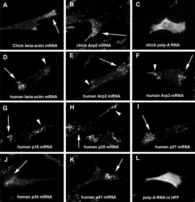
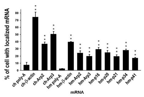
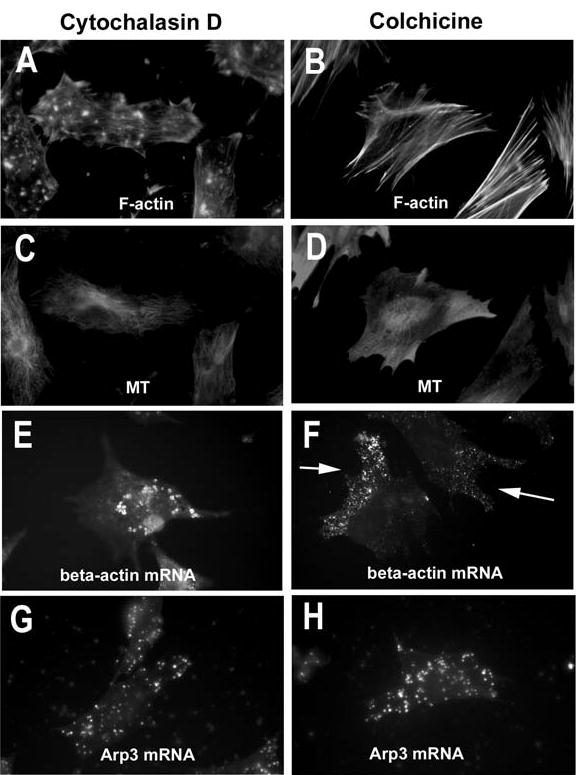
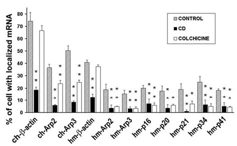
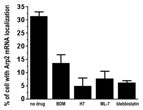
The actin-related protein 2/3 (Arp2/3) complex is a crucial actin polymerization nucleator and is localized to the leading protrusions of migrating cells. However, how the multiprotein complex is targeted to the protrusions remains unknown. Here, we demonstrate that mRNAs for the seven subunits of the Arp2/3 complex are localized to the protrusions in fibroblasts, supporting a hypothesis that the Arp2/3 complex is targeted to its site of function by mRNA localization. Depletion of serum from culture medium inhibits Arp2/3-complex mRNA localization to the protrusion, whereas serum stimulation leads to significant mRNA localization within 30 minutes. The effect of serum suggests that Arp2/3-complex mRNA localization is a cellular response to extracellular stimuli. The localization of the Arp2/3 complex mRNAs is dependent on both actin filaments and microtubules, because disruption of either cytoskeletal system (with cytochalasin D and colchicine, respectively) inhibited the localization of all seven subunit mRNAs. In addition, myosin inhibitors significantly inhibit Arp2 mRNA localization in chicken embryo fibroblasts, suggesting a myosin motor dependent mechanism for Arp2/3-complex mRNA localization.


Figures

Fig. 1
Fig. 1
The Arp2/3 complex is localized to protrusions of chicken embryo fibroblasts. The localization of Arp2 was used to infer the location of the Arp2/3 complex and confirmed in chicken embryo fibroblasts to be in a location like that seen in other cell types. Arrows point to the protrusions.
Fig. 2
Fig. 2
Effective detection of low-abundance mRNA using FISH-TSA. Images show representative chicken embryo fibroblasts processed using DIG-labelled sense (A) or antisense (B) RNA probe for chicken Arp3 mRNA. White dots in A mark the cell border. Blue indicates DAPI staining of nucleus, red indicates Arp3 mRNA in the cell and the arrow points to the mRNA localized to protrusions. Bar, 10 μm.
Fig. 3
Fig. 3
The mRNAs for the Arp2/3 complex are localized to protrusions in fibroblasts. Images show representative poly-A RNA and β-actin and Arp2/3-complex mRNA distributions in chicken embryo fibroblasts and human foreskin fibroblasts as detected with FISH-TSA. The fibroblasts have enriched Arp2/3-complex mRNA in their protrusions (arrows). In some cells, a proportion of the β-actin and Arp2/3-complex mRNA can also be found at the trailing protrusion of the cells (arrowheads). Poly-A RNA appears to be distributed uniformly.
Fig. 4
Fig. 4
Quantification of Arp2/3-complex mRNA localization in fibroblasts. The proportion of the cells with localized mRNA was quantified by counting all the cells in randomly selected fields in the fluorescence microscope. 300–500 cells were scored for each mRNA from three independent experiments. Error bars indicate s.e.m. **, statistically significant (P<0.01) differences from control basal level of poly-A RNA.
Fig. 5
Fig. 5
Arp2/3-complex and β-actin mRNAs are localized to protrusions in the cell but do not colocalize. Double detection of two types of mRNAs in the same cell was performed by using both DIG-labelled and fluorescein-labelled probe for FISH. TSA was performed sequentially. (A) Chicken Arp3 mRNA. (B) Chicken β-actin mRNA. (C) Merge of A and B. (D) Chicken Arp3 mRNA. (E) Chicken Arp2 mRNA. (F) Merge of D and E. Bar, 10 μm.
Fig. 6
Fig. 6
Arp2 mRNA expression and localization are serum dependent. (A) Representative cells during serum starvation and stimulation. (a) Cells continuously exposed to serum. (b-d) Cells after 4, 8 or 16 hours of serum starvation (without stimulation), respectively. (e-g) Cells after 16 hours of serum starvation, stimulated with 10% of FBS for 0.5, 1 and 2 hours, respectively. Scale bar, 10 μm. (B) Expression of Arp2 mRNA in CEFs after serum starvation and stimulation. The number of Arp2 mRNA fluorescence spots per cell was counted. At least 120 cells were counted for each time point from at least three independent experiments. (C) Semiquantification of Arp2 expression by RT-PCR. CEFs were cultured in 150 mm dishes and then serum starved and stimulated as in A and B. Total RNA was isolated and reverse transcribed into cDNA. The cDNA from equal numbers of cells was amplified by PCR for chicken Arp2. Data represent three independent experiments and are all normalized to time zero (AU, arbitrate unit). Error bars indicate s.e.m. (Inset) Representative image of the RT-PCR product. (D) Localization of Arp2 mRNA to protrusions of CEFs after serum depletion and stimulation. Localization of mRNA was scored using the criteria described in Materials and Methods. For each time point, 300–600 cells were scored from three experiments. Error bars indicate s.e.m. (B-D) 1, always in medium with 10% FBS; 2, 4-hour serum starvation; 3, 8-hour serum starvation; 4, 16-hour serum starvation; 5, 0.5-hour FBS stimulation; 6, 1-hour FBS stimulation; 7, 2-hour FBS stimulation.
Fig. 7
Fig. 7
Localization of Arp2/3-complex mRNA is dependent on both actin filaments and microtubules. Cytochalasin D (CD) and colchicine were used to disrupt actin filaments and microtubules in the fibroblasts, respectively. (A,C,E,G) Cells treated with 0.4 μM cytochalasin D; (B,D,F,H) cells treated with 5 μM colchicine. (A,B) Cells stained for actin filaments with fluorescein-phalloidin. (C,D) Cells stained for microtubules with mouse anti-α-tubulin antibody. (E,F) Localization of β-actin mRNA is dependent on actin filaments but not on microtubules. By contrast, localization of Arp2/3- complex mRNA is dependent on both actin filaments and microtubules (represented by Arp3 mRNA in G,H).
Fig. 8
Fig. 8
Quantification of Arp2/3-complex and actin mRNA localization in fibroblasts treated with cytochalasin D or colchicine. Cytochalasin D (CD) and colchicine were used to disrupt actin filaments and microtubules in the fibroblasts, respectively. The proportion of the cells with localized mRNA was quantified by counting all the cells in randomly selected fields in the fluorescence microscope. 300–500 cells were counted for each type of mRNA from at least three independent experiments. Prefixes: ch, chicken; hm, human. Error bar indicates s.e.m. *, statistically significant (P<0.05) differences from control cells treated with no drug. **, statistically significant differences from control cells treated with no drug (P<0.01).
Fig. 9
Fig. 9
Myosin inhibitors decrease Arp2 mRNA localization in the CEFs. CEFs were serum starved for 16 hours and then stimulated with 10% FBS alone (no drug) or 10% FBS plus 20 mM BDM, 100 μM H7, 40 μM ML-7 or 100 μM blebbistatin for 2 hours before fixation for FISH-TSA for Arp2 mRNA. Data are from at least 300 cells that derived from at least three independent experiments with error bar indicating s.e.m. All the data from drug-treated samples are significantly different from the no drug control (P<0.01).

Comment in

  • Localizing cellular housekeeping.
    Hordijk PL. Hordijk PL. Nat Rev Mol Cell Biol. 2011 Dec;12(12):771. doi: 10.1038/nrm3206. Nat Rev Mol Cell Biol. 2011. PMID: 21971042 No abstract available.

References

    1. Bailly M, Macaluso F, Cammer M, Chan A, Segall JE, Condeelis JS. Relationship between Arp2/3 complex and the barbed ends of actin filaments at the leading edge of carcinoma cells after epidermal growth factor stimulation. J Cell Biol. 1999;145:331–345. - PMC - PubMed
    1. Bailly M, Ichetovkin I, Grant W, Zebda N, Machesky LM, Segall JE, Condeelis J. The F-actin side binding activity of the Arp2/3 complex is essential for actin nucleation and lamellipod extension. Curr Biol. 2001;11:620–625. - PubMed
    1. Barbarese E, Koppel DE, Deutscher MP, Smith CL, Ainger K, Morgan F, Carson JH. Protein translation components are colocalized in granules in oligodendrocytes. J Cell Sci. 1995;108:2781–2790. - PubMed
    1. Bassell GJ, Oleynikov Y, Singer RH. The travels of mRNAs through all cells large and small. FASEB J. 1999;13:447–454. - PubMed
    1. Berleth T, Burri M, Thoma G, Bopp D, Richstein S, Frigerio G, Noll M, Nusslein-Volhard C. The role of localization of bicoid RNA in organizing the anterior pattern of the Drosophila embryo. EMBO J. 1988;7:1749–1756. - PMC - PubMed

Publication types