. 2005 Jul;37(7):739-44.
doi: 10.1038/ng1592. Epub 2005 May 27.

TRPC6 is a glomerular slit diaphragm-associated channel required for normal renal function

Affiliations

TRPC6 is a glomerular slit diaphragm-associated channel required for normal renal function

Jochen Reiser et al. Nat Genet. 2005 Jul.

Abstract

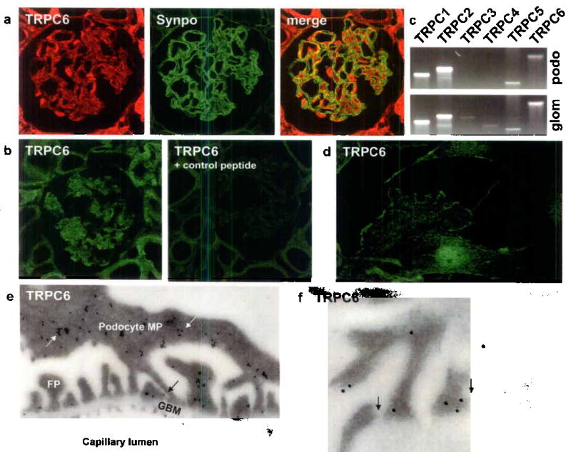
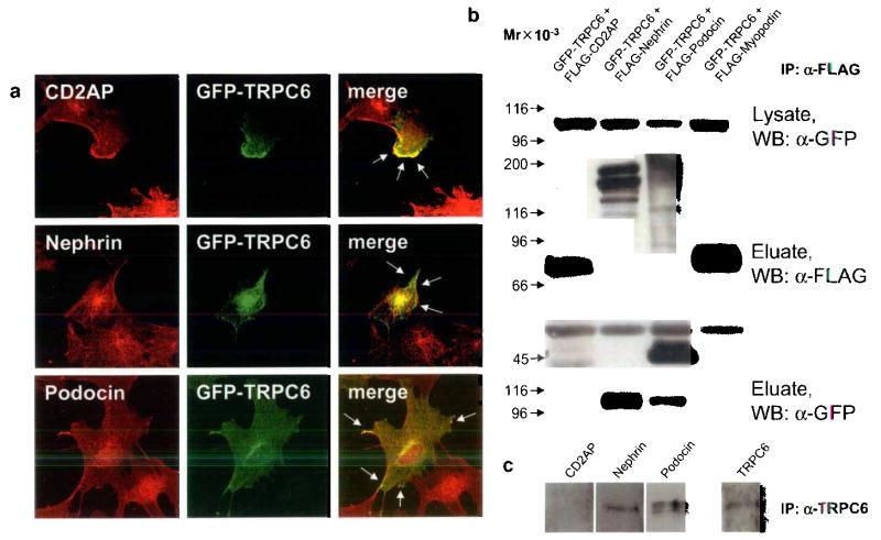
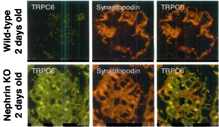
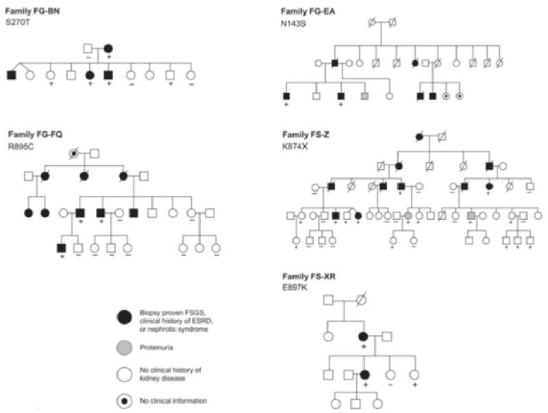
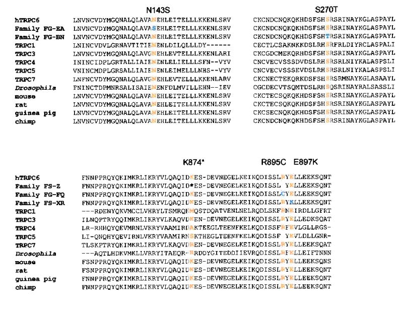
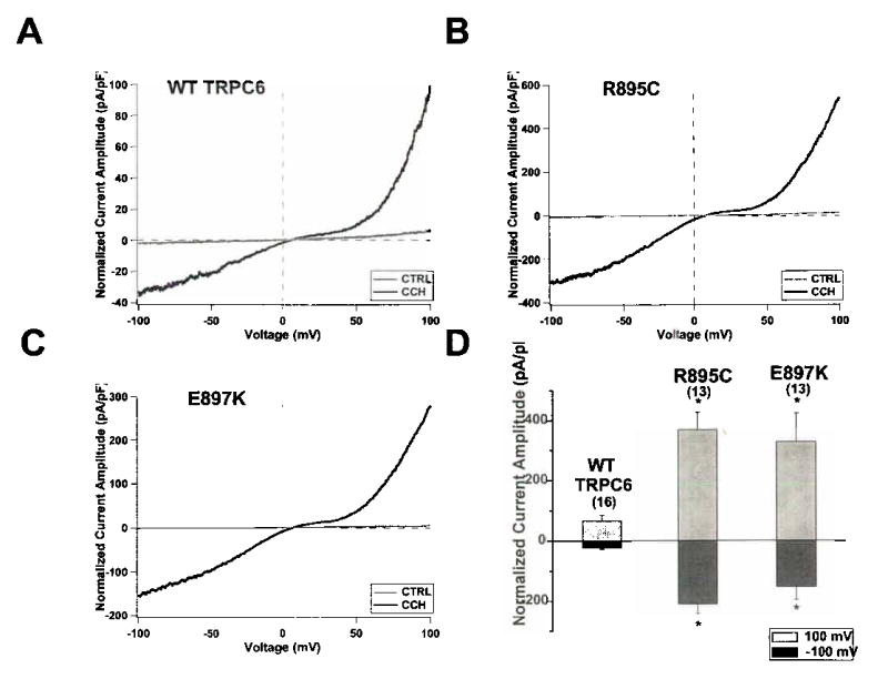
Progressive kidney failure is a genetically and clinically heterogeneous group of disorders. Podocyte foot processes and the interposed glomerular slit diaphragm are essential components of the permeability barrier in the kidney. Mutations in genes encoding structural proteins of the podocyte lead to the development of proteinuria, resulting in progressive kidney failure and focal segmental glomerulosclerosis. Here, we show that the canonical transient receptor potential 6 (TRPC6) ion channel is expressed in podocytes and is a component of the glomerular slit diaphragm. We identified five families with autosomal dominant focal segmental glomerulosclerosis in which disease segregated with mutations in the gene TRPC6 on chromosome 11q. Two of the TRPC6 mutants had increased current amplitudes. These data show that TRPC6 channel activity at the slit diaphragm is essential for proper regulation of podocyte structure and function.


Figures

FIGURE 1
FIGURE 1
TRPC6 expression in the kidney glomerulus. (a) Confocal microscopy shows TRPC6 (red) expression in the glomerulus. TRPC6 co-localizes with the podocyte marker Synaptopodin (green) resulting in a yellow overlap. (b) Compared to the TRPC6 antibody labeling, preabsorption of TRPC6 antibody with the control peptide results in a negative staining. (c) Analysis of TRPC1-6 mRNA expression in cultured podocytes by RT-PCR. In addition to TRPC6 mRNA, also TRPC1, TRPC2 and TRPC5 mRNA is detected in total podocyte RNA (podo). TRPC3 and TRPC4 mRNA is detected in total glomerular RNA (glom) but not in total podocyte RNA. (d) TRPC6 localizes in cultured podocytes to the cell membrane as shown by immunostaining. (e) Immunogold labeling displays TRPC6 localization in podocyte major processes (MP, white arrows) and foot processes (FP). Of note, TRPC6 in podocyte FP is located in close vicinity to the slit diaphragm (black arrows). (f) High-power view of slit diaphragm areas (arrows) display heavy TRPC6 immunogold labeling.
FIGURE 2
FIGURE 2
TRPC6 co-localizes and directly interacts with slit diaphragm proteins. (a) GFP-TRPC6 co-localizes with CD2AP, nephrin and podocin at the cell membrane of cultured podocytes as shown by confocal microscopy (arrows). (b) GFPTRPC6 associates with FLAG-tagged nephrin and podocin but not with CD2AP in co-transfected HEK293 cells. GFP-tagged TRPC6 was detected in total cell lysate by immunoblotting using an anti-GFP antibody (upper panel). FLAG-tagged fusion proteins were immunoprecipitated, eluted and visualized with an anti-FLAG antibody (middle panel). Co-immunoprecipitated GFP-TRPC6 was detected in eluate fractions (lower panel). FLAG-myopodin served as a negative control for TRPC6-binding. (c) Endogenous co-immunoprecipitation of TRPC6 with slit diaphragm proteins from cultured podocytes. TRPC6 interacts with nephrin and podocin but not with CD2AP. Mr, relative molecular mass; WB, primary antibody used for Western Blotting.
FIGURE 3
FIGURE 3
TRPC6 is upregulated in 2 days old nephrin-KO mouse glomeruli as shown by fluorescence microscopy and immunogold labeling. Weak TRPC6 expression was detected in glomeruli of 2 days old wt-mice (upper panel), TRPC6 was found to be upregulated in glomeruli of 2 days old nephrin-KO mice (lower panel). Of note, TRPC6 forms aggregates within the glomerulus. Double labeling of TRPC6 with the podocyte marker synaptopodin demonstrates the localization of TRPC6 in kidney podocytes resulting in a yellow staining pattern.
FIGURE 4
FIGURE 4
Familial FSGS pedigrees. TRPC6 variants segregating within each family are indicated. Genotyped individuals are indicated as carrying (+) or not carrying (–) the variant identified in each family.
FIGURE 5
FIGURE 5
Sequence alterations. TRPC channel sequence alignments (using T-COFFEE). Mutated residues all occur in highly conserved amino acid residues.
FIGURE 6
FIGURE 6
Representative whole-cell currents measured from HEK293-M1 cells transiently transfected with WT TRPC6 (a), R895C (b) or E897K (c) cDNA. Current traces were recorded as cells were perfused with control bath solution (CTRL, gray traces) or 100 μM carbachol (CCh, black traces). Voltage ramps from –100 mV to 100 mV over 150 ms were applied every 3.45 seconds from a holding potential of 0 mV. Current amplitude was normalized for cell capacitance. (d) Average normalized current amplitude measured at –100 mV (dark bars) and 100 mV (light bars) from cells expressing WT TRPC6, R895C or E897K. R895C and E897K current amplitudes were significantly increased at both –100 mV and 100 mV compared to WT current amplitudes (*, p < 0.01). The number of experiments for each is shown in parentheses, and the error bars show the S.E.M. for each measurement.

Comment in

  • A new TRP to kidney disease.
    Gudermann T. Gudermann T. Nat Genet. 2005 Jul;37(7):663-4. doi: 10.1038/ng0705-663. Nat Genet. 2005. PMID: 15990884 No abstract available.

References

    1. Zandi-Nejad K, Eddy AA, Glassock RJ, Brenner BM. Why is proteinuria an ominous biomarker of progressive kidney disease? Kidney Int Suppl. 2004:S76–89. - PubMed
    1. Somlo S, Mundel P. Getting a foothold in nephrotic syndrome. Nat Genet. 2000;24:333–5. - PubMed
    1. Pavenstadt H, Kriz W, Kretzler M. Cell biology of the glomerular podocyte. Physiol Rev. 2003;83:253–307. - PubMed
    1. Pollak MR. Inherited podocytopathies: FSGS and nephrotic syndrome from a genetic viewpoint. J Am Soc Nephrol. 2002;13:3016–23. - PubMed
    1. Winn MP, et al. Mutation in TRPC6 causes familial focal segmental glomeruloslcerosis. J Am Soc Nephrol. 2004;15:33A. - PubMed

Publication types