Review
. 2005 Jun;26(6):294-301.
doi: 10.1016/j.tips.2005.04.006.

Cutting back on pro-protein convertases: the latest approaches to pharmacological inhibition

Affiliations
Review

Cutting back on pro-protein convertases: the latest approaches to pharmacological inhibition

Martin Fugère et al. Trends Pharmacol Sci. 2005 Jun.

Abstract

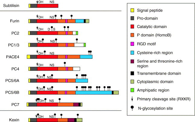
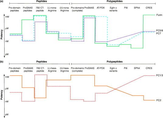
The secretory pathway in cells possesses an elaborate set of endoproteolytic enzymes that carry out a crucial step in protein precursor maturation. This step is proteolytic activation by cleavage at specific pairs of basic residues. These enzymes, named pro-protein convertases (PCs), are responsible for generating bioactive peptides and activating several enzymes and growth factors that are implicated in many important physiological events. PCs have roles in several pathologies including viral infections and cancers and, thus, are promising targets for therapeutic applications. Recent structural and homology-modeling studies demonstrate more similarity than expected at the catalytic site of the seven PCs, which makes the development of selective drugs to target individual PCs frustrating. Based on this information, we review the latest strategies to inhibit PCs, which might lead to the development of specific compounds.


Figures

Figure 1
Figure 1
PC architecture. The PCs have a highly homologous architecture that is composed of: (i) an N-terminal signal peptide, which is responsible for directing proteins into the secretory pathway; (ii) a pro-domain, which acts as a putative intramolecular chaperone for facilitated transportation, folding and regulation of enzymatic activity; (iii) a catalytic domain, which contains an active site that is responsible for substrate-specific interactions and cleavage; (iv) a P domain, which is an independent folding moiety with the conformation of a barrel-like roll that is essential for structural cohesion of the enzyme and activity by regulating stability, and Ca2+- and pH-dependence; and (v) a divergent C-terminal domain that can contain membrane attachment sequences, Cys-rich regions and intracellular sorting signals. Additionally, furin contains two functional groups for internal ionic interactions (Ca2+-binding sites), one of which is beneath the active site and provides stability. D (aspartate), H (histidine) and S (serine) are part of the catalytic triad. N (asparagine) is located in the oxyanion hole and stabilizes, through its NH2 group, the negative charge that is developed in the peptide bond as a result of the nucleophilic attack from the catalytic serine. At least two isoforms of PC5/6 (A and B) have been identified, both of which are encoded by the same gene.
Figure 2
Figure 2
Potency and specificity of PC inhibitors. Schematic representation of the inhibitory potency of peptide and polypeptide inhibitors of PCs. Both PCs of the constitutive secretory pathway (furin, PC5/6 and PC7) (a) and PCs of the regulated secretory pathway (PC2 and PC1/3) (b) are shown. Inhibitory potency (logarithmic scale) is represented by the average of the Ki values determined in vitro in studies cited in this review. For example, most pro-domain peptides inhibit furin, PC1/3, PC5/6 and PC7 at mid-nanomolar concentrations, but are ineffective at PC2. The lower the value, the higher the inhibition potency. Diagonal traces mean that inhibition potency is undetermined.
Figure 3
Figure 3
Structure aspects of PCs. (a) Ribbon representation of the furin crystal structure complexed with the permanent inhibitor dec-RVKR-cmk showing the three-dimensional arrangement of the catalytic and P domains, which are essential for PC activity. Dec-RVKR-cmk is shown in ball and stick rendering where green is carbon, blue is nitrogen and red is oxygen. The catalytic triad (Asp153, His194 and Ser368) is shown in yellow. α-Helical regions are shown in red, β-sheets are shown in blue, and the two Ca2+-binding sites are shown as purple spheres. The image is rendered using iMol 0.3 (http://www.pirx.com/iMol/). (b) Representation of the electrostatic surface potential of the active site of furin with a modeled sequence of six residues from the C-terminal of the furin pro-domain (KRRTKR). White annotations indicate the inhibitor residues and black annotations show the catalytic triad and the subsite residues (S1–S6). (c,d) Representation of the S1 subsite with a P1 arginine residue for furin (c) and PC7 (d). The amino acids of the S1 subsite (furin: Asp258 and Asp306; and PC7: Asp292 and Asp340) and those in close proximity (within 3.2 Å) are shown in a ball-and-stick representation, whereas the P1 arginine is rendered as both ball and stick and a mesh surface. This comparison demonstrates the close identity of the S1 subsites, with the exception of Thr309 for furin and Ala343 for PC7. Similar analysis of the S1 subsite of all other PCs reveals perfect identity with furin (not shown).

References

    1. Thomas G. Furin at the cutting edge: from protein traffic to embryogenesis and disease. Nat. Rev. Mol. Cell Biol. 2002;3:753–766. - PMC - PubMed
    1. Rockwell N.C., Thorner J.W. The kindest cuts of all: crystal structures of Kex2 and furin reveal secrets of precursor processing. Trends Biochem. Sci. 2004;29:80–87. - PubMed
    1. Bergeron F., et al. Subtilase-like pro-protein convertases: from molecular specificity to therapeutic applications. J. Mol. Endocrinol. 2000;24:1–22. - PubMed
    1. Kibler K.V., et al. Polyarginine inhibits gp160 processing by furin and suppresses productive human immunodeficiency virus type 1 infection. J. Biol. Chem. 2004;279:49055–49063. - PubMed
    1. Johannsen E., et al. Proteins of purified Epstein-Barr virus. Proc. Natl. Acad. Sci. U. S. A. 2004;101:16286–16291. - PMC - PubMed

Publication types

MeSH terms

LinkOut - more resources