. 2005 Nov 1;391(Pt 3):491-502.
doi: 10.1042/BJ20050344.

N-glycans of core2 beta(1,6)-N-acetylglucosaminyltransferase-I (C2GnT-I) but not those of alpha(1,3)-fucosyltransferase-VII (FucT-VII) are required for the synthesis of functional P-selectin glycoprotein ligand-1 (PSGL-1): effects on P-, L- and E-selectin binding

Affiliations

N-glycans of core2 beta(1,6)-N-acetylglucosaminyltransferase-I (C2GnT-I) but not those of alpha(1,3)-fucosyltransferase-VII (FucT-VII) are required for the synthesis of functional P-selectin glycoprotein ligand-1 (PSGL-1): effects on P-, L- and E-selectin binding

Maëlle Prorok-Hamon et al. Biochem J. .

Abstract

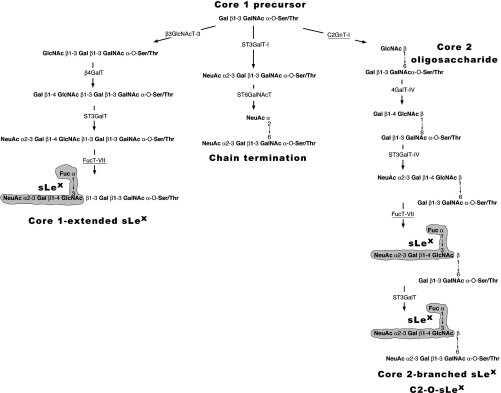
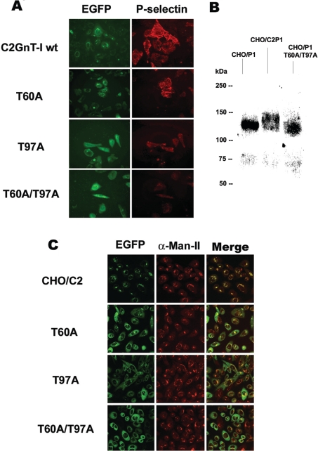
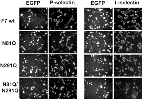
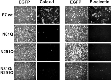
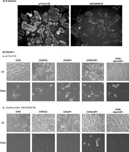
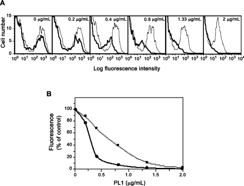
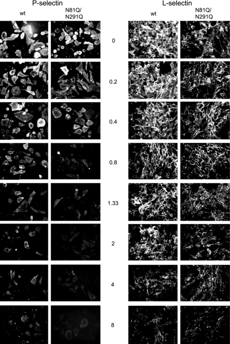
C2GnT-I [core2 beta(1,6)-N-acetyglucosaminyltransferase-I] and FucT-VII [alpha(1,3)-fucosyltransferase-VII] are the key enzymes for the biosynthesis of sialyl-Lewis x determinants on selectin ligands and therefore they represent good drug targets for the treatment of inflammatory disorders and other pathologies involving selectins. In the present study, we examined the importance of N-glycosylation for the ability of C2GnT-I and FucT-VII to generate functional selectin ligands, particularly the PSGL-1 (P-selectin glycoprotein ligand-1). We found that (i) both enzymes have their two N-glycosylation sites occupied, (ii) for C2GnT-I, the N-glycan chain linked to Asn-95 significantly contributes to the synthesis of functional PSGL-1 and is required to localize the enzyme to the cis/medial-Golgi compartment, (iii) all N-glycosylation-deficient proteins of FucT-VII displayr a dramatic impairment of their in vitro enzymatic activities, but retain their ability to fucosylate the core2-modified PSGL-I and to generate P- and L-selectin binding, and (iv) the glycomutants of FucT-VII fail to synthesize sialyl-Lewis x or to generate E-selectin binding unless core2-modified PSGL-1 is present. All combined, our results show a differential functional impact of N-glycosylation on C2GnT-1 and FucT-VII and disclose that a strongly reduced FucT-VII activity retains the ability to fucosylate PSGL-1 on the core2-based binding site(s) for the three selectins.


Figures

Figure 1
Figure 1. Involvement of C2GnT-I and FucT-VII in the biosynthesis of sLex structures
The accepted pathway for the synthesis of sLex structures on O-linked glycans starts with a common core1 precursor and follows three different directions [11]: the formation of core2-branched sLex is initiated by C2GnT-I (underlined) followed by β4GalT-IV, ST3GalT-IV and FucT-VII (underlined). The core 1 precursor can also be modified by β3GlcNAcT-3, forming an extended-core1 oligosaccharide, which is further galactosylated by β4GalT (probably the β4GalT-I) and fucosylated by FucT-VII resulting in the expression of core1-extended sLex structures. No sLex antigen is synthesized (chain termination) if the core1 precursor undergoes sialylation catalysed by ST3GalT-I and ST6GalNAcT-I.
Figure 2
Figure 2. Effects of treatment by TM or PNGase F or removal of N-glycosylation sites on the molecular masses of C2GnT-I and FucT-VII
Cells expressing the wt C2GnT-I (A) or wt FucT-VII (wt F7) (B) were cultured in the presence of 1 μg/ml TM for 72 h, extracted in SDS sample buffer and separated by SDS/PAGE (8% polyacrylamide). After blotting, proteins were probed with an anti-EGFP mAb and developed by the alkaline phosphatase-based method. For treatment with PNGase F, cells expressing C2GnT-I or FucT-VII were extracted in the PNGase F buffer, as described in the ‘Experimental’ section, treated with 20 units/ml PNGase F at 37 °C for 2 h and analysed by SDS/PAGE and immunoblotting as above (see A, a, and B, a). Cells expressing C2GnT-I or its glycomutants (A, b) or FucT-VII or its mutants (B, b) were extracted in SDS sample buffer and analysed as above. (C) Time course of PNGase F treatment (20 units/ml) of the wt FucT-VII. Otherwise conditions were the same as in (A) and (B).
Figure 3
Figure 3. Comparison of P-selectin-binding and subcellular distribution between C2GnT-I and its N-glycosylation-deficient variants
(A) CHO/F7P1 cells were transiently transfected by DNAs coding for the wt C2GnT-I or its glycomutants (EGFP) and incubated with P-selectin–IgM chimaeras. The binding was then revealed by fluorescence microscopy using an RITC (rhodamine isothiocyanate)-conjugated anti-human IgM secondary antibody. Gain and exposure time were kept constant between images. (B) Western-blot analysis of PSGL-1 synthesized in CHO cells in the absence of C2GnT-I (CHO/P1) or in the presence of the enzyme (CHO/C2P1) or the double mutant T60A/T97A (CHO/P1 T60A/T97A). Proteins were separated by SDS/PAGE followed by immunoblotting using the anti-PSGL-1 mAb PL1. (C) Confocal microscopy images of EGFP-conjugated proteins and α-Man-II immunostaining of cells expressing the wt (CHO/C2) or the mutants T60A, T97A and T60A/T97A. The images shown are the Z sections at 5 μm from the top of the cells. The results are representative of two independent experiments.
Figure 4
Figure 4. P- and L-selectin-binding activities of cells expressing the wt FucT-VII or its glycomutants
CHO/C2P1 cells were transiently transfected by DNAs coding for the wt FucT-VII (F7 wt) or its glycomutants (N81Q, N291 and N81Q/N291Q) and incubated with the human IgM-conjugated P-selectin (left panel) or L-selectin (right panel). The binding was then revealed by fluorescence microscopy as in Figure 3(A). Gain and exposure time were kept constant between images.
Figure 5
Figure 5. Altered sLex and E-selectin binding to cells expressing the N-glycosylation-deficient variants of FucT-VII
Cells expressing either the F7 wt or the mutated FucT-VII (N81Q, N291Q and N81Q/N291Q) were assayed for anti-sLex immunostaining using CSLEX-1 mAb (left panel) or E-selectin binding (right panel). The binding of CSLEX-1 mAb was revealed by an RITC (rhodamine isothiocyanate)-conjugated anti-mouse IgM and the binding of E-selectin–IgM chimaeras was revealed by an RITC-conjugated anti-human IgM. Immunostaining was then analysed by fluorescence microscopy. Gain and exposure time were kept constant between images.
Figure 6
Figure 6. Core2-modified PSGL-1 restores sLex expression and E-selectin binding activities of the mutated FucT-VII
(A) CHO/C2P1 cells were transiently transfected with the wt FucT-VII or its double mutant N81Q/N291Q and analysed for E-selectin binding as in Figure 5. (B) Cells expressing C2GnT-I alone (CHO/C2), PSGL-1 alone (CHO/P1), or both molecules (CHO/C2P1), as well as those expressing PSGL-1 with the double mutant of C2GnT-I (CHO/dbmC2P1) were transiently transfected with the wt FucT-VII or its double mutant N81Q/N291Q and analysed for sLex expression as in Figure 5. The parental CHO cells (CHO) expressing none of these molecules were chosen as control. Gain and exposure time were kept constant between images. Cf, phase contrast.
Figure 7
Figure 7. Flow cytometric analyses of P-selectin binding to cells transfected with the wt or the doubly mutated FucT-VII
CHO/C2P1 cells were transfected with constructs coding for the wt FucT-VII or its N81Q/N291Q double mutant, pretreated with increasing concentrations of the anti-PSGL-1 mAb PL1 and assayed for P-selectin binding as described in the Experimental section. (A) Dose-dependent inhibition by PL1 of P-selectin binding to cells expressing the wt FucT-VII (dotted grey lines) and to those expressing the N81Q/N291Q mutant (bold solid lines). (B) The dose–response curves deduced from the fluorescence data in (A) expressed as the percentage of the binding obtained in the absence of PL1. ■, The dose–response inhibition of P-selectin binding to cells expressing FucT-VII; ●, values from the double mutant.
Figure 8
Figure 8. PL1-mediated dose-dependent inhibition of P- and L-selectin binding
Cells expressing the wild-type FucT-VII (wt) or the double mutant N81Q/N291Q were incubated with the human IgM-conjugated P-selectin (left panel) or L-selectin (right panel) in the absence (0 μg/ml) or in the presence of increasing concentrations of PL1 (from 0.2 to 8 μg/ml). The binding of the selectins was then examined by fluorescence microscopy as in Figure 4. Gain and exposure time were kept constant between images.

References

    1. Springer T. A. Traffic signals for lymphocyte recirculation and leukocyte emigration: the multistep paradigm. Cell (Cambridge, Mass.) 1994;76:301–331. - PubMed
    1. Maksimowicz-McKinnon K., Bhatt D. L., Calabrese L. H. Recent advances in vascular inflammation: C-reactive protein and other inflammatory biomarkers. Curr. Opin. Rheumatol. 2004;16:18–24. - PubMed
    1. Lowe J. B. Glycan-dependent leukocyte adhesion and recruitment in inflammation. Curr. Opin. Cell Biol. 2003;15:531–538. - PubMed
    1. Varki A. Selectin ligands: will the real ones please stand up? J. Clin. Invest. 1997;99:158–162. - PMC - PubMed
    1. McEver R. P., Cummings R. D. Perspectives series: cell adhesion in vascular biology. Role of PSGL-1 binding to selectins in leukocyte recruitment. J. Clin. Invest. 1997;100:485–491. - PMC - PubMed

Publication types

MeSH terms