. 2005 Aug 5;280(31):28601-9.
doi: 10.1074/jbc.M501484200. Epub 2005 May 31.

Triadins are not triad-specific proteins: two new skeletal muscle triadins possibly involved in the architecture of sarcoplasmic reticulum

Affiliations

Triadins are not triad-specific proteins: two new skeletal muscle triadins possibly involved in the architecture of sarcoplasmic reticulum

Stéphane Vassilopoulos et al. J Biol Chem. .

Abstract

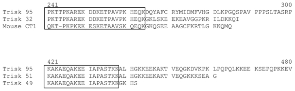
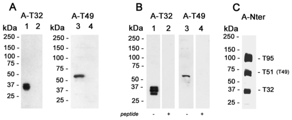
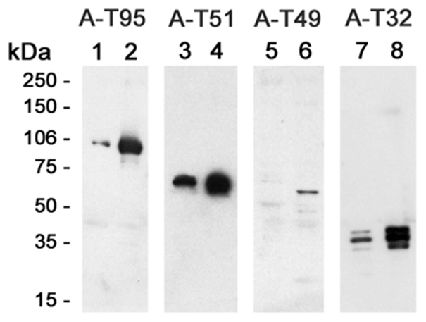
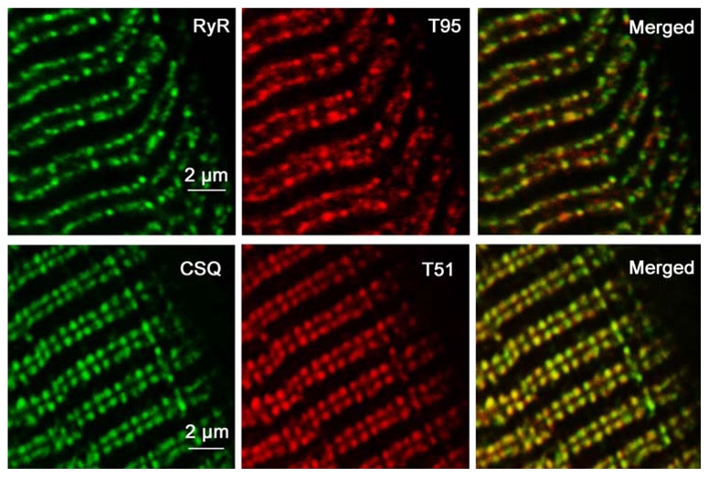
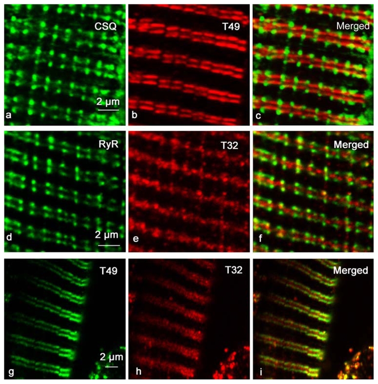
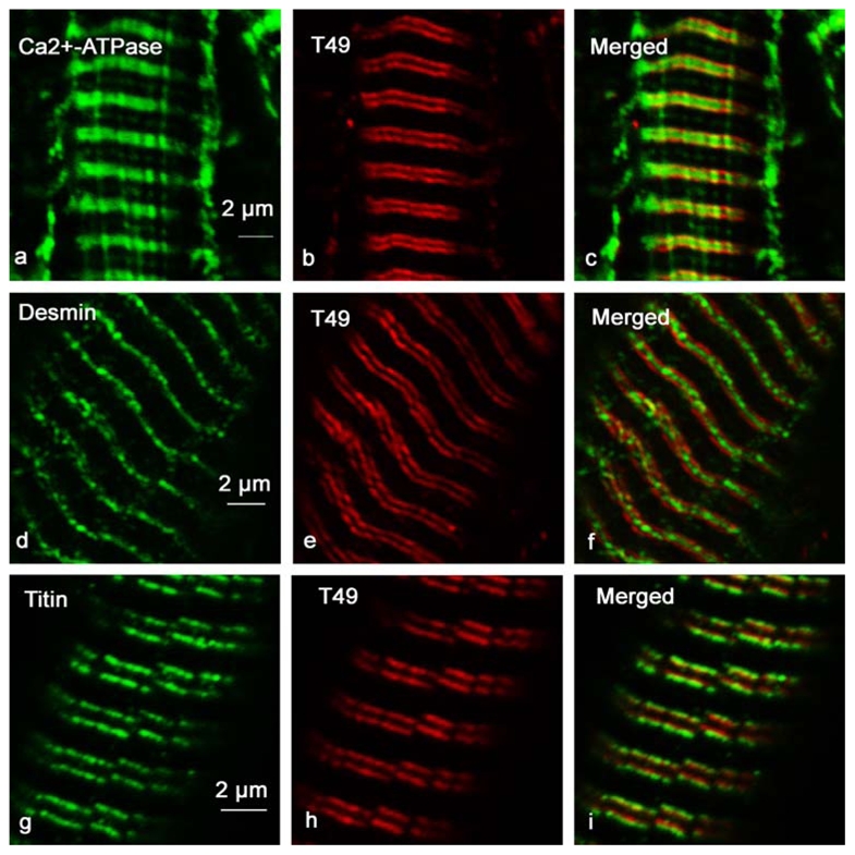
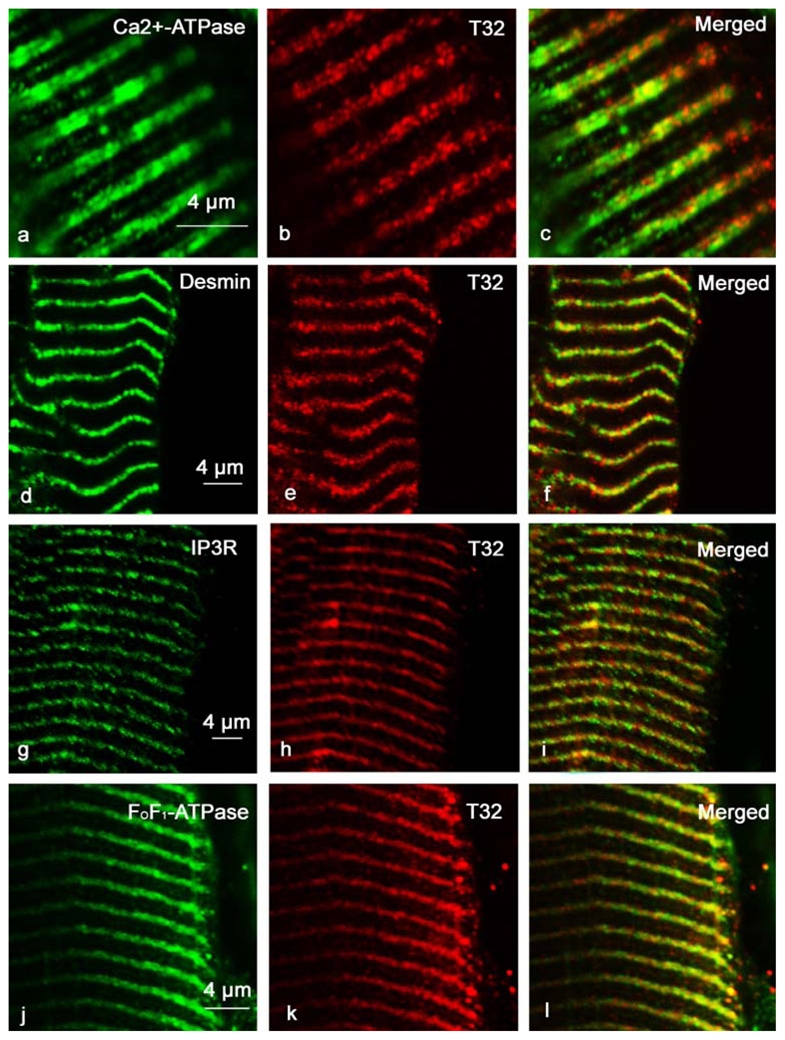
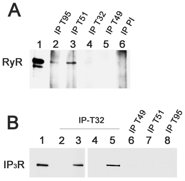
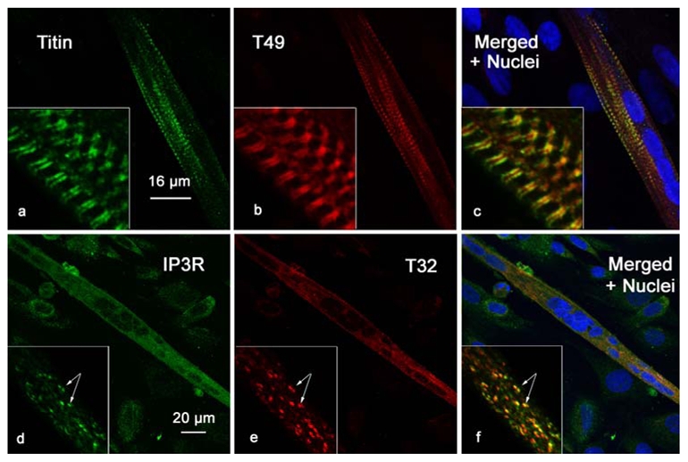
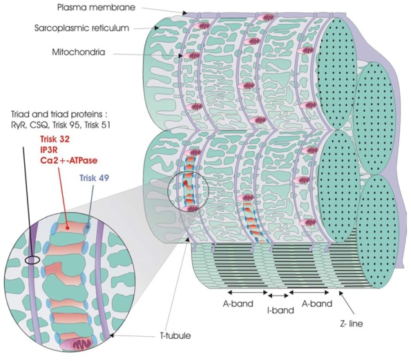
We have cloned two new triadin isoforms from rat skeletal muscle, Trisk 49 and Trisk 32, which were named according to their theoretical molecular masses (49 and 32 kDa, respectively). Specific antibodies directed against each protein were produced to characterize both new triadins. Both are expressed in adult rat skeletal muscle, and their expression in slow twitch muscle is lower than that in fast twitch muscle. Using double immunofluorescent labeling, the localization of these two triadins was studied in comparison to well-characterized proteins such as ryanodine receptor, calsequestrin, desmin, Ca(2+)-ATPase, and titin. None of these two triadins are localized within the rat skeletal muscle triad. Both are instead found in different parts of the longitudinal sarcoplasmic reticulum. We attempted to identify partners for each isoform: neither is associated with ryanodine receptor; Trisk 49 could be associated with titin or another sarcomeric protein; and Trisk 32 could be associated with IP(3) receptor. These results open further fields of research concerning the functions of these two proteins; in particular, they could be involved in the set up and maintenance of a precise sarcoplasmic reticulum structure.


Figures

Figure 1
Figure 1
Sequence alignment of the C-terminal specific sequences of Trisk 32 and Trisk 49 with Trisk 95, Trisk 51 and mouse CT1 (cardiac isoform). Amino acids within the box represent the conserved sequences between the different isoforms. Upper numbers correspond to amino acid positions in the Trisk 95 sequence.
Figure 2
Figure 2. Western blot analysis of Trisk 32 and Trisk 49
Panel A: L6 cells transfected with Trisk 32 or Trisk 49 cDNA. Fifty micrograms of cell lysates were loaded in each lane. Lane 1: L6 transfected with Trisk 32. Lanes 2 and 4: non transfected L6 cells. Lane 3: L6 transfected with Trisk 49. Western blot analysis was performed with anti-Trisk 32 (lanes 1 and 2) or anti-Trisk 49 (lanes 3 and 4). Panel B: Identification of Trisk 32 and Trisk 49 in rat skeletal muscle. Forty micrograms of rat skeletal muscle microsomes were loaded in each lane. Western blot analysis was performed with anti-Trisk 32, without (lane 1) and with (lane 2) pre-incubation of the antibody with the corresponding peptide (10 μg/ml). Western blot analysis in lanes 3 and 4 was performed with anti-Trisk 49, without (lane 3) and with (lane 4) pre-incubation with the corresponding peptide (10 μg/ml). Panel C: Western blot analysis of rat skeletal muscle with an antibody directed against the N-terminal end of triadin, common to all triadin isoforms. Five micrograms of rat skeletal microsomes were loaded. The three major bands correspond to Trisk 95, Trisk 51 and Trisk 32.
Figure 3
Figure 3. Expression of triadin isoforms in slow twitch and fast twitch muscle
The relative amount of each triadin isoform was assessed by Western blot analysis on slow twitch muscle (soleus, lanes 1, 3, 5, 7) and on fast twitch muscle (EDL, lanes 2, 4, 6, 8). Twenty micrograms microsomes from both muscle types were loaded in each lane, except for lane 5, where 40 μg of soleus microsomes were loaded. Analysis was performed with antibodies against Trisk 95 (A-T95, lanes 1 and 2), against Trisk 51 (A-T51, lanes 3 and 4), against Trisk 49 (A-T49, lanes 5 and 6), and against Trisk 32 (A-T32, lanes 7 and 8).
Figure 4
Figure 4. Co-localization of RyR and T95, and of CSQ and T51 in rat skeletal muscle sections
Longitudinal sections of adult rat EDL were immunolabeled with specific antibodies against RyR and Trisk 95 (T95), and CSQ and Trisk 51 (T51). They presented the typical triad protein labeling pattern: double rows of fluorescent dots corresponding to triads pairs on either side of the Z-line.
Figure 5
Figure 5. Trisk 49 and Trisk 32 are not in rat skeletal muscle triad
Longitudinal sections of adult rat EDL were immunolabeled with specific antibodies against CSQ and Trisk 49 (T49) (panels a, b, c), RyR and Trisk 32 (T32) (panels d, e, f) or Trisk 49 and Trisk 32 (panels g, h, i). Trisk 49 labeling (panel b) is presented as a double row of rods, closer to the Z-line than the triads are (represented by CSQ labeling, panel a). Trisk 32 labeling (panel e) appears as a row of dots in the middle of the triad double rows.
Figure 6
Figure 6. Trisk 49 is localized within the longitudinal sarcoplasmic reticulum, at the interface of the A and I bands
Longitudinal sections of adult rat EDL were immunolabeled with specific antibodies against Ca2+-ATPase and Trisk 49 (panels a, b and c), desmin and Trisk 49 (panels d, e and f), and titin and Trisk 49 (panels g, h and i). Ca2+-ATPase is a longitudinal sarcoplasmic reticulum marker, desmin labels the Z-line, and antibodies used against titin are specific to the A-I interface.
Figure 7
Figure 7. Trisk 32 is located within the longitudinal sarcoplasmic reticulum, co-localized with IP3R and mitochondria
Longitudinal sections of adult rat EDL were immunolabeled with specific antibodies against Ca2+-ATPase and Trisk 32 (panels a, b and c), desmin and Trisk 32 (panels d, e and f), IP3R and Trisk 32 (panels g, h and i), and FoF1-ATPase and Trisk 32 (panels j, k and l). Ca2+-ATPase is a longitudinal sarcoplasmic reticulum marker, desmin labels the Z-line, IP3R is located in the longitudinal sarcoplasmic reticulum, and FoF1-ATPase is a mitochondria marker.
Figure 8
Figure 8
A -Trisk 95 and Trisk 51, and not Trisk 32 nor Trisk 49, are associated with RyR. Immunoprecipitations were performed on rat skeletal muscle with antibodies against Trisk 95 (IP-T95, lane 2), against Trisk 51 (IP-T51, lane 3), against Trisk 32 (IP-T32), against Trisk 49 (IP-T49), and with non-immune serum (pre-immune: IP-PI). The presence of RyR in the immunoprecipitated proteins was analyzed by western blot. Lane 1: 5 μg of rat skeletal muscle microsomes. The two bands with high molecular weights are characteristic of RyR, the lower one, present in smaller amount, being a proteolytic degradation of RyR. B- Trisk 32, but not Trisk 49, Trisk 51 or Trisk 95, is associated with IP3R in L6 cells. L6 cells were transfected (lanes 3 and 5) or not (lanes 2 and 4) with Trisk 32 cDNA, or transfected with the cDNA of Trisk 49 (lane 6), Trisk 51 (lane 7) or Trisk 95 (lane 8). Immunoprecipitations were then performed with the corresponding anti-Trisk antibody (anti-Trisk 32 developed in guinea pig, lanes 2–3; anti-Trisk 32 developed in rabbit, lanes 4–5; anti-Trisk 49, lane 6; anti-Trisk 51, lane 7 and anti-Trisk 95, lane 8). The immunoprecipitated proteins were analyzed by western blot using anti-IP3R antibodies. Lane 1: 60 μg of non-transfected L6 cells, for control of IP3R-type III expression levels
Figure 9
Figure 9. Trisk 49 and Trisk 32 expression during differentiation of myoblasts
Primary cultures of satellite cells were induced in differentiation. When myotubes formed, the cells were fixed and labeled with antibodies against titin and Trisk 49 (panels a, b and c), or IP3R and Trisk 32 (panels d, e and f). The nuclei were stained with TO-PRO 3 (blue labeling in panels c and f). Titin and Trisk 49 antibodies labeled only myotubes (aligned nuclei) and not myoblasts (isolated nuclei), and both presented a striated pattern. IP3R and Trisk 32 antibodies label the myotubes (multinucleated cells) and the myoblasts (isolated nuclei), with a diffuse punctuated pattern.
Figure 10
Figure 10. Schematic organization of the triad area and localization of Trisk 49 and Trisk 32
Possible organizations of the sarcoplasmic reticulum and triad, and localization of Trisk 49 and Trisk 32 compared to other known proteins are represented on this figure. Both Trisk 49 and Trisk 32 are located at slightly different places within the non-junctional sarcoplasmic reticulum surrounding the Z-line, while triad proteins are found around the T-tubules.

References

    1. Flucher BE. Dev Biol. 1992;154:245–260. - PubMed
    1. Franzini-Armstrong C. In: Myology. Engel AE, Franzini-Armstrong C, editors. McGraw-Hill Inc; New York: 1994. pp. 176–199.
    1. Flucher BE, Franzini-Armstrong C. Proc Natl Acad Sci USA. 1996;93:8101–8106. - PMC - PubMed
    1. Marty I, Robert M, Villaz M, Lai Y, De Jongh KS, Catterall WA, Ronjat M. Proc Natl Acad Sci USA. 1994;91:2270–2274. - PMC - PubMed
    1. Brandt NR, Caswell AH, Wen SR, Talvenheimo JA. J Membr Biol. 1990;113:237–251. - PubMed

Publication types

MeSH terms

Associated data