. 2005 Jun 7;102(23):8132-7.
doi: 10.1073/pnas.0500269102. Epub 2005 May 31.

The target of ezetimibe is Niemann-Pick C1-Like 1 (NPC1L1)

Affiliations

The target of ezetimibe is Niemann-Pick C1-Like 1 (NPC1L1)

Margarita Garcia-Calvo et al. Proc Natl Acad Sci U S A. .

Abstract

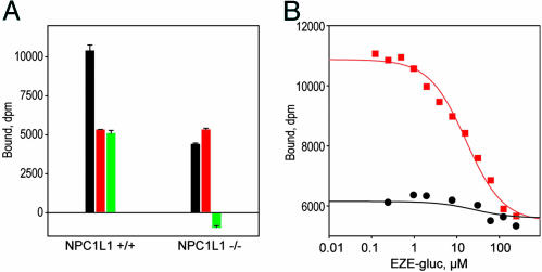
Ezetimibe is a potent inhibitor of cholesterol absorption that has been approved for the treatment of hypercholesterolemia, but its molecular target has been elusive. Using a genetic approach, we recently identified Niemann-Pick C1-Like 1 (NPC1L1) as a critical mediator of cholesterol absorption and an essential component of the ezetimibe-sensitive pathway. To determine whether NPC1L1 is the direct molecular target of ezetimibe, we have developed a binding assay and shown that labeled ezetimibe glucuronide binds specifically to a single site in brush border membranes and to human embryonic kidney 293 cells expressing NPC1L1. Moreover, the binding affinities of ezetimibe and several key analogs to recombinant NPC1L1 are virtually identical to those observed for native enterocyte membranes. KD values of ezetimibe glucuronide for mouse, rat, rhesus monkey, and human NPC1L1 are 12,000, 540, 40, and 220 nM, respectively. Last, ezetimibe no longer binds to membranes from NPC1L1 knockout mice. These results unequivocally establish NPC1L1 as the direct target of ezetimibe and should facilitate efforts to identify the molecular mechanism of cholesterol transport.


Figures

Fig. 1.
Fig. 1.
Influence of taurocholate and digitonin on [3H]EZE-gluc binding. Equal amounts (25 μg of protein) of rat BBM, or membranes from HEK 293 cells transiently expressing recombinant rat and human NPC1L1, were incubated with 25 nM 1 in a final volume of 20 μl until equilibrium was achieved. The incubation conditions were buffer A with and without sodium taurocholate and digitonin to a final concentration of 0.03% and 0.05%, respectively. Total binding (black), nonspecific binding in the presence of 100 μM unlabeled EZE-gluc (red), and specific binding (green) are shown.
Structure 1.
Structure 1.
[3H]EZE-gluc 1.
Fig. 2.
Fig. 2.
Scatchard analyses, kinetics studies, and competition studies for [3H]EZE-gluc 1 binding to rat and monkey enterocyte BBMs. (A) Saturation binding of 1 to rat BBMs. Observed total (filled circles) and nonspecific (open circles) binding, determined in the presence of 100 μM unlabeled EZE-gluc, are shown; specific binding (red squares) was assessed from the difference between total and nonspecific binding. Binding was measured at 2.5 mg/ml protein in a volume of 100 μl after 1 h of incubation. Data were fit by nonlinear regression as described in Methods. (B) Linear Scatchard representation, showing that the binding data identify a single high-affinity site with KD = 542 nM and Bmax = 20.7 pmol/mg protein. (C) Plot shows apparent rate of specific binding of 1 to rat BBM vesicles. Conditions were 25 nM 1 and3mg/ml protein at 25°C. The second-order rate constant kon (0.55 × 10⁻⁴ M-1 s-1) was calculated from kobs (0.004 s-1) as described in Methods.(D) Plot shows rate of dissociation of 1 from the same preparation. After the complex was formed by incubating 25 nM 1 and3mg/ml protein for 1 h, dissociation was initiated by competition with 100 μM unlabeled EZE-gluc. The curve is theoretical for koff = 0.0024 s-1. (E) Equilibrium determination of KD for EZE-gluc by competition of unlabeled compound against 1. Membranes (1.5 mg/ml protein) were incubated with 1 (50 nM) and the indicated concentrations of EZE-gluc for 1 h to ensure equilibrium. KD at equilibrium is 600 nM. (F-J) Corresponding measurements for rhesus monkey, which were conducted with 0.5-1.25 mg/ml protein and 22-50 nM 1, with incubation times of >3 h. Corresponding constants were KD = 41 nM, Bmax = 5.5 pmol/mg protein, kobs = 0.00028 s-1, kon = 3.9 × 10³ M-1 s-1, koff = 1.23 × 10⁻⁴ s-1, and equilibrium KD = 38.6 nM.
Fig. 3.
Fig. 3.
Intestinal distribution of ezetimibe binding sites. (A) Rhesus BBMV. The last 10 cm (ileum) of one small intestine was separated and the remaining intestine was divided into three segments (proximal, middle, and distal) of equal length (70 cm each). (B) Rat BBMV. The last 10 cm (containing the ileum) of small intestines from 25 rats were separated, and the remaining intestines were divided into three segments (proximal, middle, distal) of equal length (36 cm each). Aliquots of BBMs (75 or 200 μg protein per assay for rhesus or rat, respectively) were incubated with 50 nM [3H]EZE-gluc in the absence and presence of 100 μM unlabeled EZE-gluc until equilibrium was achieved.
Fig. 4.
Fig. 4.
Expression of human NPC1L1 in HEK 293 cells. (A) Detection of NPC1L1 in a stably transfected HEK 293 cell line (NPC1L1-293). Cell lysates from HEK 293 cells expressing NPC1L1 and WT cells were analyzed by gel electrophoresis and Western blotting with an anti-NPC1L1 antibody A1801 (19). An excess of NPC1L1-specific peptide was included to assess specificity of the antibody for NPC1L1. (B) Confocal microscope images of a fluorescent EZE-gluc analog (SCH354909) bound to the surface of NPC1L1-293 cells. Binding of SCH354909 to NPC1L1-293 cells (a), nonspecific binding of SCH354909 to NPC1L1-293 cells in the presence of 100μM unlabeled EZE-gluc (b), and binding of SCH354909 to WT HEK 293 cells (c), and nonspecific binding of SCH354909 to WT HEK 293 cells in the presence of 100 μM unlabeled EZE-gluc (d) are shown. In each case, plated cells were incubated in culture media with 500 nM SCH354909 (15) for 4 h at 37°C. Cells were subsequently washed with PBS and fluorescence was detected by using confocal microscopy.
Fig. 5.
Fig. 5.
Comparison of binding affinities for recombinant NPC1L1-293 cell membranes and native BBMs. Plots show determination of Ki values for selected analogs of EZE-gluc against recombinant rat and human NPC1L1 membranes prepared from transiently transfected HEK 293 cells compared with native rat and rhesus BBMs. The binding assays were conducted in a final volume of 20 μl in the presence of 0.03% sodium taurocholate and 0.05% digitonin until equilibrium was achieved. The structures and Ki values are shown in Table 2. Membrane sources are as follows: native rat (A), recombinant rat (B), native rhesus monkey (C), recombinant human (D). Conditions were as follows: 1.25 mg/ml protein and 100 nM 1 for A, B, and D, and 1.25 mg/ml protein and 20 nM 1 for C. Observed total and nonspecific binding in the absence of inhibition were 7,700 and 1,100 (A), 33,000 and 1,100 (B), 7,300 and 367 (C), and 19,200 and 1,000 (D) dpm. Analogs were 1, EZE-gluc (red squares); ent-1, the glucuronide of the enantiomer of ezetimibe (open black circles); 2 (blue circles); 3 (open red squares); 4 (open black triangles); and 5 (blue triangles), as defined in Table 2.
Fig. 6.
Fig. 6.
Loss of binding affinity in NPC1L1-deficient mice. (A) Enterocyte BBMs were prepared from NPC1L1-deficient male mice and same sex WT littermates, and they were tested for binding of 1. Conditions for binding were 5 mg/ml protein and 500 nM 1 in a volume of 20 μl and in the presence of 0.03% sodium taurocholate and 0.05% digitonin. Total (black), nonspecific in the presence of 500 μM cold EZE-gluc (red), and specific (green) binding are indicated, respectively, and error bars represent triplicate measurements. Membranes from WT mice are given on the left, and membranes from NPC1L1-deficient mice are given on the right. Although specific binding is readily detectable in WT mice, it is absent in NPC1L1 deficient mice. (B) Competition of unlabeled EZE-gluc against 1. Membranes from WT mice (red squares) gave Ki = 12,000 nM, whereas specific binding was virtually undetectable in membranes from the knockout animals (black circles). Conditions were those described in A.

References

    1. Grundy, S. M. (1983) Annu. Rev. Nutr. 3, 71-96. - PubMed
    1. Clader, J. W. (2004) J. Med. Chem. 47, 1-9. - PubMed
    1. Davis, H. R., Jr., Zhu, L. J., Hoos, L. M., Tetzloff, G., Maguire, M., Liu, J., Yao, X., Iyer, S. P., Lam, M. H., Lund, E. G., et al. (2004) J. Biol. Chem. 279, 33586-33592. - PubMed
    1. Bays, H. E., Moore, P. B., Drehobl, M. A., Rosenblatt, S., Toth, P. D., Dujovne, C. A., Knopp, R. H., Lipka, L. J., Lebeaut, A. P., Yang, B., et al. (2001) Clin. Ther. 23, 1209-1230. - PubMed
    1. Dujovne, C. A., Ettinger, M. P., McNeer, J. F., Lipka, L. J., LeBeaut, A. P., Suresh, R., Yang, B. & Veltri, E. P. (2002) Am. J. Cardiol. 90, 1092-1097. - PubMed

MeSH terms