. 2005 Jun 6;201(11):1781-91.
doi: 10.1084/jem.20041992. Epub 2005 May 31.

Osteopontin is a hematopoietic stem cell niche component that negatively regulates stem cell pool size

Affiliations

Osteopontin is a hematopoietic stem cell niche component that negatively regulates stem cell pool size

Sebastian Stier et al. J Exp Med. .

Abstract

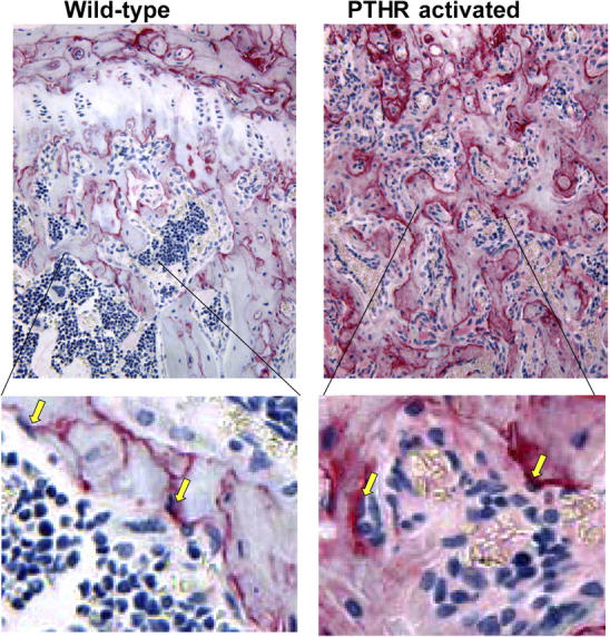
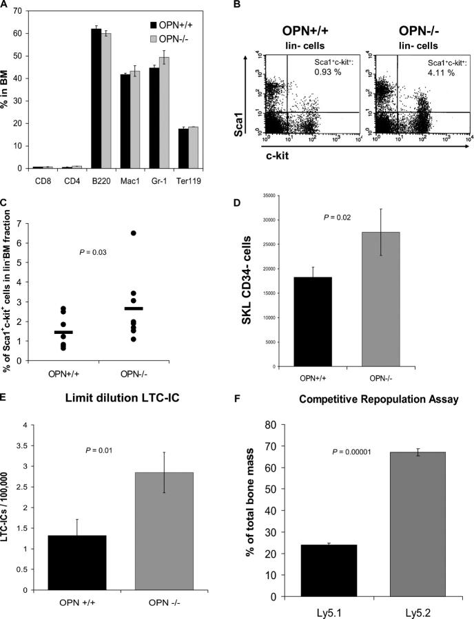
Stem cells reside in a specialized niche that regulates their abundance and fate. Components of the niche have generally been defined in terms of cells and signaling pathways. We define a role for a matrix glycoprotein, osteopontin (OPN), as a constraining factor on hematopoietic stem cells within the bone marrow microenvironment. Osteoblasts that participate in the niche produce varying amounts of OPN in response to stimulation. Using studies that combine OPN-deficient mice and exogenous OPN, we demonstrate that OPN modifies primitive hematopoietic cell number and function in a stem cell-nonautonomous manner. The OPN-null microenvironment was sufficient to increase the number of stem cells associated with increased stromal Jagged1 and Angiopoietin-1 expression and reduced primitive hematopoietic cell apoptosis. The activation of the stem cell microenvironment with parathyroid hormone induced a superphysiologic increase in stem cells in the absence of OPN. Therefore, OPN is a negative regulatory element of the stem cell niche that limits the size of the stem cell pool and may provide a mechanism for restricting excess stem cell expansion under conditions of niche stimulation.


Figures

Figure 1.
Figure 1.
OPN is increased in bone marrow with the activation of osteoblasts. Immunohistochemistry of tibia sections from wild-type (left) or littermate transgenic (right) mice with a constitutively activated PTH/parathyroid related peptide receptor driven by a 2.3-kb fragment of the collagenα1(I) promoter as previously described (reference 4). Sections were stained with antibody to osteopontin (red) and counterstained as described (reference 4), and photographed at 200× (top) with ∼4× image zoom (bottom). Yellow arrows indicate OPN-rich spindle shaped cells lining the trabecular bone that is consistent with an osteoblast morphology.
Figure 2.
Figure 2.
Primitive hematopoietic cells are increased in the bone marrow of OPN/ mice, whereas mature cells are not. (A) Bone marrow cells of OPN+/+ (littermate control) and OPN−/− mice were harvested, counted, and stained with the lineage-specific markers CD8, CD4, B220, Mac1 (CD11b), Gr-1, and Ter119 before flow cytometry. The graph shows the mean percentage ± SEM (n = 3). Bone marrow cells of OPN+/+ and OPN−/− mice were stained with Sca1, c-kit, and lineage markers (CD3, CD4, CD8, B220, Gr-1, CD11b, and Ter119) for flow cytometry. The dot plots show the Sca1+c-kit+ cells (top right) gated on lin bone marrow cells for a single experiment (B) and for a summary of six mice in each group (C). (D) The highly stem cell–enriched CD34 portion of the Sca1+c-kit+lin cells was evaluated in eight pairs of OPN+/+ (littermate control) and OPN−/− mice by flow cytometry. The absolute number was calculated and is shown. (E) To confirm the immuno-phenotypic findings, we performed LTC-IC assays at limiting dilution and calculated the frequency of LTC-ICs. The data shown are the mean frequency ± SEM of LTC-ICs per 100,000 bone marrow cells (P = 0.01, n = 5 pairs). (F) To confirm the LTC-IC data, we transplanted equal numbers of OPN-deficient (Ly5.2) and wild-type (Ly5.1) bone marrow of congenic mice into lethally irradiated wild-type recipients in a CRA. 12 wk after transplantation, the bone marrows of the recipient mice were analyzed for the contribution of Ly5.2 and Ly5.1 cells by flow cytometry with results shown (n = 8).
Figure 3.
Figure 3.
OPN/ hematopoietic stem cell increase is not cell autonomous, but stroma dependent. (A) In a serial transplantation experiment using C57BL/6 wild-type mice (Ly5.1) as recipients for either OPN−/− or OPN+/+ bone marrow (Ly5.2), OPN−/− hematopoietic stem cells lost their advantage in numbers by the second transplantation, reverting to the OPN+/+ phenotype. Data are presented as the ratio of OPN−/−/OPN+/+ with the mean absolute number of Sca1+c-kit+lin cells from five mice in each genotype at each transplantation. (B) OPN−/− primitive hematopoietic cells have no advantage in homing to the bone marrow. Whole bone marrow cells of male OPN−/− and OPN+/+ mice (Ly5.2) were transplanted into lethally irradiated female recipients (Ly5.1), and 16 h after transplantation, the bone marrow of the recipients was analyzed for Ly5.1 and Ly5.2 and differentiation markers. The graph shows that the approximately twofold increase in donor OPN−/− Sca1+c-kit+lin cells was preserved in the marrow of recipients. (C–E) Primitive cell expansion in OPN−/− mice is stroma dependent. In C, Sca1+lin hematopoietic stem cells were isolated from wild-type bone marrow and plated on either wild-type or OPN-deficient stroma in limiting dilution LTC-IC assays (n = 7). In D and E, wild-type bone marrow was transplanted into lethally irradiated OPN+/+ or OPN−/− recipients. 12 wk after transplantation, the bone marrow cells of the recipient mice were analyzed by flow cytometric analyses and functional LTC-IC assays (n = 4 for each assay). Error bars represent SEM.
Figure 4.
Figure 4.
OPN/ bone marrow has unaltered cell cycle profiles associated with increased stromal Jagged1 and Angiopoietin-1 expression and reduced primitive cell apoptosis. (A) Bone marrow Sca1+c-kit+lin cells with a bright staining for Hoechst 33342, representing cells in the G2/M phase of the cell cycle (n = 3 pairs). (B) BrdU incorporation in Sca1+c-kit+lin cells at the specified time points in OPN+/+ and OPN−/− bone marrow. Data are the result of two independent experiments with four mice per group in each experiment. A Student's t test comparison revealed no P < 0.05. (C) Bone marrow adherent stromal cells were evaluated for Jagged1, Angiopoietin (Ang)-1, and N-cadherin expression by RT-PCR (n = 6 for each). Data were normalized to an intrasample GAPDH standard, and the results of the OPN−/− vs. OPN+/+ cells were compared by ratio. (D) Bone marrow cells of OPN+/+ and OPN−/− mice were stained with antibodies to differentiation markers, the apoptosis marker Annexin V, and the DNA dye 7-AAD. Annexin V–positive/7-AAD–negative apoptotic Sca1+c-kit+lin cells are shown (n = 4 pairs). (E) Stroma-dependent apoptotic rate demonstrated by reduced apoptosis of wild type primitive hematopoietic cells when transplanted into OPN−/− mice compared with OPN+/+ recipient mice. Analyses were performed on the lin fraction 12 wk after transplantation (n = 4). Error bars represent SEM.
Figure 5.
Figure 5.
Soluble OPN induces apoptosis of primitive hematopoietic cells. Sca1+lin cells were isolated from the bone marrow of C57BL/6 mice and cultured in IMDM containing 10% FCS, SCF, Flt-3, TPO, and IL-3 with or without 1 μg/ml OPN. After 7 d, the cells were counted and analyzed in functional hematopoietic assays. (A) Soluble OPN did not alter the absolute number of CFCs per well in comparison with controls. Chart shows the total number of CFCs per well of five independent experiments (continuous lines) and the mean of all experiments (dotted line). (B) A decreased primitive cell activity could be detected in cells stimulated with OPN in comparison to controls. Chart shows the total number of LTC-ICs per well of five independent experiments (continuous lines) and the mean of all experiments (dashed line). (C) Cultured cells were stained with lineage markers, Annexin V, and 7-AAD. The chart shows the mean percentage ± SEM of lin7-AADAnnexin V+ cells representing apoptotic primitive hematopoietic cells.
Figure 6.
Figure 6.
OPN deficiency permits increased primitive hematopoietic cell compartment expansion after niche activation by PTH. OPN+/+ and OPN−/− mice were treated with PTH by daily injection for 4 wk. The bone marrow was analyzed by flow cytometry. The graph shows the average of absolute numbers of Sca1+c-kit+lin stem cells per mouse without and with PTH stimulation in OPN+/+ (littermate control) and OPN−/− mice (n = 3 or 4). Numbers indicate the difference in absolute numbers of bone marrow LKS cells between the two sets of mice.

Similar articles

Cited by

References

    1. Kiger, A.A., D.L. Jones, C. Schulz, M.B. Rogers, and M.T. Fuller. 2001. Stem cell self-renewal specified by JAK-STAT activation in response to a support cell cue. Science. 294:2542–2545. - PubMed
    1. Xie, T., and A.C. Spradling. 2000. A niche maintaining germ line stem cells in the Drosophila ovary. Science. 290:328–330. - PubMed
    1. Zhang, J., C. Niu, L. Ye, H. Huang, X. He, W.G. Tong, J. Ross, J. Haug, T. Johnson, J.Q. Feng, et al. 2003. Identification of the haematopoietic stem cell niche and control of the niche size. Nature. 425:836–841. - PubMed
    1. Calvi, L.M., G.B. Adams, K.W. Weibrecht, J.M. Weber, D.P. Olson, M.C. Knight, R.P. Martin, E. Schipani, P. Divieti, F.R. Bringhurst, et al. 2003. Osteoblastic cells regulate the haematopoietic stem cell niche. Nature. 425:841–846. - PubMed
    1. Denhardt, D.T., M. Noda, A.W. O'Regan, D. Pavlin, and J.S. Berman. 2001. Osteopontin as a means to cope with environmental insults: regulation of inflammation, tissue remodeling, and cell survival. J. Clin. Invest. 107:1055–1061. - PMC - PubMed

Publication types

MeSH terms