. 2005 Jun 6;201(11):1761-9.
doi: 10.1084/jem.20050054. Epub 2005 May 31.

Recruitment of Gr-1+ monocytes is essential for control of acute toxoplasmosis

Affiliations

Recruitment of Gr-1+ monocytes is essential for control of acute toxoplasmosis

Paul M Robben et al. J Exp Med. .

Abstract

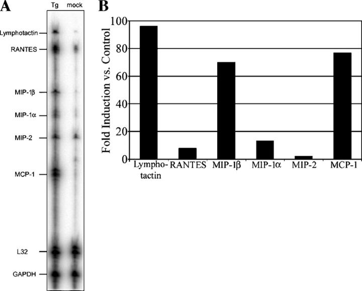
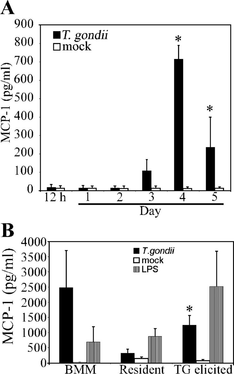
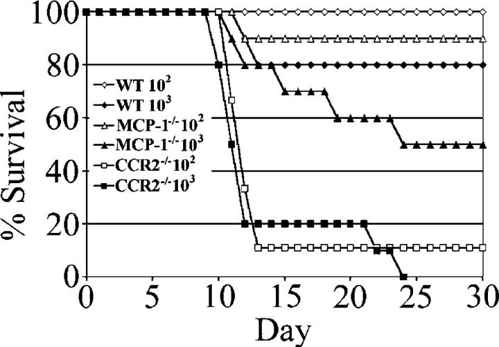
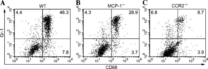
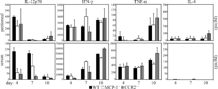
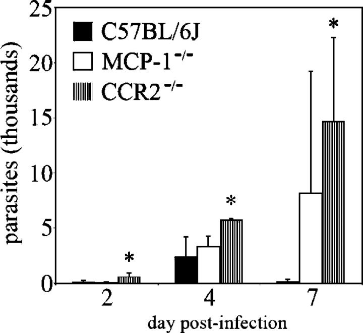
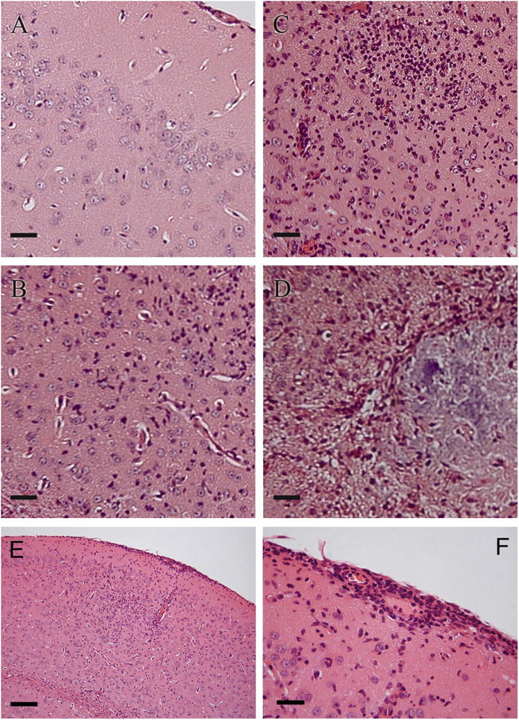
Circulating murine monocytes comprise two largely exclusive subpopulations that are responsible for seeding normal tissues (Gr-1-/CCR2-/CX3CR1high) or responding to sites of inflammation (Gr-1+/CCR2+/CX3CR1lo). Gr-1+ monocytes are recruited to the site of infection during the early stages of immune response to the intracellular pathogen Toxoplasma gondii. A murine model of toxoplasmosis was thus used to examine the importance of Gr-1+ monocytes in the control of disseminated parasitic infection in vivo. The recruitment of Gr-1+ monocytes was intimately associated with the ability to suppress early parasite replication at the site of inoculation. Infection of CCR2-/- and MCP-1-/- mice with typically nonlethal, low doses of T. gondii resulted in the abrogated recruitment of Gr-1+ monocytes. The failure to recruit Gr-1+ monocytes resulted in greatly enhanced mortality despite the induction of normal Th1 cell responses leading to high levels of IL-12, TNF-alpha, and IFN-gamma. The profound susceptibility of CCR2-/- mice establishes Gr-1+ monocytes as necessary effector cells in the resistance to acute toxoplasmosis and suggests that the CCR2-dependent recruitment of Gr-1+ monocytes may be an important general mechanism for resistance to intracellular pathogens.


Figures

Figure 1.
Figure 1.
Chemokines are up-regulated in response to an infection with a nonlethal challenge of T. gondii. (A) Peritoneal cells were collected 72 h after i.p. inoculation of mice with 102 T. gondii (Tg) or media diluent (mock). Harvested total RNA was subjected to RNase protection assay for the indicated chemokine genes. (B) Relative levels of expression during infection for chemokine genes assayed when compared with a mock infection control were determined by phosphor image analysis. Data shown are representative of two independent experiments with similar results.
Figure 2.
Figure 2.
Kinetics of MCP-1 release in peritoneum. (A) C57BL/6J mice were i.p. infected with 102 parasites of T. gondii. The concentration of MCP-1 present in peritoneal lavage fluid was determined by ELISA at the times indicated after infection (*, significantly different from mock infection; P < 0.01). (B) Naive BMM, resident peritoneal macrophages, or thioglycolate (TG)-elicited macrophages were challenged in vitro with live parasites (1:1, cells/parasites). MCP-1 concentrations in supernatant media following 24 h of infection were determined by ELISA (*, significantly different from resident peritoneal macrophages; P < 0.05). Data shown are means ± SE for two independent experiments (using data from three individual mice per time point in A).
Figure 3.
Figure 3.
Survival of CCR2−/− and MCP-1−/− mice after infection with T. gondii. Female CCR2−/−, MCP-1−/−, and wild-type C57BL/6J (WT) mice were infected i.p. with doses of 102 and 103 tachyzoites, and their survival was monitored for 30 d. Data are expressed as a cumulative percentage of two experiments (four to five mice per group).
Figure 4.
Figure 4.
Recruitment of inflammatory monocytes (Gr-1+/CD68+) is abrogated in CCR2−/− mice, but less so in MCP-1−/− mice. Peritoneal cells from (A) wild-type C57BL/6J (WT), (B) MCP-1−/−, and (C) CCR2−/− mice were harvested 4 d after i.p. inoculation of a dose of 102 T. gondii and analyzed by flow cytometry. Results shown are representative of individual mice. Numbers represent the percentage of gated cells present within the quadrant. The experiment was repeated twice with similar results, using a total of six mice per genotype.
Figure 5.
Figure 5.
CCR2−/− and MCP-1−/− mice produce high levels of IFN-γ. Peritoneal lavage fluid and serum were collected on days 4, 7, and 10 after infection from C57BL/6J (WT), MCP-1−/−, and CCR2−/− mice that had received a dose of 102 T. gondii (i.p.). Concentrations of IL-12p70, IFN-γ, TNF-α, and IL-4 were determined by ELISA. Data shown are means ± SE from two independent experiments (n = 3 individual mice per genotype) at each time point.
Figure 6.
Figure 6.
CCR2−/− and MCP-1−/− mice fail to control early parasite replication, allowing systemic dissemination. CCR2−/−, MCP-1−/−, and wild-type C57BL/6J mice were i.p. infected with a dose of 102 T. gondii. Parasites were enumerated by performing plaque assays using recovered intraperitoneal contents at the indicated times after infection. Data shown are means ± SD (from two mice per genotype) from a representative experiment of two with highly similar outcomes (*, significantly different from WT; P < 0.05).
Figure 7.
Figure 7.
Patterns of CNS inflammation observed during T. gondii infection in MCP-1−/− mice. (A–D) Hematoxylin and eosin–stained sections from brains harvested at day 35 after infection from MCP-1−/− and wild-type C57BL/6J mice infected i.p. with a dose of 102 T. gondii were examined and graded for the severity of inflammation in a blinded fashion. Representative images of the different grades of neuropathology that were observed. (A) Normal control (uninfected); (B) least severe (+); (C) intermediate (++); and (D) most severe (+++) are shown. (E) Example of encephalitis and meningitis with the infiltration of mononuclear cells. (F) Enlarged region of the meninges showing infiltration and perivascular infiltration of mononuclear cells. Similar pathology was found in a second experiment. Bars: (A–D and F) 5 μm; (E) 20 μm.

References

    1. Muller, W.A., and G.J. Randolph. 1999. Migration of leukocytes across endothelium and beyond: molecules involved in the transmigration and fate of monocytes. J. Leukoc. Biol. 66:698–704. - PubMed
    1. Passlick, B., D. Flieger, and H.W. Ziegler-Heitbrock. 1989. Identification and characterization of a novel monocytes subpopulation in human peripheral blood. Blood. 74:2527–2534. - PubMed
    1. Geissmann, F., S. Jung, and D.R. Littman. 2003. Blood monocytes consist of two principle subsets with distinct migratory properties. Immunity. 19:71–82. - PubMed
    1. Henderson, R.B., J.A.R. Hobbs, M. Mathies, and N. Hogg. 2003. Rapid recruitment of inflammatory monocytes is independent of neutrophil migration. Blood. 102:328–335. - PubMed
    1. Taylor, P.R., and S. Gordon. 2003. Pattern recognition receptors and differentiation antigens define murine myeloid cell heterogeneity ex vivo. Eur. J. Immunol. 33:2090–2097. - PubMed

Publication types

MeSH terms