. 2005 Jun 6;169(5):789-99.
doi: 10.1083/jcb.200408005. Epub 2005 May 31.

Class II phosphoinositide 3-kinase defines a novel signaling pathway in cell migration

Affiliations

Class II phosphoinositide 3-kinase defines a novel signaling pathway in cell migration

Tania Maffucci et al. J Cell Biol. .

Abstract

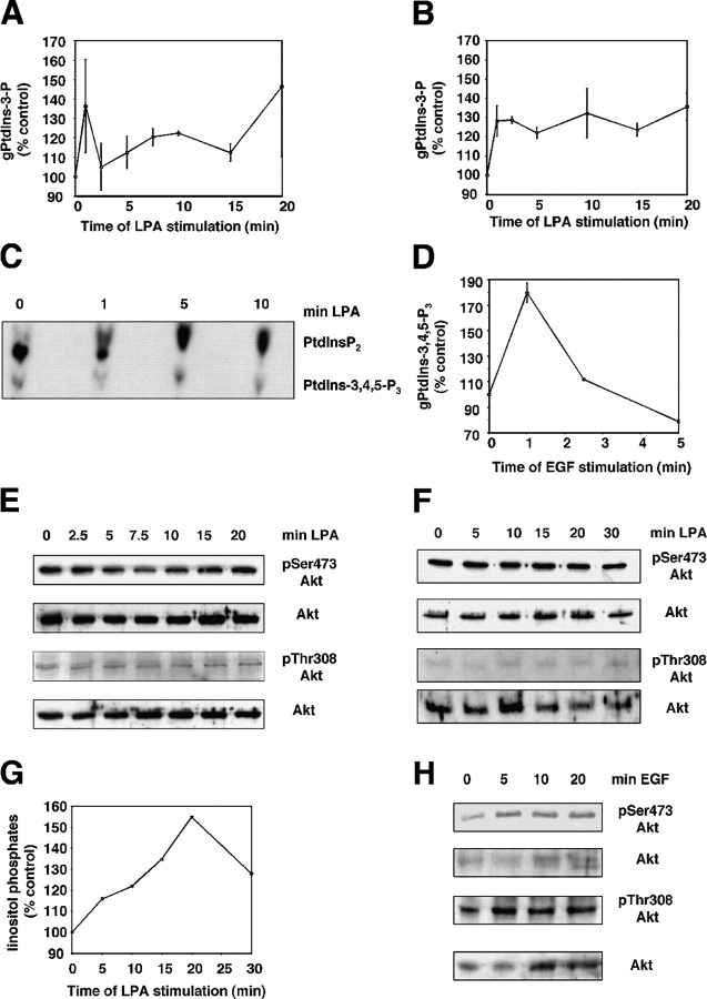
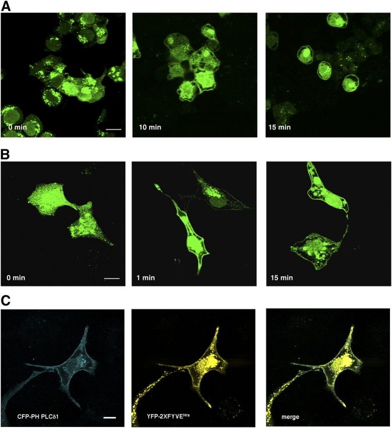
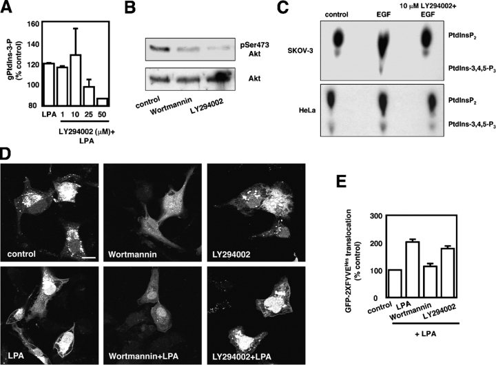
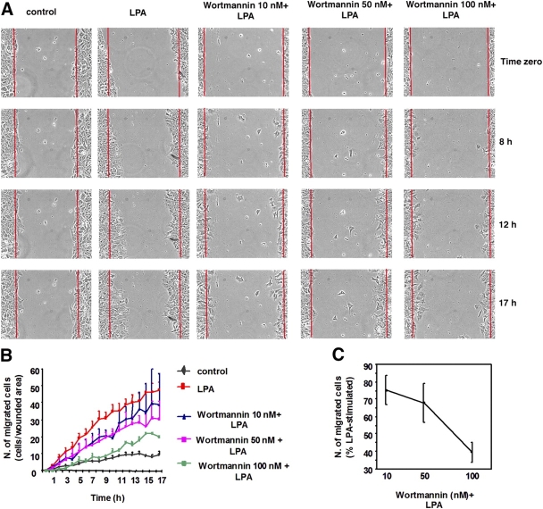
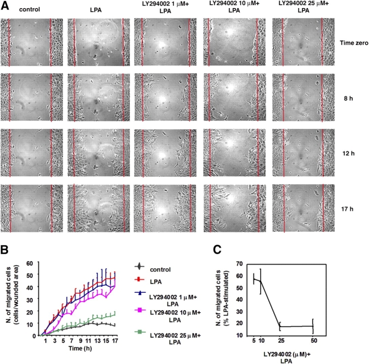
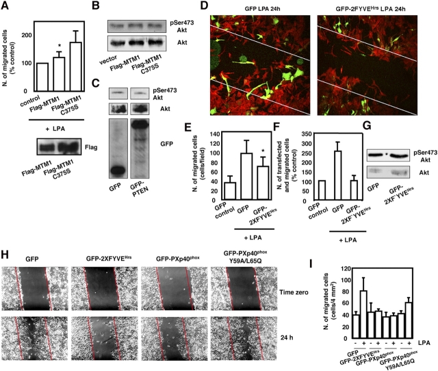
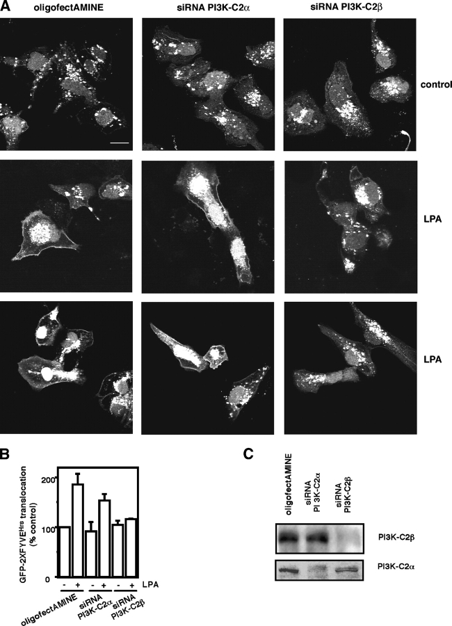
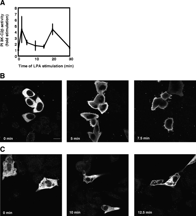
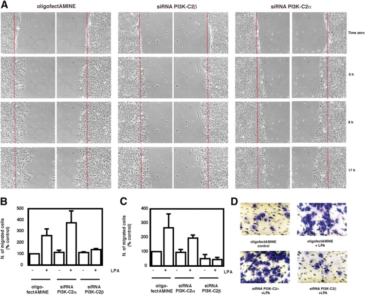
The lipid products of phosphoinositide 3-kinase (PI3K) are involved in many cellular responses such as proliferation, migration, and survival. Disregulation of PI3K-activated pathways is implicated in different diseases including cancer and diabetes. Among the three classes of PI3Ks, class I is the best characterized, whereas class II has received increasing attention only recently and the precise role of these isoforms is unclear. Similarly, the role of phosphatidylinositol-3-phosphate (PtdIns-3-P) as an intracellular second messenger is only just beginning to be appreciated. Here, we show that lysophosphatidic acid (LPA) stimulates the production of PtdIns-3-P through activation of a class II PI3K (PI3K-C2beta). Both PtdIns-3-P and PI3K-C2beta are involved in LPA-mediated cell migration. This study is the first identification of PtdIns-3-P and PI3K-C2beta as downstream effectors in LPA signaling and demonstration of an intracellular role for a class II PI3K. Defining this novel PI3K-C2beta-PtdIns-3-P signaling pathway may help clarify the process of cell migration and may shed new light on PI3K-mediated intracellular events.


Figures

Figure 1.
Figure 1.
LPA generates specifically PtdIns-3-P in HeLa and SKOV-3. HeLa (A) and SKOV-3 (B) were labeled with [3H]-myo-inositol for 24 h and stimulated with 25 μM LPA for the indicated times. Phospholipids were then extracted, deacylated, and analyzed by HPLC. Levels of PtdIns-3-P are shown as a percentage of basal levels. Data are mean ± SEM of three to four (HeLa) and three to eight (SKOV-3) independent experiments. (C) Serum-starved HeLa were labeled with [32P]phosphoric acid for 3 h and stimulated with 25 μM LPA for the indicated times. Phospholipids were then extracted and analyzed by TLC. (D) Levels of PtdIns-3,4,5-P3 in 3H-labeled HeLa assessed at different times of EGF stimulation by HPLC analysis. Data are mean ± SEM of three independent experiments. (E) Western blotting analysis of lysates from serum-starved HeLa stimulated with 25 μM LPA for the indicated times. Phosphorylation of Akt on residue Ser473 and Thr308 was assessed by using specific antibodies. Filters were stripped and reprobed with an anti-Akt antibody. (F) Western blotting analysis of lysates from serum-starved SKOV-3 stimulated with 25 μM LPA for the indicated times. Phosphorylation of Akt and total Akt levels were assessed by using specific antibodies. (G) LPA-dependent activation of PLC was assessed by monitoring the levels of total inositol phosphates generated. Data are from the experiment performed in parallel with the experiment shown in F. (H) Western blotting analysis of lysates from serum-starved SKOV-3 stimulated with 20 ng/ml EGF for the indicated times. Phosphorylation of Akt and total Akt levels were assessed by using specific antibodies.
Figure 2.
Figure 2.
The LPA-dependent pool of PtdIns-3-P is generated at the plasma membrane of HeLa and SKOV-3. HeLa (A) and SKOV-3 (B) were transfected with a cDNA encoding GFP-2XFYVEHrs. After 24 h, cells were serum deprived and stimulated with 25 μM LPA for the indicated times before fixing for confocal microscopy analysis. (C) Confocal microscopy analysis of SKOV-3 transfected with cDNAs encoding YFP-2XFYVEHrs and CFP-PH PLCδ1 and stimulated with 25 μM LPA for 10 min. Bars, 10 μm.
Figure 3.
Figure 3.
Effect of PI3K inhibitors on the LPA-mediated PtdIns-3-P formation. (A) SKOV-3 were labeled with [3H]-myo-inositol for 24 h and left untreated or pretreated with the indicated concentrations of LY294002 before stimulation with 25 μM LPA for 10 min. Phospholipids were then extracted, deacylated, and analyzed by HPLC. Levels of PtdIns-3-P are shown as a percentage of basal levels. (B) Western blotting analysis of lysates from serum-starved SKOV-3 treated with 100 nM wortmannin or 10 μM LY294002 for 30 min. Phosphorylation of Akt on residue Ser473 was assessed by using a specific antibody. Filter was stripped and reprobed with an anti-Akt antibody. (C) SKOV-3 (top) and HeLa (bottom) were labeled with [32P]phosphoric acid for 3 h and left untreated or pretreated with 10 μM LY294002 before stimulation with 20 ng/ml EGF for 5 (SKOV-3) or 1 min (HeLa). Phospholipids were extracted and analyzed by TLC. (D) GFP-2XFYVEHrs-expressing SKOV-3 cells were pretreated with 100 nM wortmannin or 10 μM LY294002. After 30 min, cells were stimulated with 25 μM LPA for 10 min before fixing for confocal microscopy analysis. Bar, 10 μm. (E) Quantitative analysis of experiments as in D. Data are mean ± SEM of five independent experiments.
Figure 4.
Figure 4.
Effect of wortmannin on the LPA-mediated migration. (A and B) Time-lapse microscopy of wound healing assay in SKOV-3 pretreated with the indicated concentrations of wortmannin for 30 min. Images in A show the wounded cell monolayers at time 0 and after the indicated hours in the absence (control) or presence of 25 μM LPA with or without the inhibitor. (B) Quantitative analysis was performed as described in Materials and methods. Data are mean ± SEM of three to four independent experiments. (C) Serum-starved SKOV-3 were pretreated with the indicated concentrations of wortmannin for 30 min. Migration was assessed by Transwell assay in the presence of 25 μM LPA and the indicated concentrations of the inhibitor. Data are mean ± SEM of five independent experiments and are expressed as a percentage of LPA-stimulated cells. In these experiments, LPA induced a three- to fourfold increase in cell migration.
Figure 5.
Figure 5.
Effect of LY294002 on the LPA-mediated migration. (A and B) Time-lapse microscopy of wound healing assay in SKOV-3 pretreated with the indicated concentrations of LY294002 for 30 min. Images in A show the wounded cell monolayers at time 0 and after the indicated hours in the absence (control) or presence of 25 μM LPA with or without the inhibitor. (B) Quantitative analysis was performed as described in Materials and methods. Data are mean ± SEM of three to five independent experiments. (C) Serum-starved SKOV-3 were pretreated with the indicated concentrations of LY294002 for 30 min. Migration was assessed by Transwell assay in the presence of 25 μM LPA and the indicated concentrations of the inhibitor. Data are mean ± SEM of five independent experiments and are expressed as a percentage of LPA-stimulated cells. In these experiments, LPA induced a three- to fourfold increase in cell migration.
Figure 6.
Figure 6.
PtdIns-3-P is involved in the LPA-mediated migration of SKOV-3. (A-G) SKOV-3 were transiently transfected with the indicated cDNAs. Transfection efficiency in these cells was ∼50%. (A, top) SKOV-3 were transfected with cDNAs encoding Flag-MTM1 and Flag-MTM1 C375S. After 24 h, cells were serum deprived overnight and migration was assessed by Transwell assay in the presence of 25 μM LPA. The total number of migrated cells was counted after staining the membranes of the Transwell chambers with crystal violet. Data are mean ± SEM of three independent experiments. *, P < 0.05. (bottom) Representative blot monitoring the levels of expression of Flag-MTM1 and Flag-MTM1 C375S by using an anti-Flag antibody. (B) Phosphorylation of Akt on residue Ser473 assessed in lysates from SKOV-3 transfected with an empty vector or cDNAs encoding Flag-MTM1 and Flag-MTM1 C375S. Filters were stripped and reprobed with an anti-Akt antibody. (C) Phosphorylation of Akt on residue Ser473 assessed in lysates from SKOV-3 transfected with cDNAs encoding GFP or GFP-PTEN. Filters were stripped and reprobed with an anti-Akt antibody. Expression of the exogenous proteins was assessed by using an anti-GFP antibody. Images are from different lanes in the same blot. (D) SKOV-3 grown on glass coverslips were transfected with cDNAs encoding GFP or GFP-2XFYVEHrs. After 24 h, cells were serum deprived overnight and migration was assessed by wound healing assay. After wounding and further 24 h in the presence of 25 μM LPA, cells were fixed and analyzed by confocal microscopy. Actin was stained by using Alexa 594 phalloidin and the merged images are shown. Diagonal lines specify the position of the original wound. (E and F) SKOV-3 were transfected as in D. Migration was then assessed by Transwell assay in the presence of 25 μM LPA. For data shown in E, the total number of cells that had migrated was counted after staining the membranes of the Transwell chambers with crystal violet. Data are mean ± SEM from three independent experiments performed in duplicate. *, P < 0.05. For data shown in F, the membranes of the Transwell chambers were fixed and analyzed by fluorescent microscopy to visualize and count only transfected (green) cells that had migrated. Data are mean ± SEM from four independent experiments. (G) Phosphorylation of Akt on residue Ser473 assessed in lysates from SKOV-3 transfected with cDNAs encoding GFP or GFP-2XFYVEHrs. Filters were stripped and reprobed with an anti-Akt antibody. (H–I) HeLa were transfected with cDNAs encoding the indicated fusion proteins (transfection efficiency was >80%). After 24 h, cells were serum deprived overnight and wound healing was performed in the presence of 25 μM LPA. (H) Representative phase-contrast images of the wounded cell monolayers at time 0 and after 24 h of migration. (I) Quantitative analysis of wound healing as in H. Data are mean ± SEM of three independent experiments.
Figure 7.
Figure 7.
Knockdown of PI3K-C2β completely inhibits the LPA-mediated PtdIns-3-P formation. (A) SKOV-3 were transfected with oligofectAMINE alone or with siRNAs corresponding to PI3K-C2α and PI3K-C2β and further transfected with a cDNA encoding GFP-2XFYVEHrs. After 24 h, cells were serum deprived overnight and stimulated with 25 μM LPA for 10 min before fixing for confocal microscopy analysis. Bar, 10 μm. (B) Quantitative analysis of GFP-2XFYVEHrs translocation to the plasma membrane in SKOV-3 as in A. Data are mean ± SEM from three independent experiments. (C) Representative blots of lysates from SKOV-3 transfected with oligofectAMINE or the indicated siRNAs. Levels of PI3K-C2α and PI3K-C2β were assessed by using specific antibodies.
Figure 8.
Figure 8.
PI3K-C2β is a downstream effector of LPA. (A) PI3K-C2β activity was assessed in LPA-stimulated HeLa by in vitro kinase assay as described in Materials and methods. Data are mean ± SEM of three independent experiments. (B and C) HeLa (B) and SKOV-3 (C) were transfected with a cDNA encoding a myc-tagged PI3K-C2β. After 24 h, cells were serum deprived overnight and stimulated with 25 μM LPA for the indicated times before fixing for confocal microscopy analysis. Bar, 10 μm.
Figure 9.
Figure 9.
PI3K-C2β is involved in LPA-mediated cell migration. (A) Time-lapse microscopy of wound healing assay in serum-deprived SKOV-3 transfected with oligofectAMINE or the indicated siRNAs. Images show two opposite sides of the same representative fields of the recorded wounded area at time 0 and at the indicated hours in the presence of 25 μM LPA. (B) Transwell assay of serum-deprived SKOV-3 transfected with oligofectAMINE or with the indicated siRNAs. Data are mean ± SEM of four independent experiments performed in duplicate. (C) Transwell assay of serum-deprived HeLa transfected with oligofectAMINE or with the indicated siRNAs. Data are mean ± SEM of three independent experiments. (D) Representative phase-contrast images of migrated HeLa in Transwell assay as in C obtained by staining the membranes of the Transwell chambers with crystal violet.

References

    1. Anderson, N.G., T. Ahmad, K. Chan, R. Dobson, and N.J. Bundred. 2001. ZD1839 (Iressa), a novel epidermal growth factor receptor (EGFR) tyrosine kinase inhibitor, potently inhibits the growth of EGFR-positive cancer cell lines with or without erbB2 overexpression. Int. J. Cancer. 94:774–782. - PubMed
    1. Arcaro, A., S. Volinia, M.J. Zvelebil, R. Stein, S.J. Watton, M.J. Layton, I. Gout, K. Ahmadi, J. Downward, and M.D. Waterfield. 1998. Human phosphoinositide 3-kinase C2beta, the role of calcium and the C2 domain in enzyme activity. J. Biol. Chem. 273:33082–33090. - PubMed
    1. Arcaro, A., M.J. Zvelebil, C. Wallasch, A. Ullrich, M.D. Waterfield, and J. Domin. 2000. Class II phosphoinositide 3-kinases are downstream targets of activated polypeptide growth factor receptors. Mol. Cell. Biol. 20:3817–3830. - PMC - PubMed
    1. Arcaro, A., U.K. Khanzada, B. Vanhaesebroeck, T.D. Tetley, M.D. Waterfield, and M.J. Seckl. 2002. Two distinct phosphoinositide 3-kinases mediate polypeptide growth factor-stimulated PKB activation. EMBO J. 21:5097–5108. - PMC - PubMed
    1. Bian, D., S. Su, C. Mahanivong, R.K. Cheng, Q. Han, Z.K. Pan, P. Sun, and S. Huang. 2004. Lysophosphatidic acid stimulates ovarian cancer cell migration via a Ras-MEK Kinase 1 pathway. Cancer Res. 64:4209–4217. - PubMed

Publication types

MeSH terms

Substances