

Real-time analysis of the role of Ca(2+) in flagellar movement and motility in single sea urchin sperm
- PMID: 15928204
- PMCID: PMC2171626
- DOI: 10.1083/jcb.200411001
Real-time analysis of the role of Ca(2+) in flagellar movement and motility in single sea urchin sperm
Abstract
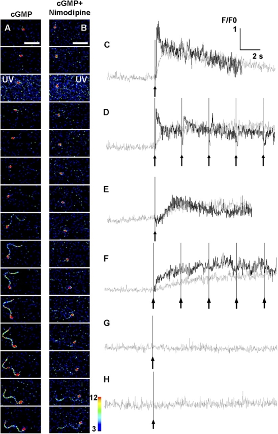
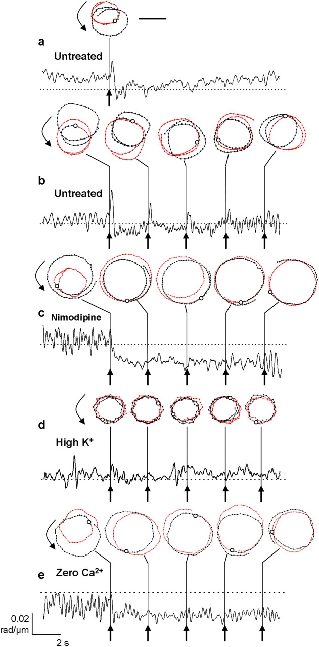
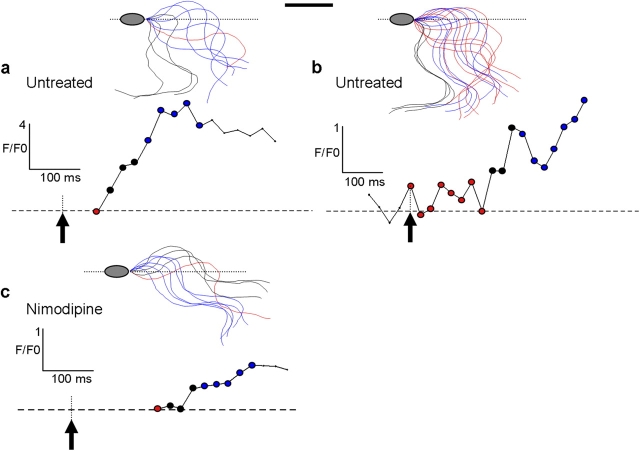
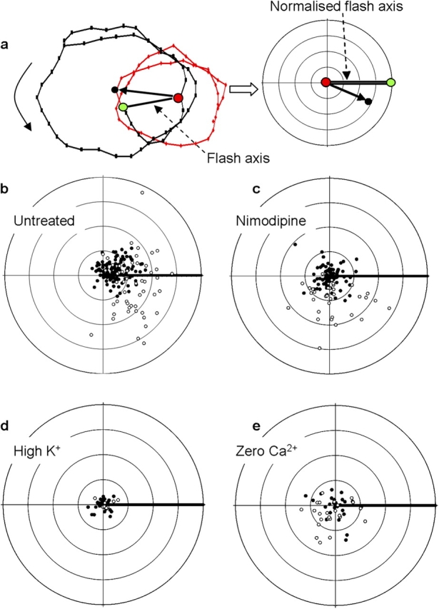
Eggs of many marine and mammalian species attract sperm by releasing chemoattractants that modify the bending properties of flagella to redirect sperm paths toward the egg. This process, called chemotaxis, is dependent on extracellular Ca(2+). We used stroboscopic fluorescence imaging to measure intracellular Ca(2+) concentration ([Ca(2+)]i) in the flagella of swimming sea urchin sperm. Uncaging of cyclic GMP induced Ca(2+) entry via at least two distinct pathways, and we identified a nimodipine-sensitive pathway, compartmentalized in the flagella, as a key regulator of flagellar bending and directed motility changes. We found that, contrary to current models, the degree of flagellar bending does not vary in proportion to the overall [Ca(2+)]i. Instead we propose a new model whereby flagella bending is increased by Ca(2+) flux through the nimodipine-sensitive pathway, and is unaffected by [Ca(2+)]i increases through alternative pathways.
Figures
Similar articles
-
Niflumic acid disrupts marine spermatozoan chemotaxis without impairing the spatiotemporal detection of chemoattractant gradients.J Cell Sci. 2013 Mar 15;126(Pt 6):1477-87. doi: 10.1242/jcs.121442. Epub 2013 Feb 15. J Cell Sci. 2013. PMID: 23418354
-
Network model predicts that CatSper is the main Ca2+ channel in the regulation of sea urchin sperm motility.Sci Rep. 2017 Jun 26;7(1):4236. doi: 10.1038/s41598-017-03857-9. Sci Rep. 2017. PMID: 28652586 Free PMC article.
-
Tuning sperm chemotaxis.Biochem Soc Trans. 2010 Oct;38(5):1270-4. doi: 10.1042/BST0381270. Biochem Soc Trans. 2010. PMID: 20863297 Review.
-
The rate of change in Ca(2+) concentration controls sperm chemotaxis.J Cell Biol. 2012 Mar 5;196(5):653-63. doi: 10.1083/jcb.201106096. Epub 2012 Feb 27. J Cell Biol. 2012. PMID: 22371558 Free PMC article.
-
Sperm chemotaxis and regulation of flagellar movement by Ca2+.Mol Hum Reprod. 2011 Aug;17(8):457-65. doi: 10.1093/molehr/gar041. Epub 2011 May 24. Mol Hum Reprod. 2011. PMID: 21610215 Review.
Cited by
-
Dual Sensing of Physiologic pH and Calcium by EFCAB9 Regulates Sperm Motility.Cell. 2019 May 30;177(6):1480-1494.e19. doi: 10.1016/j.cell.2019.03.047. Epub 2019 May 2. Cell. 2019. PMID: 31056283 Free PMC article.
-
The CatSper channel controls chemosensation in sea urchin sperm.EMBO J. 2015 Feb 3;34(3):379-92. doi: 10.15252/embj.201489376. Epub 2014 Dec 22. EMBO J. 2015. PMID: 25535245 Free PMC article.
-
Ca2+ bursts occur around a local minimal concentration of attractant and trigger sperm chemotactic response.Proc Natl Acad Sci U S A. 2008 Dec 9;105(49):19312-7. doi: 10.1073/pnas.0808580105. Epub 2008 Dec 1. Proc Natl Acad Sci U S A. 2008. PMID: 19047630 Free PMC article.
-
Chemosensory Ca2+ dynamics correlate with diverse behavioral phenotypes in human sperm.J Biol Chem. 2011 May 13;286(19):17311-25. doi: 10.1074/jbc.M110.211524. Epub 2011 Mar 21. J Biol Chem. 2011. PMID: 21454470 Free PMC article.
-
The role of alkalinization-induced Ca2+ influx in sperm motility activation of a viviparous fish Redtail Splitfin (Xenotoca eiseni).Biol Reprod. 2018 Dec 1;99(6):1159-1170. doi: 10.1093/biolre/ioy150. Biol Reprod. 2018. PMID: 29982498 Free PMC article.
References
-
- Baba, S.A., and Y. Mogami. 1985. An approach to digital image analysis of bending shapes of eukaryotic flagella and cilia. Cell Motil. Cytoskeleton. 5:475–489.
-
- Brokaw, C.J. 1974. Calcium and flagellar response during the chemotaxis of bracken spermatozoids. J. Cell. Physiol. 83:151–158. - PubMed
-
- Chung, M.K., and H. Kim. 2002. Volume-activated chloride currents from human fibroblasts: blockade by nimodipine. Gen. Physiol. Biophys. 21:85–101. - PubMed
Publication types
MeSH terms
Substances
Grants and funding
LinkOut - more resources
Full Text Sources
Miscellaneous
