. 2005 May 31;33(10):3095-108.
doi: 10.1093/nar/gki611. Print 2005.

UNR translation can be driven by an IRES element that is negatively regulated by polypyrimidine tract binding protein

Affiliations

UNR translation can be driven by an IRES element that is negatively regulated by polypyrimidine tract binding protein

Sigrid Cornelis et al. Nucleic Acids Res. .

Abstract

Upstream of N-ras (Unr) has been described as an internal initiation trans-acting factor (ITAF) in the cap-independent translation of some particular viral and cellular mRNAs. Two factors led us to hypothesize that the UNR 5'-untranslated region (5'-UTR) may contain an internal ribosome entry site (IRES). The first was the requirement for persisting Unr expression under conditions that correlate with cap-independent translation. The other was the observation that the primary UNR transcript contains a 447 nt long 5'-UTR including two upstream AUGs that may restrict translation initiation via cap-dependent ribosome scanning. Here we report that the UNR 5'-UTR allows IRES-dependent translation, as revealed by a dicistronic reporter assay. Various controls ruled out the contribution of leaky scanning, cryptic promoter sequences or RNA processing events to the ability of the UNR 5'-UTR to mediate internal initiation of translation. Ultraviolet cross-linking analysis and RNA affinity chromatography revealed the binding of polypyrimidine tract binding protein (PTB) to the UNR IRES, requiring a pyrimidine-rich region (nucleotides 335-355). Whereas overexpression of PTB in several cell lines inhibited UNR IRES activity and UNR protein expression, depletion of endogenous PTB using RNAi increased UNR IRES activity. Moreover, a mutant version of the UNR IRES lacking the PTB binding site was more efficient at directing IRES-mediated translation. In conclusion, our results demonstrate that translation of the ITAF Unr can itself be regulated by an IRES that is downregulated by PTB.


Figures
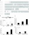
Figure 1
Figure 1
UNR 5′-UTR directs internal ribosome entry. (A) Homology between the sequences Mus musculus, Gallus gallus, Bos taurus and human UNR 5′-UTRs. Sequences were aligned from the ATG sequence encoding the initiating methionine. Identical nucleotides in three of the four sequences are indicated by gray boxes. Two pyrimidine-rich sequences are underlined. (B) Upper panel: schematic representation of a dicistronic mRNA containing the UNR-specific sequence cloned as an intercistronic sequence between the coding regions for firefly luciferase (Fluc) and Renilla luciferase (Rluc). Middle panel: comparison of the IRES activity mediated by the UNR 5′-UTR and the c-myc IRES. The dicistronic expression vectors Di-pRF, Di-pRF-UNR and Di-pRF-cMYC were transfected in HEK293T cells, and the activities of Fluc and Rluc were measured after 24 h. The bars represent the average (n = 3) ± SD of Fluc (hatched bars) and Rluc (open bars) activities. Lower panel: representation of the IRES activities calculated as the ratio between Fluc and Rluc. (C) Comparison of the efficiency of internal initiation mediated by the UNR 5′-UTR to cap-dependent initiation of translation. The dicistronic expression vectors Di-pRF-UNR and Di-pRF-cMYC and a mixture of monocistronic pSV-Sport Renilla luciferase and pSV-Sport firefly luciferase plasmids were transfected in HEK293T cells, and the activities of Fluc and Rluc were measured after 24 h. Bars represent the average (n = 3) ± SD of the ratio between Fluc and Rluc activities, and are representative of three independent transfection experiments. (D) UNR 5′-UTR driven translation of the downstream cistron is not sensitive to cap analogue. In vitro synthesized capped transcript derived from Di-pRF-UNR was translated in HeLa S3 cell extract in the absence or presence of 1 mM m7GpppG cap analogue and Rluc and Fluc activities were measured as described in Materials and Methods.
Figure 2
Figure 2
Translation of the second cistron is not due to leaky scanning, readthrough, reinitiation or aberrant mRNA species. (A) Influence of a stable hairpin (ΔG = −56 kcal/mol at 37°C) inserted upstream of the first cistron. Dicistronic expression vector Di-pFR-UNR or Di-phpFR-UNR was cotransfected with the pSV-Sport β-galactosidase plasmid in HEK293T cells. Fluc and Rluc activity was normalized to the β-galactosidase activity. The hatched and open bars represent the average (n = 3) ± SD of relative Fluc activities and relative Rluc activities, respectively. Bars are representative of three independent transfection experiments. (B) Analysis of potential cryptic promoter activity present in the UNR 5′-UTR. pGL3-UNR, pGL3-SV40 and pGL3-basic were cotransfected with the pSV-Sport Renilla luciferase plasmid in HEK293T cells. Bars represent the average (n = 3) ± SD of the ratio between Fluc and Rluc activities and are representative of three independent transfection experiments. (C) The dicistronic expression vector Di-pRF-UNR and a mixture of monocistronic pSV-Sport Renilla luciferase and pSV-Sport firefly luciferase plasmids were transfected in HEK293T cells. Poly(A)+ mRNA was isolated as described in Materials and Methods and protein extracts were prepared in parallel. Upper panel: northern blot analysis of a serial dilution (1/4) of poly(A)+ RNA prepared from monocistronic Fluc/Rluc transfectants starting from 4 μg down to 0.25 μg. On the same gel 1 μg poly(A)+ mRNA from dicistronic Di-UNR transfectants was loaded. Dicistronic and monocistronic mRNA expression in the HEK293T transfectants were revealed with a cDNA probe corresponding to the firefly luciferase open reading frame. Numbers on the left indicate the length of RNA markers (New England Biolabs). Lower panel: Fluc protein expression levels of a serial dilution (1/2) starting from 10 μg down to 1.25 μg protein extract from mono-Fluc transfectants. On the same gel a 10 μg cell extract from the Di-pRF-UNR transfectants was loaded.
Figure 3
Figure 3
PTB interacts with the UNR 5′-UTR. (A) Recombinant His-tagged PTB protein interacts with the UNR 5′-UTR. UV cross-linking assays were performed by pre-incubating 400 ng of His-tagged PTB protein with 32P-labeled probes corresponding to the UNR 5′-UTR, the EMCV IRES (EM) or the PITSLRE IRES (PIT), as indicated. In the case of the UNR 5′-UTR, specificity was determined by prior incubation of the UNR 5′-UTR probe in the absence or presence of 10–500 molar excess of unlabeled UNR IRES transcript, EMCV IRES transcript (EM) or PITSLRE IRES transcript (PIT), respectively. After RNA binding and UV-irradiation, samples were treated with an RNase cocktail and resolved by SDS–PAGE. The arrow depicts the position of cross-linked His-tagged PTB. (B) Cytoplasmic extracts from parental HEK293T cells (−) or from HEK293T cells overexpressing E-tagged PTB (PTB-E) were prepared as described in Materials and Methods. After incubation and UV-irradiation with the RNA probes corresponding to the UNR 5′-UTR or the EMCV IRES (EM), samples were treated with an RNase cocktail. One part of the sample was analyzed by SDS–PAGE (left panel), the other part was further mixed with anti-E-tag antibody for immunoprecipitation of labeled E-tagged PTB. Bound proteins were analyzed by SDS–PAGE. The arrow depicts the position of cross-linked E-tagged PTB (right panel). (C) Biotinylated UNR, EMCV (EM) and PITSLRE (PIT) IRES RNAs bound to streptavidin beads were incubated with cytoplasmic extracts from parental HEK293T cells as described in Materials and Methods. After extensive washing, RNA bound proteins were analyzed by western blotting using anti-PTB antibodies. The arrow depicts the position of endogenous PTB.
Figure 4
Figure 4
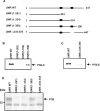
Deletion of the second pyrimidine-rich sequence in the UNR 5′-UTR RNA strongly reduces its binding to PTB. (A) Schematic representation of the UNR 5′-UTR fragments used in UV cross-linking reactions. Numbers indicate the positions of the nucleotides based on the human UNR cDNA. The black ellipses represent the CU-rich regions at positions (257–270) and (335–355) in the UNR 5′-UTR element. (B and C) UV cross-linking of 32P-labeled UNR fragments with cell extracts derived from HEK293T cells overexpressing PTB-E, followed by immunoprecipitation of labeled E-tagged PTB and SDS–PAGE. (D) UV cross-linking of 32P-labeled UNR fragments with HEK293T cell extracts, followed by SDS–PAGE. The arrow indicates the position of endogenous PTB.
Figure 5
Figure 5
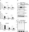
PTB overexpression inhibits IRES-mediated translation of UNR in cultured cells. (A) Effect of PTB on UNR IRES-driven expression in a dicistronic reporter assay. Cos-1, CHO and HEK293T cells were cotransfected with the dicistronic reporter plasmid, Di-pRF-UNR and the expression plasmid for PTB or PTBΔnls. Fluc and Rluc activities were measured in the corresponding cell extracts 24 h later. The bars represent the average activities (n = 3) ± SD of Fluc (hatched bars) and Rluc (open bars). Bars are representative of three independent experiments. (B) Overexpression of PTB suppresses Unr protein expression. Cell lysates from HEK293T cells cotransfected with Di-pRF-UNR and PTB or PTBΔnls were analyzed for PTB, Fluc or Unr protein expression by western blotting and detection with anti-PTB anti-E-tag, anti-Fluc or anti-Unr antibodies, respectively. β-Actin expression levels were analyzed as a loading control. (C) Overexpression of PTB has no effect on UNR mRNA levels as revealed by northern blotting. A cDNA corresponding to the unr open reading frame was used as a probe. Detection of β-actin mRNA levels served as a loading control. Numbers on the left indicate the length of RNA markers (New England Biolabs).
Figure 6
Figure 6
RNAi-mediated depletion of PTB in HEK293T cells stimulates UNR IRES activity. Di-pRF-UNR was transfected in HEK293T cells treated with PTB siRNA as described in Materials and Methods. Cells were analyzed 24 and 48 h after the last siRNA transfection for UNR IRES activity by measuring Rluc and Fluc activities, which are expressed as a ratio of Fluc to Rluc (n = 2) ± SD. Bars are representative of two independent experiments. Western blot analysis of HEK293T cells treated with PTB siRNA or with a nonspecific RNAi duplex (NT siRNA) revealed that PTB expression was strongly reduced upon PTB siRNA transfection. Actin was used as an internal control.
Figure 7
Figure 7
Deletion of a PTB binding site in the UNR 5′-UTR enhances UNR IRES activity. (A) The dicistronic expression vectors Di-pRF-UNR and Di-pRF-UNRΔ355–375 were cotransfected in HEK293Tcells with the empty pCAGGS vector or with one containing PTBΔnls. Cells were analyzed 24 h later for Rluc and Fluc activities, which are expressed as a ratio of Fluc to Rluc (n = 3) ± SD. Bars are representative of three independent experiments. (B) Investigation of potential cryptic promoter activity present in the UNR 5′-UTRΔ335–355 mutant. pGL3-UNR, pGL3-UNRΔ335–355, pGL3-SV40 and pGL3-basic were cotransfected with the pSV-Sport Renilla luciferase plasmid in HEK293T cells. Bars represent the average (n = 3) ± SD of the ratio between the Fluc and Rluc activities. Bars are representative of three independent experiments.

Similar articles

Cited by

References

    1. Pestova T.V., Kolupaeva V.G., Lomakin I.B., Pilipenko E.V., Shatsky I.N., Agol V.I., Hellen C.U. Molecular mechanisms of translation initiation in eukaryotes. Proc. Natl Acad. Sci. USA. 2001;98:7029–7036. - PMC - PubMed
    1. Stoneley M., Willis A.E. Cellular internal ribosome entry segments: structures, trans-acting factors and regulation of gene expression. Oncogene. 2004;23:3200–3207. - PubMed
    1. Vagner S., Galy B., Pyronnet S. Irresistible IRES. Attracting the translation machinery to internal ribosome entry sites. EMBO Rep. 2001;2:893–898. - PMC - PubMed
    1. Fernandez J., Yaman I., Mishra R., Merrick W.C., Snider M.D., Lamers W.H., Hatzoglou M. Internal ribosome entry site-mediated translation of a mammalian mRNA is regulated by amino acid availability. J. Biol. Chem. 2001;276:12285–12291. - PubMed
    1. Henis-Korenblit S., Strumpf N.L., Goldstaub D., Kimchi A. A novel form of DAP5 protein accumulates in apoptotic cells as a result of caspase cleavage and internal ribosome entry site-mediated translation. Mol. Cell. Biol. 2000;20:496–506. - PMC - PubMed

Publication types

MeSH terms