Comparative Study
. 2005 Jul;11(7):1141-52.
doi: 10.1261/rna.2550105. Epub 2005 May 31.

The pseudouridine synthase RluD is required for normal ribosome assembly and function in Escherichia coli

Affiliations
Comparative Study

The pseudouridine synthase RluD is required for normal ribosome assembly and function in Escherichia coli

Nancy S Gutgsell et al. RNA. 2005 Jul.

Abstract

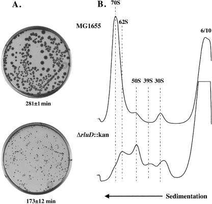
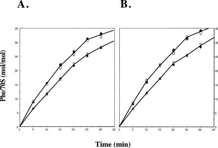
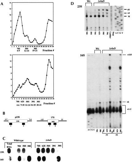
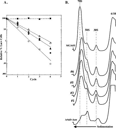
RluD is the pseudouridine synthase responsible for the formation of Psi1911, Psi1915, and Psi1917 in Escherichia coli 23S rRNA. Previous work from our laboratory demonstrated that disruption of the rluD gene and/or loss of the pseudouridine residues for which it is responsible resulted in a severe growth phenotype. In the current work we have examined further the effect of the loss of the RluD protein and its product pseudouridine residues in a deletion strain lacking the rluD gene. This strain exhibits defects in ribosome assembly, biogenesis, and function. Specifically, there is a deficit of 70S ribosomes, an increase in 50S and 30S subunits, and the appearance of new 62S and 39S particles. Analysis of the 39S particles indicates that they are immature precursors of the 50S subunits, whereas the 62S particles are derived from the breakdown of unstable 70S ribosomes. In addition, purified mutant 70S ribosomes were found to be somewhat less efficient than wild type in protein synthesis. The defect in ribosome assembly and resulting growth phenotype of the mutant could be restored by expression of wild-type RluD and synthesis of Psi1911, Psi1915, and Psi1917 residues, but not by catalytically inactive mutant RluD proteins, incapable of pseudouridine formation. The data suggest that the loss of the pseudouridine residues can account for all aspects of the mutant phenotype; however, a possible second function of the RluD synthase is also discussed.


Figures

FIGURE 1.
FIGURE 1.
PCR, Southern transfer and hybridization, and Ψ sequence analysis of wild-type and ΔrluD::kan mutant E. coli strains. (A) PCR reaction products were generated using genomic DNA as template. Primers used contain sequences homologous to the 5′ and 3′ ends of the rluD gene retained in the ΔrluD::kan mutant strain (forward primer 5′-GAAGCAGTATATATGGCACAACGAGTACAG CTC and reverse primer 5′-GGGAAGCTTTCATAACCAGTCCAC TTCATC). (Lanes 1,3) wild-type MG1655; (lanes 2,4), ΔrluD::kan. (Lanes 1,2) undigested; (lanes 3,4) reaction products digested with 20 units of HindIII (NEB) at 37°C for ≥16 h. Left side of the graph, sizes (kb) of the molecular weight markers. Right side of the graph, sizes (kb) of the DNA bands for each strain. (B) Southern transfer and hybridization were performed using genomic DNA incubated with 20 units of BamHI (NEB) at 37°C for 3 h to achieve partial digestion. Probes were generated from the wild-type MG1655 PCR product by digestion with 10 units NruI (NEB) and 5 units SphI (NEB) to generate the 806-nt probe specific for the center of rluD and a mixed probe specific for the 5′ and 3′ ends of rluD, respectively. Wild-type MG1655 (lanes 1); ΔrluD::kan (lanes 2); (panel I) hybridization with the 806-nt probe; (panel II) the same filter stripped; (panel III) the filter subsequently hybridized with the mixed 5′ and 3′ probes. (C) Pseudouridine sequencing analysis of 23S rRNA from MG1655 and ΔrluD::kan. Total RNA was isolated from the indicated strains and examined for Ψ (Ofengand et al. 2001).
FIGURE 2.
FIGURE 2.
Colony morphology, exponential growth rates and ribosome profiles of wild-type and ΔrluD::kan mutant E. coli strains. (A) LB plates without drug (wild type) or with 34 μg/mL kanamycin (mutant only) were incubated 41 h at 37°C. Dilutions were prepared from suspensions of wild-type and mutant cells and 6.8 × 10⁸ cells/mL for each strain were spread on plates and incubated. Exponential growth rates were measured by monitoring cell density at 600 nm with a Perkin Elmer Lambda 25 UV/VIS Spectrophotometer. Doubling time was determined from a semilogarithmic plot of A600 versus time. Each plot consisted of five to seven time points. Doubling times were the average of two determinations (wild type) or six determinations (mutant), and are shown under the plates. (B) Ribosome profiles of wild-type and ΔrluD::kan cells were performed as described in Materials and Methods. Wild-type and mutant cells were lysed at 6 mM Mg++ and gradients were run at 10 mM Mg++. Twelve A260 units were layered onto 14%–32% sucrose gradients and centrifuged at 22,000 rpm for 19 h at 4°C.
FIGURE 3.
FIGURE 3.
Ribosome profiles of wild-type and ΔrluD::kan mutant strains analyzed at various Mg++ concentrations. Ribosome profiles were performed as described in Materials and Methods. Wild-type and mutant cells were lysed, and gradients run at the Mg++ concentrations (mM) shown for lysis/sedimentation. Twelve A260 units were layered onto 14%–32% sucrose gradients and centrifuged at 22,000 rpm for 19 h at 4°C.
FIGURE 4.
FIGURE 4.
Polyphenyalanine synthesis by wild-type and mutant 70S ribosomes. Polyphenylalanine synthesis was performed as described in Materials and Methods. (A) 70S ribosome fractions were isolated from sucrose gradients at 15 mM Mg++ concentration during lysis and sedimentation. Nine individual experiments were performed using 70S fractions from four wild-type and seven mutant ribosome preparations; MG1655/I::tet 0.5 A260 (closed squares) or ΔrluD::kan/I::tet 0.5 A260 units (closed circles). Four individual experiments were performed using 70S fractions from two wild-type and four mutant ribo-some preparations; MG1655/I::tet 1.0 A260 unit (open squares) or ΔrluD::kan/I::tet 1.0 A260 unit (open circles). (B) Three individual experiments were performed using 70S fractions from two wild-type and three mutant ribosome preparations (lysis 6 mM and sedimentation 15 mM Mg++). Symbols are the same as for A.
FIGURE 5.
FIGURE 5.
Fraction collection, slot blot, and primer extension analysis (see Materials and Methods). (A) A260 units/mL of ribosome fractions collected from sucrose gradients (Mg++ concentration 6 mM/10 mM at lysis/sedimentation) for wild-type MG1655/I-::tet (closed squares) and ΔrluD::kan/I-::tet mutant (open squares). (Numbers below each graph are the fractions pooled for the ribosomal subunits indicated.) (B) Open boxes represent mature rRNA species, lines represent precursor sequences, and solid bars indicate probes used for slot blot hybridization (2 and 4) and primer extension analysis (1 and 3). (C) Equal amounts (5 μg) of RNA from pooled ribosomal fractions were transferred to nitrocellulose membranes and probed with the indicated oligonucleotides. (D) Equal amounts (0.5 μg) of RNA from the same pooled fractions were analyzed by primer extension using the indicated oligonucleotides.
FIGURE 6.
FIGURE 6.
Competition studies, exponential growth rates, and ribo-some profiles of MG1655 (wild type) and four pseudorevertant isolates of the ΔrluD::kan mutant. (A) Growth competition studies were done as described previously (Gutgsell et al. 2000). MG1655 (wild type) (closed squares), four individual pseudorevertant isolates (open and closed triangles and circles). Exponential growth rates were done as described in Materials and Methods. Doubling times were determined as described in Figure 2. Each plot consisted of seven to nine time points. Doubling times were the average of two determinants. (B) Ribosome profiles were performed as described in Materials and Methods. Wild-type, pseudorevertant, and mutant cells were lysed at 6 mM Mg++ and gradients were run at 10 mM Mg++. Twelve A260 units were layered onto 14%–32% sucrose gradients and centrifuged at 22,000 rpm for 19 h at 4°C.

References

    1. Agrawal, R.K., Sharma, M.R., Kiel, M.C., Hirokawa, G., Booth, T.M., Spahn, C.M., Grassucci, R.A., Kaji, A., and Frank, J. 2004. Visualization of ribosome-recycling factor on the Escherichia coli 70S ribosome: functional implications. Proc. Natl. Acad. Sci. 101: 8900–8905. - PMC - PubMed
    1. Bachman, B.J., Low, K.B., and Taylor, A.L. 1976. Recalibrated linkage map of Escherichia coli K-12. Bacteriol. Rev. 40: 116–167. - PMC - PubMed
    1. Bashan, A., Agmon, I., Zarivach, R., Schluenzen, F., Harms, J., Berisio, R., Bartels, H., Franceschi, F., Auerach, T., Hansen, H.A.S., et al. 2003. Structural basis of the ribosomal machinery for peptide bond formation, translocation, and nascent chain progression. Mol. Cell 11: 91–102. - PubMed
    1. Behm-Ansmant, I., Grosjean, H., Massenet, S., and Motorin, Y. 2004. Pseudouridylation at position 32 of mitochondrial and cytoplasmic tRNAs requires two distinct enzymes in Saccharomyces cerevisiae. J. Biol. Chem. 279: 52998–53006. - PubMed
    1. Charollais, J., Pflieger, D., Vinh, J., Dreyfus, M., and Iost, I. 2003. The DEAD-box RNA helicase SrmB is involved in the assembly of 50S ribosomal subunits in Escherichia coli. Mol. Microbiol. 48: 1253–1265. - PubMed

Publication types

MeSH terms

LinkOut - more resources