Comparative Study
. 2005 Jul 1;81(1):102-9.
doi: 10.1002/jnr.20542.

Deafness-related decreases in glycine-immunoreactive labeling in the rat cochlear nucleus

Affiliations
Comparative Study

Deafness-related decreases in glycine-immunoreactive labeling in the rat cochlear nucleus

Mikiya Asako et al. J Neurosci Res. .

Abstract

There is increasing evidence of activity-related plasticity in auditory pathways. The present study examined the effects of decreased activity on immunolocalization of the inhibitory neurotransmitter glycine in the cochlear nucleus of the rat after bilateral cochlear ablation. Specifically, glycine-immunoreactive puncta adjacent to somatic profiles were compared in normal hearing animals and animals deafened for 14 days. The number of glycine-immunoreactive puncta surrounding somatic profiles of spherical and globular bushy cells, glycine-immunoreactive type I stellate multipolar cells, radiate neurons (type II stellate multipolar cells), and fusiform cells decreased significantly. In addition, the number of glycine immunopositive tuberculoventral (vertical or corn) cells in the deep layer of the dorsal cochlear nucleus also decreased significantly. These results suggest that decreased inhibition reported in cochlear nucleus after deafness may be due to decreases in glycine.


Figures

Fig. 1
Fig. 1
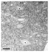
Glycine immunoreactivity in the dorsal cochlear nucleus (DCN). The molecular, fusiform and deep layers of the DCN contain many glycine-immunolabeled puncta. As expected, the cartwheel cells in the molecular layer are glycine immunopositive. The fusiform neurons in the fusiform layer are immunonegative for glycine. Mol, molecular layer; Fus, fusiform layer; Deep, deep layer. Scale bar = 25 μm.
Fig. 2
Fig. 2
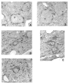
Glycine immunoreactivity in the ventral cochlear nucleus in normal hearing rats (A–C) and in rats 14 days after bilateral deafening (D, E). Glycine immunolabeling decreases in intensity and in the number of axosomatic puncta on most cell types 14 days after deafness. Scale bar = 5 μm.
Fig. 3
Fig. 3
Differences in the number of glycine-immunoreactive axosomatic puncta on different cell types in the cochlear nucleus (CN) between normal hearing rats (white bars) and rats 14 days after bilateral deafening (black bars). Data are expressed as the number of labeled puncta on a somatic profile (top) and as the number of puncta per 10 μm of somatic profile (bottom). Significant (*P ≤ 0.05) decreases are seen for glycine-immunoreactive type I stellate multipolar cells in posteroventral CN (PVCN), spherical bushy cells, globular bushy cells, radiate cells (type II stellate multipolar cells) and fusiform cells, but not glycine-immunoreactive type I stellate multipolar cells in anteroventral CN (AVCN). Error bars represent ±1 standard deviation.
Fig. 4
Fig. 4
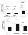
Changes in cell size in the cochlear nucleus (CN) of normal hearing rats (white bars) vs. that in rats 14 days after deafness (black bars). Significant (*P ≤ 0.05) decreases are seen in glycine-immunoreactive type I stellate multipolar cells, spherical bushy cells and globular bushy cells, but not radiate cells (type II stellate multipolar cells) or fusiform cells. Error bars represent ±1 standard deviation. AVCN, anteroventral CN; PVCN, posteroventral CN; VCN, ventral CN; DCN, dorsal CN.
Fig. 5
Fig. 5
Differences in the size of tuberculoventral cells (TBC) in the deep layer of the dorsal cochlear nucleus (DCN) when comparing normal hearing rats (white bars) and rats 14 days after bilateral deafening (black bars), as well as the number of glycine-immunoreactive TBC per 1,000 μm2. A significant decrease was seen both in cell size and in the number of glycine-immunopositive cells per 1,000 μm2. Error bars represent ±1 standard deviation. *P ≤ 0.05.

Similar articles

Cited by

References

    1. Adams JC, Mugnaini E. Immunocytochemical evidence for inhibitory and disinhibitory circuits in the superior olive. Hear Res. 1990;49:281–298. - PubMed
    1. Altschuler RA, Betz H, Parakkal MH, Reeks KA, Wenthold RJ. Identification of glycinergic synapses in the cochlear nucleus through immunocytochemical localization of the postsynaptic receptor. Brain Res. 1986;369:316–320. - PubMed
    1. Babalian AL, Jacomme AV, Doucet JR, Ryugo DK, Rouiller EM. Commissural glycinergic inhibition of bushy and stellate cells in the anteroventral cochlear nucleus. Neuroreport. 2002;13:555–558. - PubMed
    1. Bauer CA, Brozoski TJ, Holder TM, Caspary DM. Effects of chronic salicylate on GABAergic activity in rat inferior colliculus. Hear Res. 2000;147:175–182. - PubMed
    1. Bendiske J, Morest DK. Plastic changes in synaptic vesicle distribution in the cochlear nucleus following noise exposure in mice. Assoc Res Otolaryngol Abs. 2004:300.

Publication types