. 2005 Oct 1;391(Pt 1):105-14.
doi: 10.1042/BJ20050328.

Peroxin 5-peroxin 14 association in the protozoan Leishmania donovani involves a novel protein-protein interaction motif

Affiliations

Peroxin 5-peroxin 14 association in the protozoan Leishmania donovani involves a novel protein-protein interaction motif

Kleber P Madrid et al. Biochem J. .

Abstract

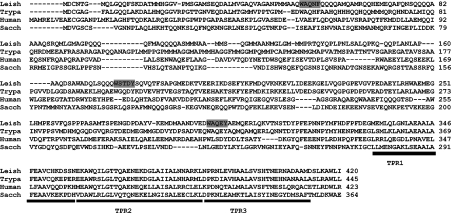
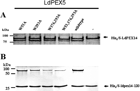
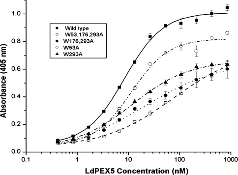
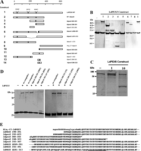
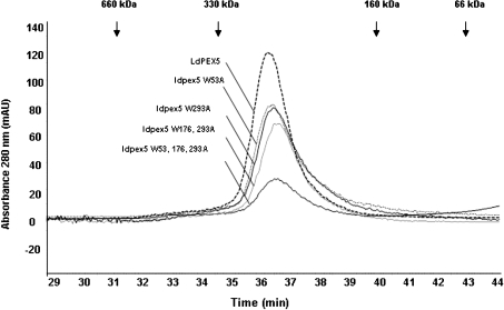
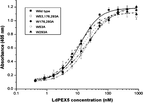
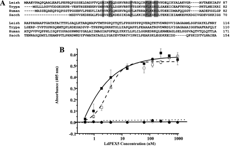
Import of proteins with a PTS1 (peroxisomal targeting signal 1) into the Leishmania glycosomal organelle involves docking of a PTS1-laden LdPEX5 [Leishmania donovani PEX5 (peroxin 5)] receptor to LdPEX14 on the surface of the glycosomal membrane. In higher eukaryotes, the PEX5-PEX14 interaction is mediated by a conserved diaromatic WXXXY/F motif. Site-directed and deletion mutageneses of the three WXXXY/F repeats in LdPEX5 did not abolish the LdPEX5-LdPEX14 association. Analysis of the equilibrium dissociation constant (K(d)) revealed that ldpex5-W53A (Trp53-->Ala), ldpex5-W293A, ldpex5-W176,293A and ldpex5-W53,176,293A mutant receptors were capable of binding LdPEX14 with affinities comparable with wild-type LdPEX5. That the diaromatic motifs were not required for the LdPEX5-LdPEX14 interaction was further verified by deletion analysis that showed that ldpex5 deletion mutants or ldpex5 fragments lacking the WXXXY/F motifs retained LdPEX14 binding activity. Mapping studies of LdPEX5 indicated that the necessary elements required for LdPEX14 association were localized to a region between residues 290 and 323. Finally, mutational analysis of LdPEX14 confirmed that residues 23-63, which encompass the conserved signature sequence AX2FLX7SPX6FLKGKGL/V present in all PEX14 proteins, are essential for LdPEX5 binding.


Figures

Figure 1
Figure 1. Multiple sequence alignment of PEX5 proteins
A partial sequence alignment of the N-terminal region of the L. donovani (Leish), T. brucei (Trypa), human and S. cerevisiae (Sacch) PEX5 was performed using the CLUSTAL_X computer program [35]. The WXXXY/F motifs in the LdPEX5 are designated by the grey shaded boxes. The black bar delineates the first three TPRs that form part of the PTS1-binding pocket.
Figure 2
Figure 2. LdPEX14–LdPEX5 interaction
The interaction of His6/S–LdPEX14 (A) or His6/S–ldpex14-(1–120) (His6/S–ldpex14-120) (B) with either the wild-type LdPEX5 or ldpex5 WXXXY/F site-directed mutants was assessed by pull-down assays using S-protein agarose beads. LdPEX14 proteins were mixed with 10 μg of purified, ldpex5-W53A, ldpex5-W293A, ldpex5-W176,293A, ldpex5-W53,176,293A or wild-type LdPEX5 or with no LdPEX5 and the mixture was added to S-protein agarose. Beads were stringently washed with 1% Triton X-100 and 500 mM NaCl in TBS (Tris-buffered saline; 50 mM Tris/HCl, pH 8.0 and 150 mM NaCl) and bound proteins were analysed by Coomassie Blue-stained SDS/PAGE.
Figure 3
Figure 3. Determination of the LdPEX14–LdPEX5 equilibrium dissociation constant
Microtitre plates were coated with His6/S–LdPEX14 and then incubated with increasing amounts of LdPEX5 (■), ldpex5-W53A (○), ldpex5-W293A (▲), ldpex5-W176,293A (●), or ldpex5-W53,176,293A (□). Bound LdPEX5 or ldpex5 proteins were quantified by an indirect ELISA using anti-LdPEX5 antisera. Each assay was performed in triplicate and the average absorbance values were plotted as a function of the log of the LdPEX5 or ldpex5 concentration using the ORIGIN 7.0 software. Kd values were determined as the protein concentration that gave half the maximal LdPEX5 or ldpex5 binding.
Figure 4
Figure 4. Mapping of the LdPEX14-binding domain
(A) A schematic representation of the ldpex5 constructs that were overexpressed, purified and tested in pull-down or ELISA-based assays in order to map the LdPEX14 interaction motif. Constructs designated in boldface were shown to have LdPEX14 binding activity. (B) Coomassie Blue-stained SDS/PAGE analysis of S-protein agarose pull-down assays performed with His6/S–ldpex14-(1–120). For these assays, 10 μg of His6/S–ldpex14-(1–120) was mixed with no LdPEX5 or 10 μg of LdPEX5, NT-ldpex5, ldpex5-(203–391), ldpex5-(290–391), ldpex5-(203–269), Δ269–291-ldpex5, Δ181–313-ldpex5 and ldpex5-(1–270). The LdPEX5 constructs indicated by the numbers above each gel lane correspond to the LdPEX5 structures shown in (A). (C) For these assays, 10 μg of His6/S–LdPEX14 was mixed with no LdPEX5, 10 μg of LdPEX5 or 10 μg of ldpex5-(283–625). Numbers above each lane correspond to the LdPEX5 constructs illustrated in (A). (D) LdPEX5 competition assays. S-protein agarose beads were incubated with 10 μg of His6/S–ldpex14-(1–120) alone or with 4 μg of LdPEX5 in the presence and absence of increasing concentrations of competitor peptides ldpex5-(268–303), ldpex5-(290–391), ldpex5-(203–347) and ldpex5-(268–323) at molar excess ranging from 1- to 200-fold. Proteins bound to the S-protein beads were analysed by SDS/PAGE. (E) Partial alignment of overlapping sequences of ldpex5 mutants and ldpex5 fragments used to map the LdPEX14-binding motif. The amino acid sequence retained in the ldpex5 constructs with LdPEX14 binding activity is designated by the underline. Sequences in lower-case letters are derived from the expression vector. Broken lines preceding and following the sequence indicate that this portion of the protein sequence extends beyond the residues shown. The asterisk indicates the C-terminus of the ldpex5 fragment.
Figure 5
Figure 5. Size-exclusion chromatography analysis of the ldpex5 mutants
Wild-type LdPEX5 and site-directed mutant ldpex5-W53A, ldpex5-W293A, ldpex5-W176,293A, ldpex5-W53,176,293A proteins were purified to homogeneity as fusion proteins using the New England Biolabs IMPACT system. Intein fusion proteins were cleaved using DTT, dialysed and concentrated. The oligomeric state of these proteins was determined by loading 25–50 μg of purified protein on to a Bio-Sil 250 column (7.8 mm×600 mm) in 25 mM Tris/HCl (pH 7.5) and 120 mM NaCl at a flow rate of 0.25 ml/min. Column eluate was monitored spectrophotometrically at 280 nm. Arrows indicate the elution positions of the standard proteins.
Figure 6
Figure 6. LdPEX5–XPRT interaction
Microtitre plates were coated with recombinant L. donovani XPRT and then incubated with increasing concentrations of one among wild-type LdPEX5 (■), ldpex5-W53A (○), ldpex5-W293A (▲), ldpex5-W176,293A (●) and ldpex5-W53,176,293A (□). The amount of LdPEX5 bound to the XPRT was determined by indirect ELISA using anti-LdPEX5 antisera (see the Materials and methods section). Binding experiments were performed in triplicate and absorbance values were averaged and plotted as a function of the log of the LdPEX5 or ldpex5 concentration. Data were fitted to a sigmoidal curve using the ORIGIN 7.0 computer program and Kd values are the LdPEX5 concentrations that gave half the maximal LdPEX5 or ldpex5 binding.
Figure 7
Figure 7. Mapping the LdPEX5 binding domain on LdPEX14
(A) The multiple sequence alignment of the N-terminal region of the L. donovani (Leish), T. brucei (Trypa), human and S. cerevisiae (Sacch) PEX14 protein illustrates the conserved signature sequence (boldface). (B) Binding assay for the interaction of LdPEX5 with LdPEX14 N-terminal truncation mutants. Microtitre plates were coated with 1 μg/well wild-type LdPEX14 (□), ldpex14-(24–464) (■), ldpex14-(44–464) (○) or ldpex14-(64–464) (●), blocked with 2% milk powder in PBS 0.05% Tween 20 and incubated with various concentrations of LdPEX5 (0.4–860 nM). Amount of bound LdPEX5 was determined by indirect ELISA using anti-LdPEX5 rabbit antisera and goat anti-rabbit horseradish peroxidase-conjugated secondary antibody.

References

    1. Dacks J. B., Doolittle W. F. Reconstructing/deconstructing the earliest eukaryotes: how comparative genomics can help. Cell (Cambridge, Mass.) 2001;107:419–425. - PubMed
    1. Zeiner G. M., Sturm N. R., Campbell D. A. The Leishmania tarentolae spliced leader contains determinants for association with polysomes. J. Biol. Chem. 2003;278:38269–38275. - PubMed
    1. Muhich M. L., Boothroyd J. C. Polycistronic transcripts in trypanosomes and their accumulation during heat shock: evidence for a precursor role in mRNA synthesis. Mol. Cell. Biol. 1988;8:3837–3846. - PMC - PubMed
    1. Kable M. L., Heidmann S., Stuart K. D. RNA editing: getting U into RNA. Trends Biochem. Sci. 1997;22:162–168. - PubMed
    1. Englund P. T., Hajduk S. L., Marini J. C. The molecular biology of trypanosomes. Annu. Rev. Biochem. 1982;51:695–726. - PubMed

Publication types

MeSH terms

Substances