. 2005 Jun;113(6):749-55.
doi: 10.1289/ehp.7596.

Lead exposure inhibits fracture healing and is associated with increased chondrogenesis, delay in cartilage mineralization, and a decrease in osteoprogenitor frequency

Affiliations

Lead exposure inhibits fracture healing and is associated with increased chondrogenesis, delay in cartilage mineralization, and a decrease in osteoprogenitor frequency

Jonathan J Carmouche et al. Environ Health Perspect. 2005 Jun.

Abstract

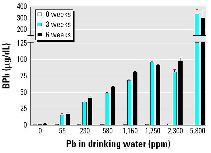
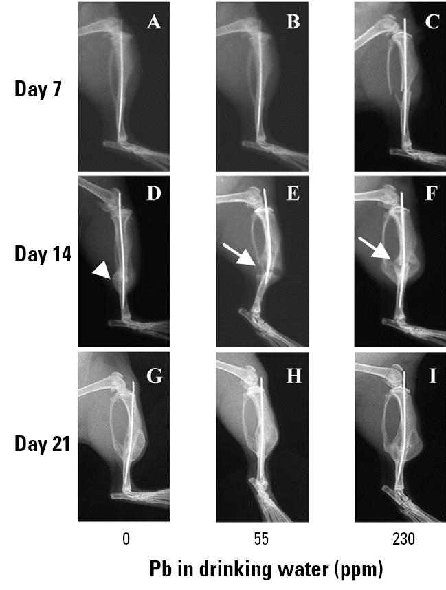
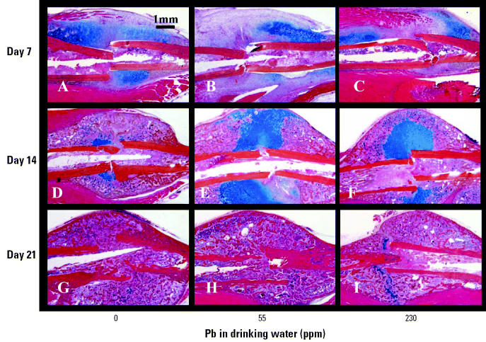
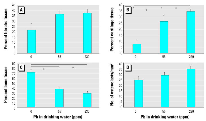
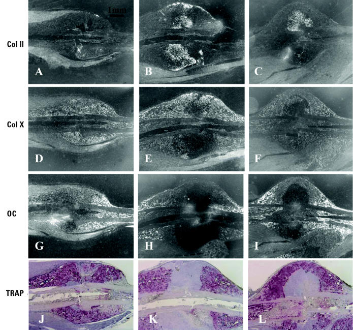
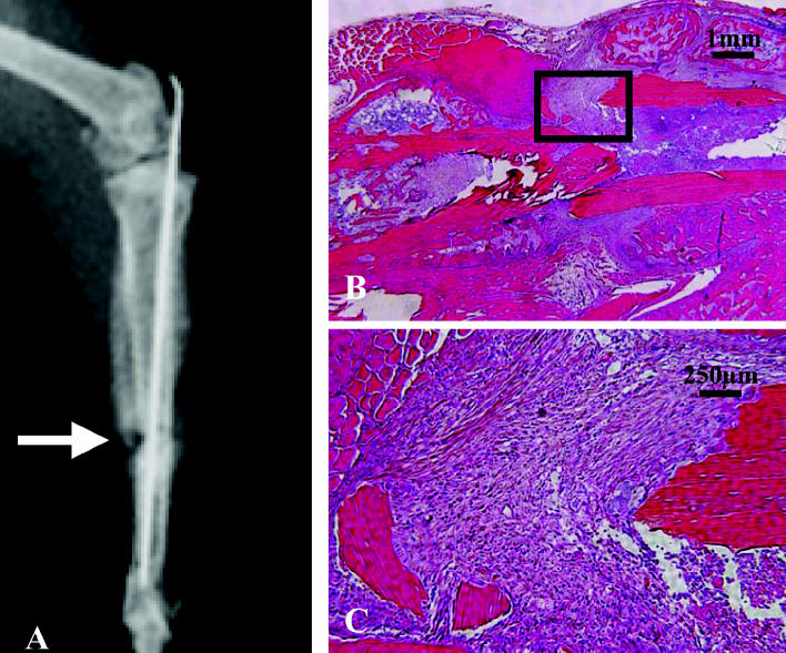
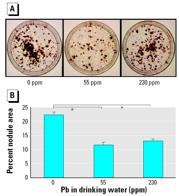
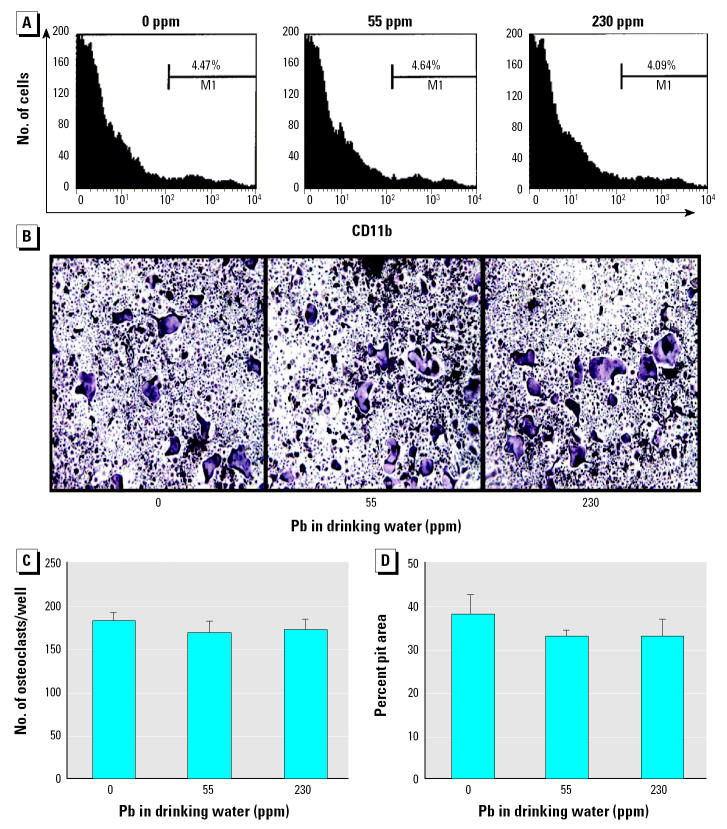
Lead exposure continues to be a significant public health problem. In addition to acute toxicity, Pb has an extremely long half-life in bone. Individuals with past exposure develop increased blood Pb levels during periods of high bone turnover or resorption. Pb is known to affect osteoblasts, osteoclasts, and chondrocytes and has been associated with osteoporosis. However, its effects on skeletal repair have not been studied. We exposed C57/B6 mice to various concentrations of Pb acetate in their drinking water to achieve environmentally relevant blood Pb levels, measured by atomic absorption. After exposure for 6 weeks, each mouse underwent closed tibia fracture. Radiographs were followed and histologic analysis was performed at 7, 14, and 21 days. In mice exposed to low Pb concentrations, fracture healing was characterized by a delay in bridging cartilage formation, decreased collagen type II and type X expression at 7 days, a 5-fold increase in cartilage formation at day 14 associated with delayed maturation and calcification, and a persistence of cartilage at day 21. Fibrous nonunions at 21 days were prevalent in mice receiving very high Pb exposures. Pb significantly inhibited ex vivo bone nodule formation but had no effect on osteoclasts isolated from Pb-exposed animals. No significant effects on osteoclast number or activity were observed. We conclude that Pb delays fracture healing at environmentally relevant doses and induces fibrous nonunions at higher doses by inhibiting the progression of endochondral ossification.


Figures

Figure 1
Figure 1. In vivo Pb exposures in group A mice (n = 4/group). Mice were exposed continuously to Pb in drinking water for the indicated time; see “Materials and Methods” for details. Error bars indicate SE.
Figure 2
Figure 2. Radiographic analysis of the Pb effects on fracture healing in group B mice (n = 6/group). Mice were continuously exposed to Pb in drinking water for 6 weeks, with X rays from representative mice taken at the indicated time after fracture. See “Materials and Methods” for details. Arrows indicate the radiolucency in the day-14 fracture callus of Pb-treated mice (E, F), which is absent in the unexposed animals (D).
Figure 3
Figure 3. Histologic analysis of the Pb effects on fracture healing in group B mice continuously exposed to Pb in drinking water for 6 weeks (n = 6/group). See “Materials and Methods” for details. ABH/OG histology sections are shown at 10× magnification. Note the large amount of Alcian blue–stained cartilage in the day-14 fracture callus of Pb-treated mice (E, F). The immature fracture callus has less cartilage and exhibits a more advanced stage of remodeling in the unexposed animals (D).
Figure 4
Figure 4. Histomorphometry of the fracture callus of group B mice continuously exposed to Pb in drinking water for 6 weeks (n = 6/group). See “Materials and Methods” for details. No significant differences were found between the groups at 7 and 21 days (data not shown) or between the amount of fibrotic tissue (A) and osteoclast numbers (D) between the groups. However, Pb significantly increased the amount of cartilage (B) and decreased the amount of bone (C) present in the day-14 fracture calluses of exposed mice. Error bars indicate SE. : *p < 0.05 determined using ANOVA.
Figure 5
Figure 5. Inhibition of cartilage maturation in day-14 fracture callus of group B mice continuously exposed to Pb in drinking water for 6 weeks (n = 6/group). Histology sections parallel to those presented in Figure 3D–F used for in situ hybridization to radiolabeled antisense probes for Col II (A–C), Col X (DF), or osteocalcin (OC; G–I) or stained for TRAP (J–L). There is increased Col II signal in the middle of the Pb-exposed fracture callus (B, C) compared with controls, and there is an absence of Col X (E, F), osteocalcin (H, I), and TRAP (K, L) signal in this same region.
Figure 6
Figure 6. Pb exposure in group A mice (n = 4) exposed to 2,300 ppm Pb for 6 weeks, fractured, and assessed for skeletal repair. See “Materials and Methods” for details. A day-14 X ray from a representative mouse demonstrates the limited radiographic healing (arrow) in these mice (A). ABH/OG-stained histology section of day 21 fracture callus from a representative mouse at 10× (B) and 40× (C) magnification confirms the presence of fibrotic tissue between the fractured ends of the tibia and the complete absence of endochondral bone formation. These findings indicate a fibrous nonunion.
Figure 7
Figure 7. Inhibition of osteoprogenitor cells in group B mice (n = 6/group) continuously exposed to Pb in drinking water for 6 weeks. See “Materials and Methods” for details. (A) Representative photographs of the von Kossa–stained plates. (B) Bone nodules in these plates quantified as the percentage of nodule area as described in “Materials and Methods.” : *p < 0.05 determined using ANOVA.
Figure 8
Figure 8. Lack of effects on osteoclast precursors after in vivo Pb exposure. M1, gate used to distinguish between CD11b-positive and -negative cells on the FACScalibur cytometer. Splenocytes from group B mice (n = 6/group) were used to determine the CD11b+ OCP frequency by flow cytometry analysis (A) or cultured in M-CSF and RANKL to form osteoclasts on tissue culture plates (B, C) or on cortical bone wafers (D). We observed no significant differences in OCP frequency (A), TRAP-stained osteoclast morphology at 10× magnification (B), TRAP+ multinucleated osteoclast formation (C), or bone-resorbing potential (D) between groups.

References

    1. Barnes GL, Kostenuik PJ, Gerstenfeld LC, Einhorn TA. Growth factor regulation of fracture repair. J Bone Miner Res. 1999;14:1805–1815. - PubMed
    1. Berlin K, Gerhardsson S, Borjesson J, Lindh E, Sundstrom N, Schutz A, et al. Lead intoxication caused skeletal disease. Scand J Work Environ Health. 1995;21:296–300. - PubMed
    1. Bonnarens F, Einhorn TA. Production of a standard closed fracture in laboratory animal bone. J Orthop Res. 1984;2:97–101. - PubMed
    1. Bonucci E, Barckhaus RH, Silvestrini G, Ballanti P, Di Lorenzo G. Osteoclast changes induced by lead poisoning (saturnism) Appl Pathol. 1983;1:241–250. - PubMed
    1. Campbell JR, Rosier RN, Novotny L, Puzas JE. The association between environmental lead exposure and bone density in children. Environ Health Perspect. 2004;112:1200–1203. - PMC - PubMed

Publication types

LinkOut - more resources