. 2005 Jun;14(6):1526-37.
doi: 10.1110/ps.051363105.

Structural model of the amino propeptide of collagen XI alpha1 chain with similarity to the LNS domains

Affiliations

Structural model of the amino propeptide of collagen XI alpha1 chain with similarity to the LNS domains

Arzhang Fallahi et al. Protein Sci. 2005 Jun.

Abstract

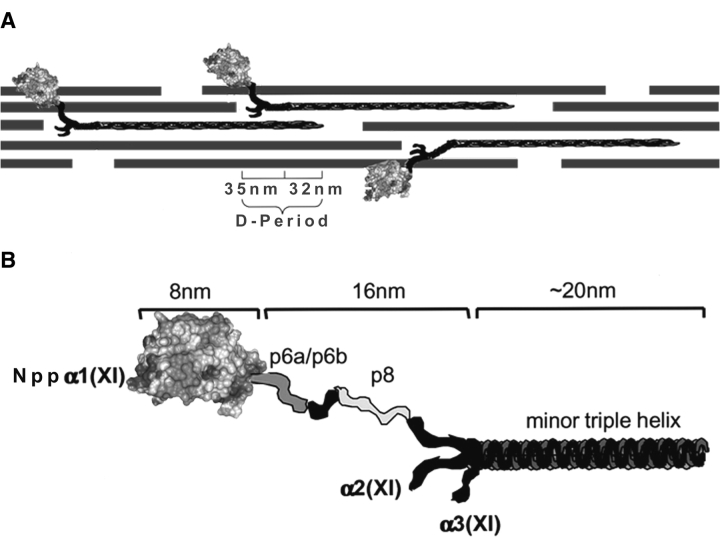
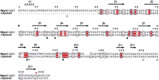
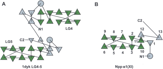
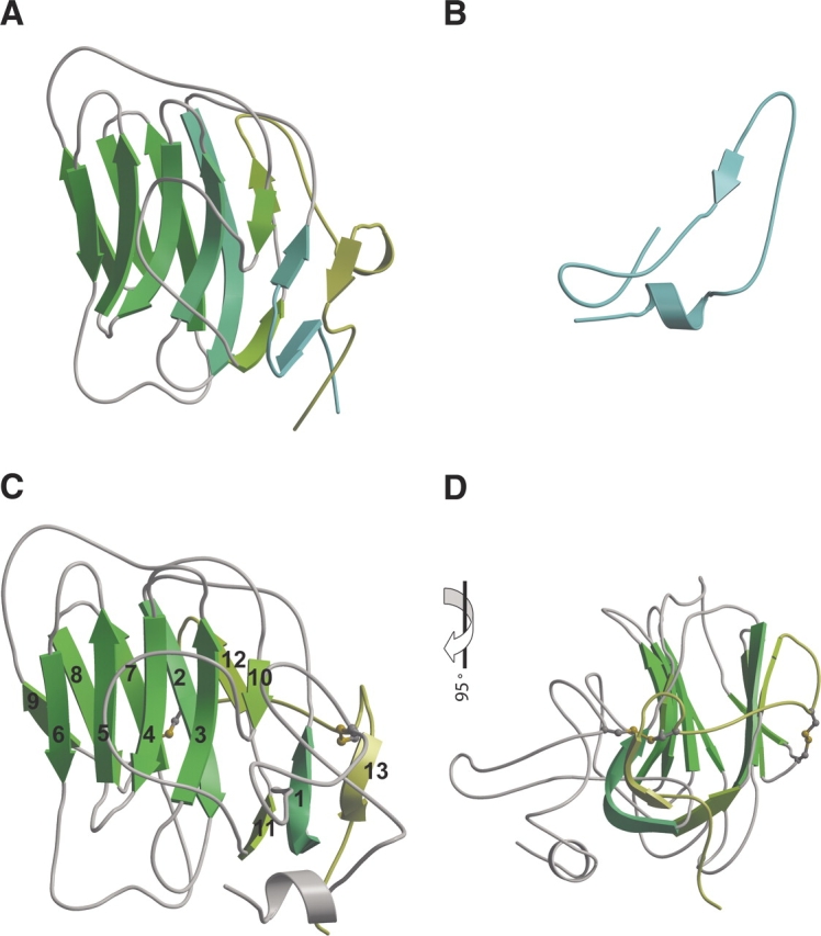
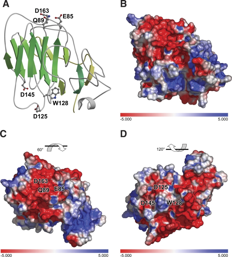
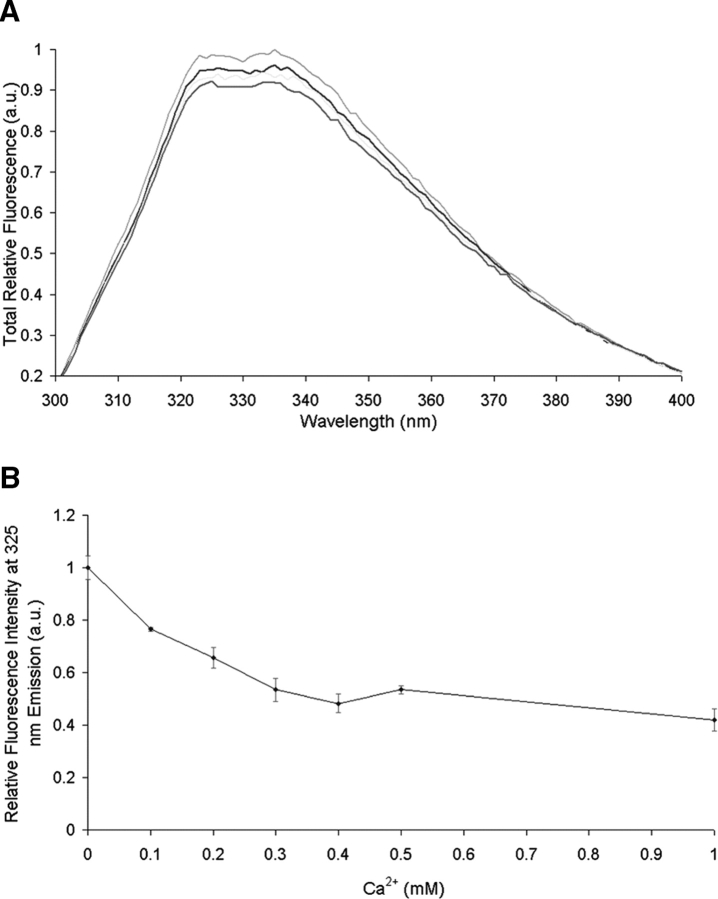
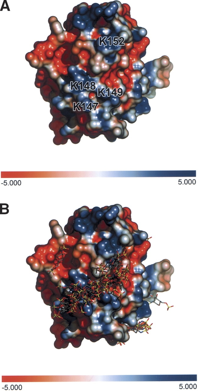
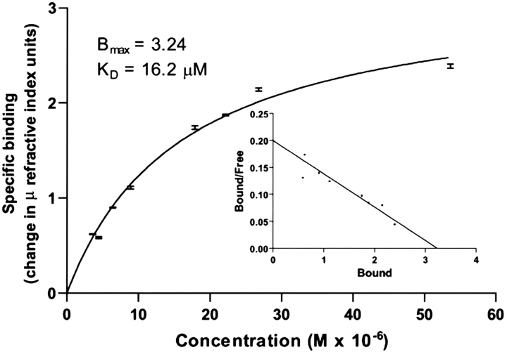
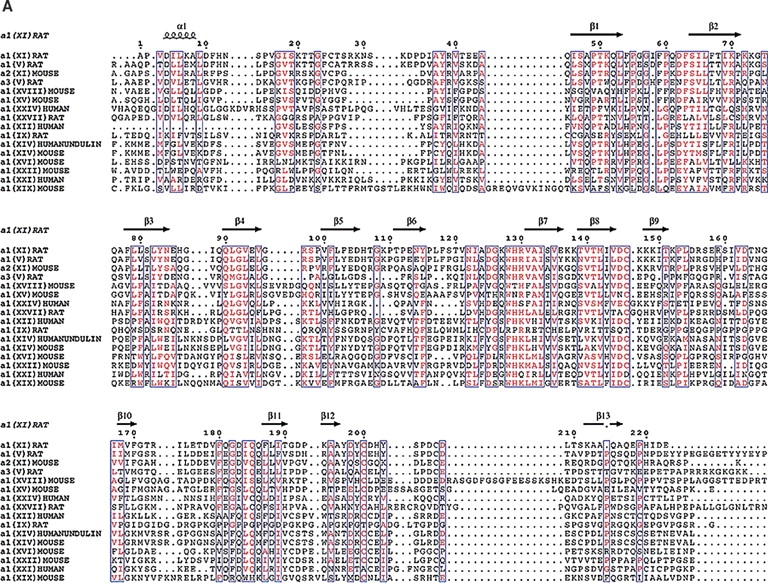
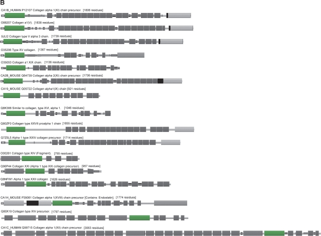
Fibrillar collagens are the principal structural molecules of connective tissues. The assembly of collagen fibrils is regulated by quantitatively minor fibrillar collagens, types V and XI. A unique amino-terminal propeptide domain of these collagens has been attributed this regulatory role. The structure of the amino terminal propeptide has yet to be determined. Low sequence similarity necessitated a secondary structure-based method to carry out homology modeling based upon the determined structure of LNS family members, named for a common structure in the laminin LG5 domain, the neurexin 1B domain and the sex hormone binding globulin. Distribution of amino acids within the model suggested glycosaminoglycan interaction and calcium binding. These activities were tested experimentally. Sequence analyses of existing genes for collagens indicate that 16 known collagen alpha chains may contain an LNS domain. A similar approach may prove useful for structure/function studies of similar domains in other collagens with similar domains. This will provide mechanistic details of the organization and assembly of the extracellular matrix and the underlying basis of structural integrity in connective tissues. The absolute requirement for collagen XI in skeletal growth is indicated by collagen XI deficiencies such as chondrodystrophies found in the cho/cho mouse and in humans with Stickler syndrome.


Figures

Figure 1.
Figure 1.
Schematic of collagen fibril and Npp α1(XI) collagen. (A) Collagen fibril shown comprised of collagen types II, IX, and XI. The globular domain Nppα1(XI) collagen is shown to extend from the surface of the collagen fibril. The dimensions of the gap-overlap region of the D-period are indicated. (B) Schematic representation of the Npp α1(XI) and adjacent variable region and minor triple helix. The dimensions as determined (Gregory et al. 2000) by transmission electron microscopy are indicated. The position of the Npp α1(XI) domain is indicated as distal with respect to the adjacent p6a/p6b/p8 variable region and minor triple helix. The α2(XI) and α3(XI) chains are also indicated in the diagram.
Figure 2.
Figure 2.
Alignment of 1dyk and Npp α1(XI) collagen. The laminin α2 LG chain was used as template for 183 amino acids of Npp α1(XI) collagen. Open circles indicate the position of four cysteine residues of Npp α1(XI) collagen; green circles indicate the position of residues of potential calcium binding sites. Conserved amino acid residues are shown as white letters on red background, while similar amino acid residues are shown as red letters on white background. Positions of predicted β-strands are indicated by arrows above the sequence. Position of predicted α-helix is indicated by a coil symbol above the sequence. Amino acids are numbered from 1 to 223, with 1 defined as the first amino acid after the signal peptide cleavage site. Bone morphogenetic protein-1 proteolytic processing site is indicated by an inverted red triangle. Putative heparin binding site is indicated by blue lettering at positions 147–152.
Figure 3.
Figure 3.
Protein structural topology. (A) Topology of 1dyk LG4-5. The conserved motif is shown in green, while the outside regions are gray. β-Strands are shown as triangles and α-helices are shown as circles. (B) Topology of Npp α1(XI) collagen. The β-strands of 1dyk LG4-5 show identical topology with Npp α1(XI) collagen as well as other LNS family members (data not shown), and is characteristic of the LNS family of proteins. Although the directions of the triangles will vary depending on the total number of β-strands, the connectivity of the β-strands is constant.
Figure 4.
Figure 4.
Ab initio and homology model. Three-dimensional structure of (A) 1dyk LG5, (B) the first 40 amino acids by ab initio modeling, (C) the model resulting from merger of ab initio and homology modeling, (D) model shown in 4-color after 90° rotation. Disulfide bonds are shown in panels C and D. β-Strands are numbered sequentially from the amino to carboxyl end of the protein.
Figure 5.
Figure 5.
Putative calcium binding sites. Sequence homology with laminin α2 LG5 domain indicates the potential for similarity in calcium binding function, utilizing amino acid residues D163 and Q89. The amino acid side chain of E85 is in close proximity is also indicated. A second potential Ca2+ binding site is indicated near amino acid residues D125 and D145. (A) Ribbon diagram. (B) Electrostatic surface, identical orientation as that shown in panel A. (C) 60° rotation of model with respect to panel B to show Ca2+ binding site D163Q89. (D) 120° rotation of model with respect to panel B to show Ca2+ binding site D125D145.
Figure 6.
Figure 6.
Effect of divalent cation on the fluorescence emission spectrum of Npp α1(XI) collagen. Protein (40 μg/mL) in 5 mM MOPS buffer (pH 7.5), containing 150 mM NaCl was excited at 285 nm. (A) The emission spectra were recorded between 300 and 400 nm. (B) A decrease in fluorescence intensity was observed as a function of increasing Ca2+ concentration.
Figure 7.
Figure 7.
Putative heparin binding site. (A) Interactions between heparin and Npp α1(XI) collagen were predicted. For each heparin ligand, one job of 100 docking runs was performed using a population of 200 individuals and an energy evaluation number of 2 × 10⁶ employing the Lamarckian Genetic Algorithm. A further refined docking was centered at the putative heparin binding site on residues 147–152 (KKKITK). (B) Amino acids 147–152 form a track of positively charged side chains on the surface of the protein.
Figure 8.
Figure 8.
Interaction between heparan sulfate and Npp α1(XI) collagen. Concentrations of heparan sulfate (0.625–0.75 mg/mL) were injected over immobilized Npp at a flow rate of 10 μL per min in phosphate buffered saline containing Tween-20 (0.05% v/v). An average of the response at equilibrium was determined for each concentration and the resulting equilibrium resonance units were plotted against concentration. The data were fit to a steady-state one-site affinity model to determine the equilibrium dissociation constant (Kd) of heparan sulfate for Npp α1(XI) collagen. Scatchard analysis was carried out and is presented in the inset.
Figure 9.
Figure 9.
A comparison of collagens containing similar LNS domains. (A) Identical residues are red while similar residues are purple. The β-strands fall in highly conserved regions suggesting that our model may extended to other collagens that contain the LNS or thrombospondin-like domain. The β-strand which contains the putative heparin binding site is unique to Npp α1(XI) collagen and not conserved among the other collagens. The highly conserved cysteine residues suggest that they may be important in the structure of this domain. (B) Pfam schematic of LNS (TSPN) domain (shown as green box) in collagens. This domain is contained in a number of collagens but may vary in position.
Figure 9.
Figure 9.
A comparison of collagens containing similar LNS domains. (A) Identical residues are red while similar residues are purple. The β-strands fall in highly conserved regions suggesting that our model may extended to other collagens that contain the LNS or thrombospondin-like domain. The β-strand which contains the putative heparin binding site is unique to Npp α1(XI) collagen and not conserved among the other collagens. The highly conserved cysteine residues suggest that they may be important in the structure of this domain. (B) Pfam schematic of LNS (TSPN) domain (shown as green box) in collagens. This domain is contained in a number of collagens but may vary in position.

References

    1. Bateman, A., Coin, L., Durbin, R., Finn, R.D., Hollich, V., Griffiths-Jones, S., Khanna, A., Marshall, M., Moxon, S., and Sonnhammer, E.L. et al. 2004. The Pfam protein families database. Nucleic Acids Res. 32 138–141. - PMC - PubMed
    1. Beckmann, G., Hanke, J., Bork, P., and Reich, J.G. 1998. Merging extracellular domains: Fold prediction for laminin G-like and amino-terminal thrombospondin-like modules based on homology to pentraxins. J. Mol. Biol. 275 725–730. - PubMed
    1. Berman, H.M., Westbrook, J., Feng, Z., Gilliland, G., Bhat, T.N., Weissig, H., Shindyalov, I.N., and Bourne, P.E. 2000. The Protein Data Bank. Nucleic Acids Res. 28 235–242. - PMC - PubMed
    1. Blaschke, U.K., Eikenberry, E.F., Hulmes, D.J.S., Galla, H.-J., and Bruckner, P. 2000. Collagen XI nucleates self-assembly and limits lateral growth of cartilage fibrils. J. Biol. Chem. 275 10370–10378. - PubMed
    1. Bork, P. 1992. The modular architecture of vertebrate collagens. FEBS Lett. 307 49–54. - PubMed

Publication types

Substances

LinkOut - more resources