. 2005 Aug;16(8):3666-77.
doi: 10.1091/mbc.e05-01-0014. Epub 2005 Jun 1.

Spindle checkpoint signaling requires the mis6 kinetochore subcomplex, which interacts with mad2 and mitotic spindles

Affiliations

Spindle checkpoint signaling requires the mis6 kinetochore subcomplex, which interacts with mad2 and mitotic spindles

Shigeaki Saitoh et al. Mol Biol Cell. 2005 Aug.

Abstract

The spindle checkpoint coordinates cell cycle progression and chromosome segregation by inhibiting anaphase promoting complex/cyclosome until all kinetochores interact with the spindle properly. During early mitosis, the spindle checkpoint proteins, such as Mad2 and Bub1, accumulate at kinetochores that do not associate with the spindle. Here, we assess the requirement of various kinetochore components for the accumulation of Mad2 and Bub1 on the kinetochore in fission yeast and show that the necessity of the Mis6-complex and the Nuf2-complex is an evolutionarily conserved feature in the loading of Mad2 onto the kinetochore. Furthermore, we demonstrated that Nuf2 is required for maintaining the Mis6-complex on the kinetochore during mitosis. The Mis6-complex physically interacts with Mad2 under the condition that the Mad2-dependent checkpoint is activated. Ectopically expressed N-terminal fragments of Mis6 localize along the mitotic spindle, highlighting the potential binding ability of Mis6 not only to the centromeric chromatin but also to the spindle microtubules. We propose that the Mis6-complex, in collaboration with the Nuf2-complex, monitors the spindle-kinetochore attachment state and acts as a platform for Mad2 to accumulate at unattached kinetochores.


Figures
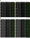
Figure 1.
Figure 1.
Mad2 accumulates on kinetochores in early mitosis and relocates to the spindle and the SPBs before the onset of anaphase. (A) Fission yeast cells expressing Mad2-GFP along with SpCENP-C-CFP, a kinetochore marker, were observed by time-lapse three-dimensional deconvolution microscopy. For CFP-tagging of SpCENP-C, a modified version of CFP, named “cerulean,” was used (Rizzo et al., 2004). These images were taken every 0.75 min. In merged images in the right column, Mad2-GFP signal was pseudocolored green and SpCENP-C-CFP signal was pseudocolored red. Dot-like Mad2-GFP signals occurring at the early stage of mitosis (time = 0.75–2.25 min) colocalized with a subset of kinetochores. Two Mad2-GFP dots occurring at the later stage (time = 6–9 min) did not colocalize with kinetochores, which moved along short linear path between these two dots. (B) Time-lapse images of a cell expressing Mad2-GFP and CFP-α-tubulin, a spindle marker, were shown. Images were taken every 0.5 min. In merged images, Mad2-GFP signal was colored green and CFP-α-tubulin was colored red. Under the imaging condition in this experiment, CFP-α-tubulin signals on cytoplasmic astral MTs were too weak to be detected. Two Mad2-GFP dots occurring at the later stage of mitosis (time = 5.5–9.5 min) located at the both poles of the spindle. Mad2-GFP also localized along the spindle at this stage. The number indicates the duration in minutes. Bar, 10 μm.
Figure 2.
Figure 2.
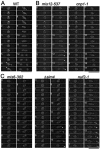
The proper kinetochore accumulation of Mad2 requires Mis6-Sim4 and Nuf2, but not Mis12 or SpCENP-A. (A) Fission yeast wild-type cells expressing Mad2-GFP were observed by time-lapse three-dimensional deconvolution microscopy. Images were taken every 0.5 min. Dot-like GFP signals occurring at the early stage of mitosis (time = 0.5–3.5 min) represent the accumulation of Mad2 at the kinetochore. Arrowheads and asterisks indicate the transient accumulation of Mad2 on the spindle and the spindle poles, respectively. (B and C) Mitotic behavior of Mad2 in the indicated mutant cells that were cultured at their restrictive conditions (6–8 h at 36°C for mis6-302 and mis12-537, 8–10 h at 33°C for cnp1-1, 1.5–4 h at 36°C for nuf2-1, and 12–16 h at 33°C with 2 μM thiamine for sim4+-shut-off (Δsim4)) is shown. The kinetochore accumulation of Mad2 was observed in the mutants in B, but it was greatly diminished or not observed in the mutants in C. In the sim4+-shut-off cells, sim4+ gene expression is placed under the control of thiamine-repressible nmt1-81 promoter. Sim4 protein was virtually undetectable, and >80% of cells showed unequal nuclear division phenotype 14 h after the addition of thiamine. Movies of the cells shown in this figure are provided as supplemental materials (Movies S1–S6).
Figure 3.
Figure 3.
The kinetochore accumulation of Bub1 in kinetochore mutants. (A) Time-lapse images of a cell expressing Bub1-GFP and SpCENP-C-CFP were shown. Images were taken every 0.75 min. Bub1-GFP localized in nuclei during interphase (time = 0 min). During early M phase (time 0.75–3 min), bright dot-like signals of Bub1-GFP were observed, and these signals colocalized with a subset of kinetochores. In the merged images, Bub1-GFP and SpCENP-C-CFP are shown in green and red, respectively. (B) The mitotic behavior of Bub1 was examined in various kinetochore-defective mutants at their restrictive conditions. Time-lapse images of the first six frames (2.5 min) of each mutant cell in mitosis are shown. The dot-like signals represent the accumulation of Mad2 on kinetochores. Bub1 localizes on the nuclear chromatin during interphase, and accumulates on kinetochores during early mitosis in mis6-302, sim4+-shut-off (Δsim4), mis12-537, nuf2-1, and cnp1-1 mutant cells.
Figure 4.
Figure 4.
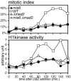
The Mad2-dependent spindle checkpoint is defective in the mis6-302 mutant. Cells with one of the following genetic backgrounds were cultured in YES medium in the presence of 50 μg/ml CBZ at 30°C: WT, Δmad2, mis6-302, or mis6-302 Δmad2 double mutant. The frequency (percentage) of cells containing hypercondensed chromosomes was determined by DAPI staining in 20-min intervals (top). Histone H1 kinase activity in the total cell lysate was also measured (bottom).
Figure 5.
Figure 5.
The depletion of Mis6-Sim4 does not impair the kinetochore localization of SpCENP-C, Mis12, or Nuf2. (A) The sim4+-shut-off cells carrying either the integrated Mis6-GFP, SpCENP-A-GFP, Mis12-GFP, SpCENP-C-GFP, or Nuf2-GFP gene were cultured in EMM2 at 33°C for 0 (Sim4 was expressed) and 14 h (Sim4 was repressed) after the addition of thiamine. The photographs of GFP fluorescence were taken after fixation with methanol pre-chilled at –80° C. (B) The time-lapse serial images of Nuf2-GFP in Sim4-depleted cells are shown.
Figure 6.
Figure 6.
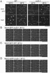
Sim4 delocalizes from the kinetochore during mitosis in nuf2-1 mutant cells. (A) Wild-type cells (left) or nuf2-1 mutant cells (right) with sim4+-GFP gene integrated were cultured at either 36°C for 4 h or 22°C (permissive condition) and fixed with methanol pre-chilled at –80° C. Three-dimensional serial images were taken and projected into a single plane after deconvolution. Arrowheads indicate dot-like signals of Sim4 observed in nuf2-1 mutant cells at the restrictive condition. (B–D) Time-lapse images of nuf2-1 mutant cells with Sim4-GFP (B) or Mis12-GFP (D) or of wild-type cells with Sim4-GFP (C) are shown. The cells were incubated at 36°C for 1.5–2 h.
Figure 7.
Figure 7.
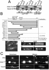
Mis6 physically interacts with Mad2. (A) Immunoprecipitation experiments were performed using crude extract from cut7-477 or cut9-665 mutant cells harboring integrated mis6+-FLAG and mad2+-GFP genes. The cut7 mutant cells were cultured at either 36°C for 3 h (restrictive condition, lanes 1–3) or 26°C (permissive condition, lanes 4–6). The cut9 mutant cells were cultured at 36°C for 3.5 h (restrictive condition, lanes 7–9). The anti-GFP mAb (Roche Diagnostics) was used for the precipitation of GFP-tagged Mad2 (lanes 3, 6, and 9). In the mock experiment, the buffer was added instead of the antibody (lanes 2, 5, and 8). Twenty percent of the inputs were loaded on lanes 1, 4, and 7. (B) A series of truncated Mis6 were fused with GFP and ectopically expressed under the nmt1-1 promoter, and their subcellular localization was determined. C.A., N, and S stand for cytoplasmic aggregate formation, nuclear localization, and spindle localization, respectively. N-terminal domains that are highly conserved among fission yeast, chicken, and human were indicated by gray boxes in the schematic drawing of Mis6 protein. The mutation site of Mis6-302 temperature-sensitive protein also was indicated by a vertical bar. Representative images of the truncated Mis6 constructs forming cytoplasmic aggregates, localizing in the nuclei or localizing along the mitotic spindle were shown in right bottom panels. An image of ectopically expressed GFP alone, which dispersed throughout the cell, also is presented (GFP). Bottom left, cells expressing Mis61-265 were stained with DAPI. Prometaphase-like cells with hypercondensed chromosomes were frequently observed. (C) The ectopically expressed Mis61-265 localized along the mitotic spindles. Wild-type cells expressing Mis61-265-GFP was immunostained with anti-α-tubulin antibody (TAT1). Chromatin DNA was stained by DAPI. Representative cells in the M phase (middle and right columns) and in the interphase (left column) are presented. GFP fluorescence of the cytoplasmic aggregates was so intense that it leaked into the DAPI channel in some samples (arrows). The position of astral MTs in the mitotic cells was indicated by arrowheads.

Similar articles

Cited by

References

    1. Appelgren, H., Kniola, B., and Ekwall, K. (2003). Distinct centromere domain structures with separate functions demonstrated in live fission yeast cells. J. Cell Sci. 116, 4035–4042. - PubMed
    1. Bernard, P., Hardwick, K., and Javerzat, J. P. (1998). Fission yeast bub1 is a mitotic centromere protein essential for the spindle checkpoint and the preservation of correct ploidy through mitosis. J. Cell Biol. 143, 1775–1787. - PMC - PubMed
    1. Cahill, D. P., Lengauer, C., Yu, J., Riggins, G. J., Willson, J. K., Markowitz, S. D., Kinzler, K. W., and Vogelstein, B. (1998). Mutations of mitotic checkpoint genes in human cancers. Nature 392, 300–303. - PubMed
    1. Cheeseman, I. M., Anderson, S., Jwa, M., Green, E. M., Kang, J., Yates, J. R., 3rd, Chan, C. S., Drubin, D. G., and Barnes, G. (2002a). Phospho-regulation of kinetochore-microtubule attachments by the Aurora kinase Ipl1p. Cell 111, 163–172. - PubMed
    1. Cheeseman, I. M., Drubin, D. G., and Barnes, G. (2002b). Simple centromere, complex kinetochore: linking spindle microtubules and centromeric DNA in budding yeast. J. Cell Biol. 157, 199–203. - PMC - PubMed

Publication types

MeSH terms