. 2005 Aug;16(8):3873-86.
doi: 10.1091/mbc.e04-12-1105. Epub 2005 Jun 1.

MLN64 is involved in actin-mediated dynamics of late endocytic organelles

Affiliations

MLN64 is involved in actin-mediated dynamics of late endocytic organelles

Maarit Hölttä-Vuori et al. Mol Biol Cell. 2005 Aug.

Abstract

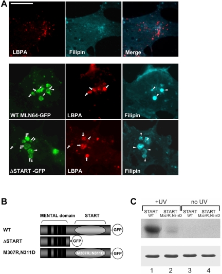
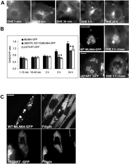
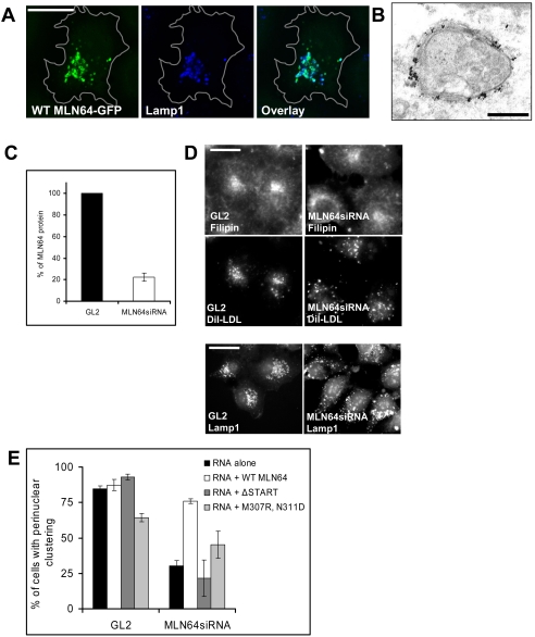
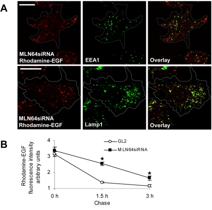
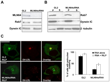
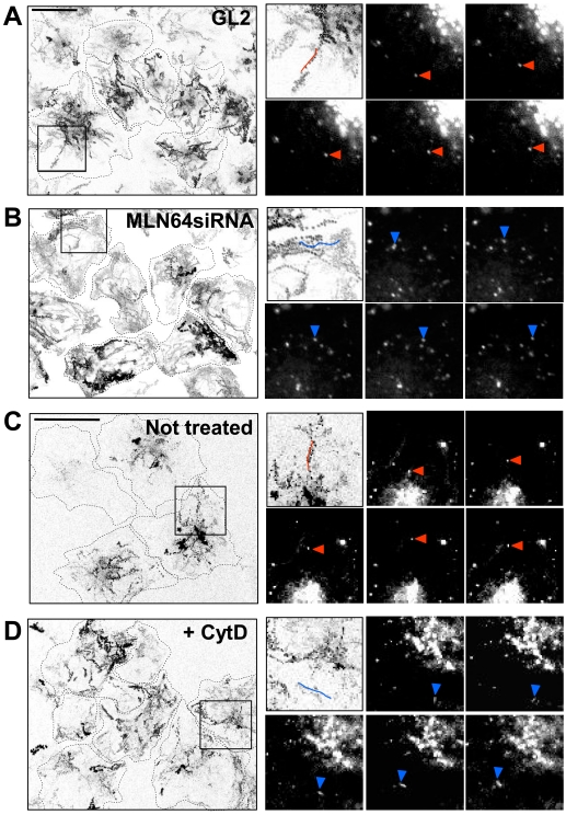
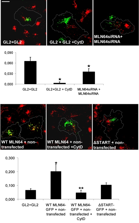
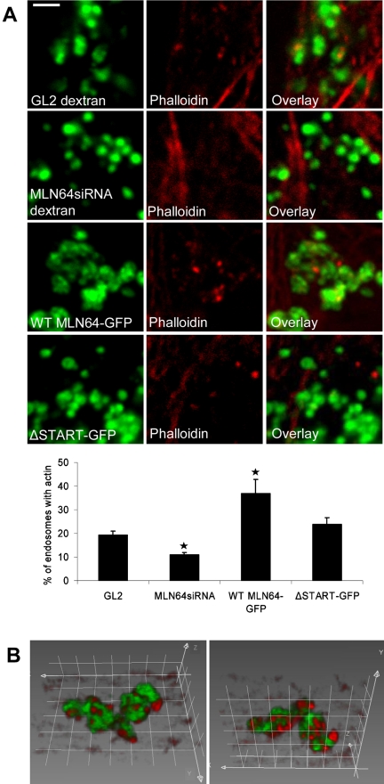
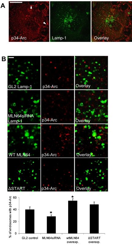
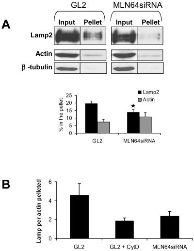
MLN64 is a late endosomal cholesterol-binding membrane protein of an unknown function. Here, we show that MLN64 depletion results in the dispersion of late endocytic organelles to the cell periphery similarly as upon pharmacological actin disruption. The dispersed organelles in MLN64 knockdown cells exhibited decreased association with actin and the Arp2/3 complex subunit p34-Arc. MLN64 depletion was accompanied by impaired fusion of late endocytic organelles and delayed cargo degradation. MLN64 overexpression increased the number of actin and p34-Arc-positive patches on late endosomes, enhanced the fusion of late endocytic organelles in an actin-dependent manner, and stimulated the deposition of sterol in late endosomes harboring the protein. Overexpression of wild-type MLN64 was capable of rescuing the endosome dispersion in MLN64-depleted cells, whereas mutants of MLN64 defective in cholesterol binding were not, suggesting a functional connection between MLN64-mediated sterol transfer and actin-dependent late endosome dynamics. We propose that local sterol enrichment by MLN64 in the late endosomal membranes facilitates their association with actin, thereby governing actin-dependent fusion and degradative activity of late endocytic organelles.


Figures

Figure 1.
Figure 1.
MLN64 overexpression in COS cells. (A) Nontransfected cells or cells overexpressing GFP-fused WT MLN64 or ΔSTART were stained with filipin and anti-LBPA antibodies. Arrows, MLN64-positive organelles containing LBPA. Arrowheads, MLN64-positive organelles containing filipin. Bar, 10 μm. (B) Schematic representation of WT and mutant MLN64 cDNAs used. (C) WT (lanes 1 and 3) and M307R, N311D mutant (lanes 2 and 4) MLN64 START domains were incubated with [3H]photocholesterol and either UV-irradiated or not. After SDS-PAGE, crosslinked [3H]cholesterol was revealed by fluorography (top). For comparison, similar amounts of recombinant proteins were stained with Coomassie Blue after SDS-PAGE (bottom).
Figure 2.
Figure 2.
MLN64 enhances sterol deposition within late endosomes. (A) COS-1 cells were labeled with DHE for 1 min and then incubated as indicated. (B) COS-1 cells overexpressing GFP-fused WT or mutant MLN64 were labeled with DHE and then incubated as indicated. The GFP and DHE signals were imaged from living cells at indicated time points and the intensity ratio of DHE versus GFP in endosomes was assessed. Bars are mean intensity ratios from 18 to 54 endosomes analyzed, *p < 0.05 between WT MLN64 and M307R,N311D or ΔSTART. Exemplary fluorescence micrographs from 5-h chase are shown. Bars, 10 μm. (C) NPC fibroblasts were electroporated with plasmids encoding GFP-fused WT or ΔSTART MLN64 and stained with filipin. Bars, 20 μm.
Figure 3.
Figure 3.
Characterization of MLN64 overexpression and knockdown in HeLa cells. (A) Cells overexpressing WT MLN64-GFP were stained with anti-Lamp1 antibodies. Images are confocal and represent a single focal plane. Bar, 20 μm. (B) WT MLN64-transfected cells were processed for immunoelectron microscopy with anti-MLN64 C-terminal antibodies. Bar, 200 nm. (C) Cells were transfected with control siRNA oligos (GL2) or oligos targeted against MLN64 (MLN64siRNA). Cell lysates (30 μg of protein) were analyzed by Western blotting using anti-MLN64 C-terminal antibodies. The amount of MLN64 protein was determined by densitometric scanning and expressed as a percentage of the protein in MLN64-depleted versus control cells (mean from 4 experiments). (D) Control RNA transfected (GL2) or MLN64-depleted cells (MLN64siRNA) were labeled with DiI-LDL, fixed, and stained with filipin and Lamp1-antibodies. Bars, 20 μm. (E) Quantification of the dispersal of DiI-LDL-positive organelles. Cells were transfected with siRNAs alone or cotransfected with plasmids encoding GFP-fused WT or mutant MLN64 cDNAs. The cells were then labeled with DiI-LDL before fixation. The bars represent percentage of cells with perinuclear clustering of DiI-LDL-positive organelles (2-10 independent experiments, n = 48-605 cells).
Figure 4.
Figure 4.
Depletion of MLN64 does not impair the sorting of EGF from early endosomes but retards EGF degradation. (A) MLN64-depleted cells (MLN64siRNA) were incubated with rhodamine-labeled EGF for 30 min, further incubated for 1.5 h, fixed, and stained with antibodies against EEA1 and Lamp1. Images are confocal and represent a single focal plane. Bars, 20 μm. (B) Control (GL2) or MLN64-depleted cells (MLN64siRNA) were fed rhodamine-labeled EGF for 1 h and further incubated for 1.5 or 3 h. The mean intensity of cell-associated EGF fluorescence was analyzed from 15 cells at each time point, *p < 0.05 between GL2 control and MLN64siRNA.
Figure 5.
Figure 5.
Rab7-RILP-dynein machinery in MLN64-depleted HeLa cells. (A) Cells were transfected with control siRNA oligos (GL2) or oligos targeted against MLN64 (MLN64siRNA). Aliquots (30 μg) of cell lysates were analyzed by Western blotting using anti-MLN64 C-terminal, anti-Rab, or anti-dynein IC antibodies. (B) PNS from control (GL2) or MLN64-depleted cells (MLN64siRNA) was pelleted at 100 000 g and total membrane pellets (P) and supernatants (S) analyzed by Western blotting using anti-Rab7, anti-dynein IC, or anti-β-tubulin antibodies. (C) Cells were transfected with siRNAs alone or cotransfected with a plasmid encoding HA-tagged RILP, labeled with DiI-LDL, fixed and stained with anti-RILP antibodies. Bars represent percentage of cells with perinuclear clustering of DiI-LDL-positive organelles from two independent experiments (111-158 cells in total). Images are confocal and represent a single focal plane. Bars, 20 μm.
Figure 6.
Figure 6.
MLN64 depletion and actin disruption have similar effects on endosome motility in HeLa cells. Control (GL2) (A) or MLN64-depleted cells (MLN64siRNA) (B) were labeled with DiI-LDL and then imaged for 4 min (1 image/s). In C and D, untransfected cells were labeled with DiI-LDL and further incubated for 2.5 h, the last 1 h with or without 0.2 μM cytochalasin D, and imaged as described in A and B. The trajectories of labeled endosomes from a field of cells are shown (left). Cell boundaries are sketched with dotted lines. Boxed areas are depicted in the top left insets. Video frames at 2-s intervals are shown, and the individual endosomes producing the tracks outlined in the top left insets are indicated with arrowheads. Examples of stellate trajectories are highlighted in red and peripheral trajectories in blue. Bars, 20 μm.
Figure 7.
Figure 7.
Effect of MLN64 depletion or overexpression in the fusion of late endocytic organelles in HeLa cells. Nontransfected and GL2 control or MLN64siRNA-transfected cells were labeled with fluorescein or rhodamine-conjugated dextrans. Alternatively, cells were transfected with GFP-fused WT MLN64 or ΔSTART. The cells were then cocultured as indicated, fused with PEG, and incubated for 1 h either in normal growth medium or medium supplemented with 0.2 μM cytochalasin D. The degree of overlay was determined by Pearson's correlation coefficient. The values are from 23 to 55 individual cells, *p < 0.05 between GL2 control and GL2 + cytochalasin D, MLN64siRNA, or WT MLN64-GFP overexpression, **p < 0.05 between WT MLN64-GFP overexpression and WT MLN64-GFP overexpression + cytochalasin D. Images are confocal and represent a single focal plane. Bar, 20 μm.
Figure 8.
Figure 8.
MLN64 modulates the association of actin with late endosomes in HeLa cells. (A) Phalloidin staining of GL2 control and MLN64siRNA-transfected cells labeled with fluorescein-conjugated dextran and HeLa cells transfected with GFP-fused WT MLN64 or ΔSTART. The percentage of late endocytic organelles decorated with actin patches was analyzed from confocal sections of four to 10 individual cells at high magnification, n of endosomes ≥358 per treatment, *p < 0.05 between GL2 control and MLN64siRNA or WT MLN64-GFP overexpression. Images are confocal and represent a single focal plane. Bars, 2 μm. (B) Three-dimensional rendering of WT MLN64-GFP-positive endosomes stained with phalloidin is shown from two angles. Grid spacing, 1 μm.
Figure 9.
Figure 9.
MLN64 modulates the association of p34-Arc with late endosomes in HeLa cells. (A) Control HeLa cells were stained with anti-p34-Arc and anti-Lamp1 antibodies. Images are confocal and represent a single focal plane. Arrowheads (left) point to the prominent lamellipodial p34-Arc staining that is only partially visible in the section. Bar, 20 μm. (B) GL2 control and MLN64siRNA transfected cells were stained with anti-p34-Arc and anti-Lamp1 antibodies. WT MLN64 or ΔSTART overexpressing cells were stained with anti-p34-Arc and anti-MLN64 (N-terminal) antibodies. The percentage of late endocytic organelles decorated with p34-Arc was calculated from 10 to 12 individual cells as in Figure 8A, n of endosomes ≥624 per treatment, *p < 0.05 between GL2 control and MLN64siRNA or WT MLN64-GFP overexpression. Images are confocal and represent a single focal plane. Bars, 2 μm.
Figure 10.
Figure 10.
MLN64 depletion decreases the sedimentation of Lamp2-positive organelles with actin. (A) PNS from control (GL2) or MLN64-depleted cells (MLN64siRNA) was incubated in the presence of 100 μM nocodazole and pelleted at 100,000 × g for 5 min. Aliquots of PNS before centrifugation (input) and pellets were immunoblotted using anti-Lamp2, anti-actin, and anti-β-tubulin antibodies. The amount of Lamp2 and actin pelleted was determined by densitometric scanning and expressed as percentage of input (mean of 12 samples). *p < 0.05 between GL2 and MLN64siRNA. (B) The fraction of Lamp2 sedimented was normalized with the fraction of actin pelleted. GL2 + CytD indicates PNS of control (GL2) cells incubated in the presence of 4 μM cytochalasin D. Bars represent mean of seven to 12 samples.

Similar articles

Cited by

References

    1. Alpy, F., Stoeckel, M. E., Dierich, A., Escola, J. M., Wendling, C., Chenard, M. P., Vanier, M. T., Gruenberg, J., Tomasetto, C., and Rio, M. C. (2001). The steroidogenic acute regulatory protein homolog MLN64, a late endosomal cholesterol-binding protein. J. Biol. Chem. 276, 4261-4269. - PubMed
    1. Alpy, F., Latchumanan, V. K., Kedinger, V., Janoshazi, A., Thiele, C., Wendling, C., Rio, M. C., and Tomasetto, C. (2005). Functional characterization of the MENTAL domain. J. Biol. Chem. 280, 17945-17952. - PubMed
    1. Alpy, F., Wendling, C., Rio, M. C., and Tomasetto, C. (2002). MENTHO, a MLN64 homologue devoid of the START domain. J. Biol. Chem. 277, 50780-50787. - PubMed
    1. Arai, R., Geffard, M., and Calas, A. (1992). Intensification of labelings of the immunogold silver staining method by gold toning. Brain Res. Bull. 28, 343-345. - PubMed
    1. Barbero, P., Bittova, L., and Pfeffer, S. R. (2002). Visualization of Rab9-mediated vesicle transport from endosomes to the trans-Golgi in living cells. J. Cell Biol. 156, 511-518. - PMC - PubMed

Publication types

MeSH terms

LinkOut - more resources