

Nef from pathogenic simian immunodeficiency virus is a negative factor for vaccinia virus
- PMID: 15930136
- PMCID: PMC1142211
- DOI: 10.1073/pnas.0503542102
Nef from pathogenic simian immunodeficiency virus is a negative factor for vaccinia virus
Abstract
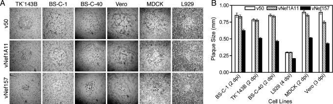
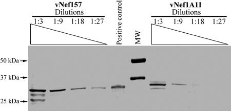
The nef gene of human and simian immunodeficiency viruses (HIV and SIV) is important for pathogenicity and maintenance of high virus loads. We previously reported that recombinant vaccinia viruses (rVVs) expressing nef from attenuated SIVmac1A11 (vNef1A11) produced typical plaques on thymidine kinase-deficient 143B cells, whereas rVVs expressing nef derived from the pathogenic SIVmac239 (vNef157) formed plaques with altered morphology. Here, we show that vNef157 is attenuated in normal and nude mice, whereas the pathogenicity of vNef1A11 is similar to that of a control virus. Thus, Nef157 is an attenuating factor in the vaccinia virus (VV) system, contrasting sharply with its function in lentiviruses. We also show that Nef157 inhibits VV cell-to-cell spread, causing formation of atypical plaques regardless of thymidine kinase deficiency, neoplasticity, and species of the infected cell line. We hypothesized that Nef157 interferes with VV spread by association with actin, but no direct colocalization of Nef and the cytoskeletal actin network was detected. Instead, higher levels of Nef157 protein were observed, although mRNAs for both nef genes were produced at comparable levels. Thus, the mechanism behind such Nef157 protein accumulation and Nef157-mediated VV attenuation could be related to the process that causes an opposite effect in its native SIV system, making SIVmac239 more pathogenic than SIVmac1A11.
Figures
Similar articles
-
Altered plaque formation by recombinant vaccinia virus expressing simian immunodeficiency virus Nef.J Virol. 1998 Jun;72(6):5291-5. doi: 10.1128/JVI.72.6.5291-5295.1998. J Virol. 1998. PMID: 9573307 Free PMC article.
-
Chronology of genetic changes in the vpu, env, and Nef genes of chimeric simian-human immunodeficiency virus (strain HXB2) during acquisition of virulence for pig-tailed macaques.Virology. 1998 Sep 1;248(2):275-83. doi: 10.1006/viro.1998.9300. Virology. 1998. PMID: 9721236
-
Properties of a chimeric simian-human immunodeficiency virus expressing an hybrid HIV-1 Nef/SIVmac Nef protein.Arch Virol. 2002 Oct;147(10):1963-75. doi: 10.1007/s00705-002-0857-8. Arch Virol. 2002. PMID: 12376757
-
Human immunodeficiency virus (HIV) type-1, HIV-2 and simian immunodeficiency virus Nef proteins.Mol Aspects Med. 2010 Oct;31(5):418-33. doi: 10.1016/j.mam.2010.05.003. Epub 2010 Jun 4. Mol Aspects Med. 2010. PMID: 20594957 Review.
-
Genetic and biological comparisons of pathogenic and nonpathogenic molecular clones of simian immunodeficiency virus (SIVmac).AIDS Res Hum Retroviruses. 1992 Mar;8(3):395-402. doi: 10.1089/aid.1992.8.395. AIDS Res Hum Retroviruses. 1992. PMID: 1571198 Review.
Cited by
-
Recombinant Rift Valley fever vaccines induce protective levels of antibody in baboons and resistance to lethal challenge in mice.Proc Natl Acad Sci U S A. 2011 Sep 6;108(36):14926-31. doi: 10.1073/pnas.1112149108. Epub 2011 Aug 23. Proc Natl Acad Sci U S A. 2011. PMID: 21873194 Free PMC article.
-
Contribution of nonneutralizing vaccine-elicited antibody activities to improved protective efficacy in rhesus macaques immunized with Tat/Env compared with multigenic vaccines.J Immunol. 2009 Mar 15;182(6):3718-27. doi: 10.4049/jimmunol.0803115. J Immunol. 2009. PMID: 19265150 Free PMC article.
References
-
- Kestler, H. W., III, Mori, K., Silva, D. P., Kodama, T., King, N. W., Daniel, M. D. & Desrosiers, R. C. (1990) J. Med. Primatol. 19, 421-429. - PubMed
-
- Arora, V. K., Fredericksen, B. L. & Garcia, J. V. (2002) Microbes Infect. 4, 189-199. - PubMed
-
- Garcia, J. V. & Miller, A. D. (1991) Nature 350, 508-511. - PubMed
-
- Lama, J., Mangasarian, A. & Trono, D. (1999) Curr. Biol. 9, 622-631. - PubMed
Publication types
MeSH terms
Substances
Grants and funding
LinkOut - more resources
Full Text Sources
