Comparative Study
. 2005 Jun 14;102(24):8501-6.
doi: 10.1073/pnas.0503285102. Epub 2005 Jun 1.

Insights into Lafora disease: malin is an E3 ubiquitin ligase that ubiquitinates and promotes the degradation of laforin

Affiliations
Comparative Study

Insights into Lafora disease: malin is an E3 ubiquitin ligase that ubiquitinates and promotes the degradation of laforin

Matthew S Gentry et al. Proc Natl Acad Sci U S A. .

Abstract

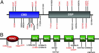
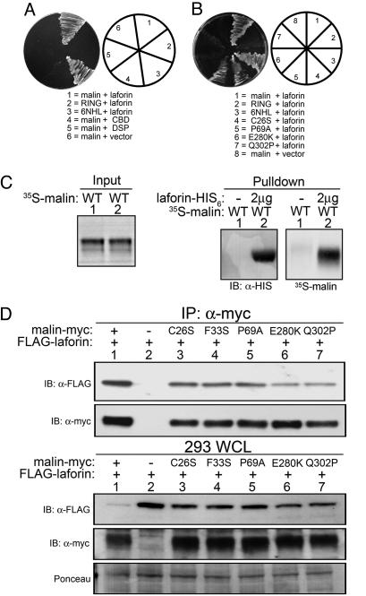
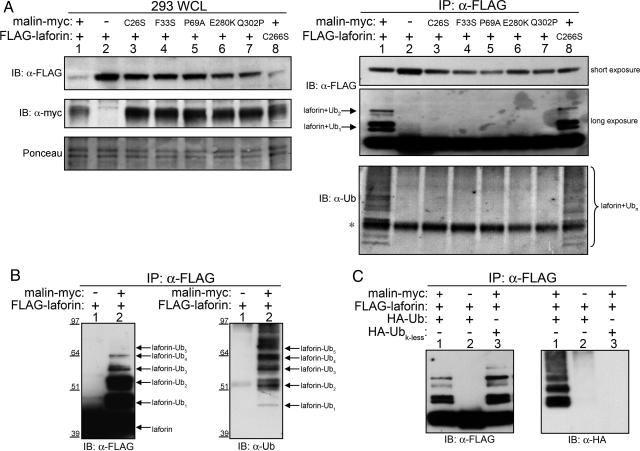
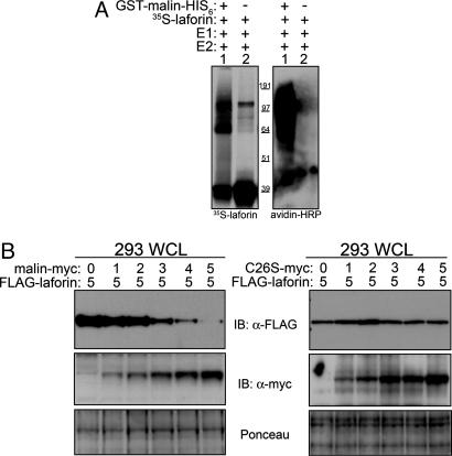
Lafora disease (LD) is a fatal form of progressive myoclonus epilepsy caused by recessive mutations in either a gene encoding a dual-specificity phosphatase, known as laforin, or a recently identified gene encoding the protein known as malin. Here, we demonstrate that malin is a single subunit E3 ubiquitin (Ub) ligase and that its RING domain is necessary and sufficient to mediate ubiquitination. Additionally, malin interacts with and polyubiquitinates laforin, leading to its degradation. Missense mutations in malin that are present in LD patients abolish its ability to polyubiquitinate and signal the degradation of laforin. Our results demonstrate that laforin is a physiologic substrate of malin, and we propose possible models to explain how recessive mutations in either malin or laforin result in LD. Furthermore, these data distinguish malin as an E3 Ub ligase whose activity is necessary to prevent a neurodegenerative disease that involves formation of nonproteinacious inclusion bodies.


Figures

Fig. 1.
Fig. 1.
A schematic of laforin and malin. LD mutations are shown for each protein, with missense mutations in red and other mutations in black. (A) Laforin contains a CBD and a DSP domain. (B) Malin contains a RING domain and six NHL repeats.
Fig. 2.
Fig. 2.
Malin provides E3 Ub ligase activity in vitro. In vitro Ub assays were performed as described in Methods with E1 enzyme, 1 of 10 E2 enzymes, ATP, biotin-Ub, and recombinant GST-malin-H6 (+) or GST alone (-).
Fig. 3.
Fig. 3.
Malin interacts with laforin. (A and B) Yeast cells were transformed with the various plasmid combinations, and the activation of the HIS and ADE reporters was assessed by growth on selective plates. (C) 35S-labeled, in vitro translated malin (35S-malin) was incubated with Ni-NTA agarose or Ni-NTA agarose bound to recombinant laforin-H6. The agarose was washed extensively, and bound proteins were analyzed by means of Western blotting with α-HIS and autoradiography for laforin and malin, respectively. Panels showing input and bound (pulldown) 35S-malin were exposed for different lengths of time. (D) HEK293T cells were cotransfected with 5 μg of malin-myc, empty vector, or mutant malin-myc and 5 μg of FLAG-laforin. WCLs were immunoblotted with α-myc and reprobed with α-FLAG. The nitrocellulose from the WCLs was stained with Ponceau S to monitor loading. Additionally, WCLs were subjected to IP with α-myc followed by α-myc immunoblotting and reprobed with α-FLAG.
Fig. 4.
Fig. 4.
Laforin is polyubiquitinated in a WT malin-dependent manner. (A) HEK293T cells were cotransfected with 5 μg of malin-myc, empty vector, or mutant malin-myc and 5 μg of FLAG-laforin or mutant FLAG-laforin. WCLs were immunoblotted with α-FLAG and reprobed with α-myc. The nitrocellulose from the WCLs was stained with Ponceau S to monitor loading. Additionally, WCLs were subjected to IP with α-FLAG followed by immunoblotting with α-FLAG and reprobed with α-Ub. Asterisk marks a nonspecific band. (B) HEK293T cells were cotransfected with 3 μg of malin-myc or empty vector and 7 μg of FLAG-laforin. WCLs were subjected to IP with α-FLAG, immunoblotted with α-FLAG, and reprobed with α-Ub. (C) HEK293T cells were cotransfected with malin-myc, FLAG-laforin, HA-Ub, and HA-Ub lacking lysines (HA-Ubk-less) as indicated. WCLs were subjected to IP with α-FLAG, immunoblotted with α-FLAG, and reprobed with α-HA.
Fig. 5.
Fig. 5.
Malin polyubiquitinates laforin and promotes its degradation. (A) In vitro ubiquitination assays were performed as described in Fig. 2 but were supplemented with 35S-labeled, in vitro translated laforin (35S-laforin). 35S-laforin ubiquitination was monitored by autoradiography, and biotin-Ub was detected with avidin-HRP. (B) HEK293T cells were cotransfected with increasing micrograms (0–5) of malin-myc or mutant malin-myc (C26S-myc), 5 μg of FLAG-laforin, and enough empty vector to make the total transfection amount 10 μg. WCLs were immunoblotted with α-FLAG and reprobed with α-myc. The nitrocellulose was stained with Ponceau S to monitor loading.

References

    1. Lehesjoki, A. E. (2002) Adv. Neurol. 89, 193-197. - PubMed
    1. Van Heycop Ten Ham, M. W. (1975) in Handbook of Clinical Neurology, eds. Vinken, P. J. & Bruyn, G. W. (North–Holland, Amsterdam), Vol. 15, pp. 382-422.
    1. Berkovic, S. F., Andermann, F., Carpenter, S. & Wolfe, L. S. (1986) N. Engl. J. Med. 315, 296-305. - PubMed
    1. Minassian, B. A. (2002) Adv. Neurol. 89, 199-210. - PubMed
    1. Lafora, G. A. B. G. (1911) Z. Ges. Neurol. Psychiatr. 6, 1-14.

Publication types

Substances