. 2005 Jun 1;65(11):4747-53.
doi: 10.1158/0008-5472.CAN-05-0018.

BRCA2 suppresses cell proliferation via stabilizing MAGE-D1

Affiliations

BRCA2 suppresses cell proliferation via stabilizing MAGE-D1

Xin-xia Tian et al. Cancer Res. .

Abstract

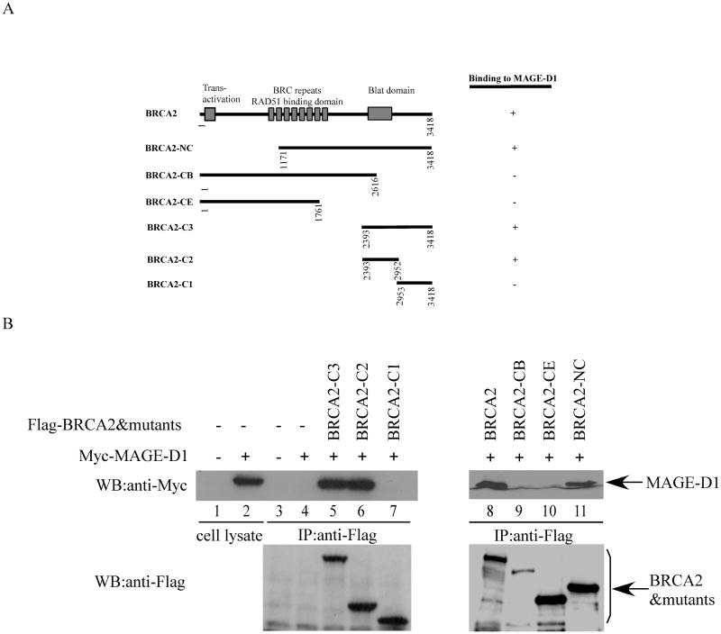
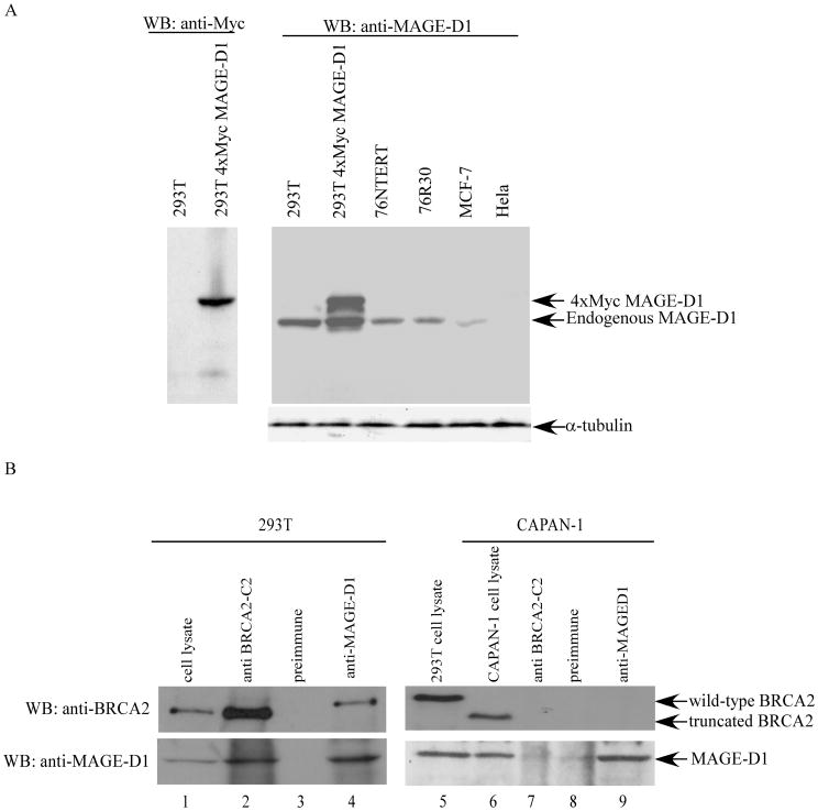
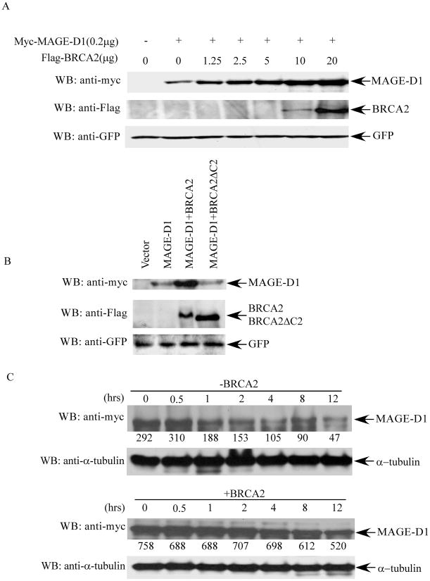
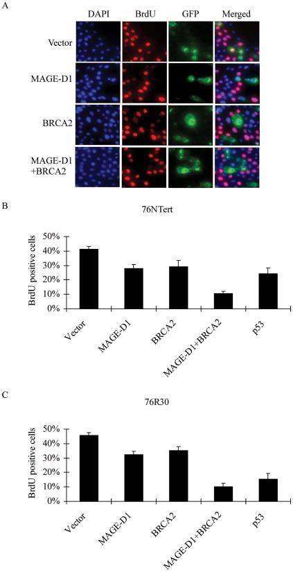
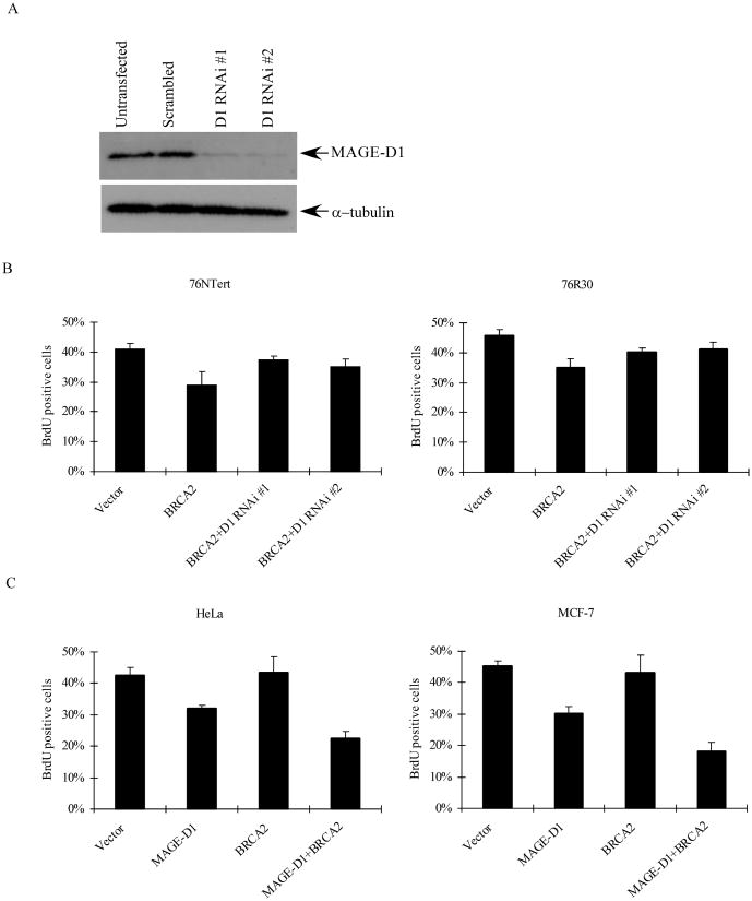
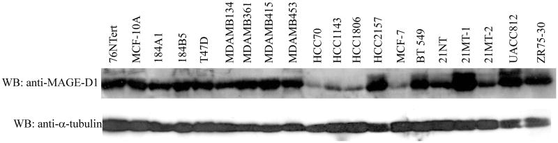
Germ line mutations in BRCA2 gene predispose women to early-onset familial breast and ovarian cancer. BRCA2 is a protein of multiple functions. In addition to its role in DNA double-strand break repair, BRCA2 also plays a role in stabilization of stalled DNA replication forks, cytokinesis, transcription regulation, mammalian gametogenesis, centrosome duplication, and suppression of cell proliferation. However, how BRCA2 mutations predispose women specifically to breast and ovarian cancer remains undefined. Here we found that BRCA2 binds and stabilizes MAGE-D1, a member of the MAGE gene family of proteins. Expression of BRCA2 and MAGE-D1 synergistically suppresses cell proliferation independently of the p53 pathway. Using two MAGE-D1 RNA interferences and two cell lines expressing low or undetectable levels of MAGE-D1, we further showed that the expression of MAGE-D1 is required for BRCA2-mediated suppression of cell proliferation, indicating that MAGE-D1 is a downstream target of BRCA2 and that BRCA2 suppresses cell proliferation via stabilizing MAGE-D1. Importantly, MAGE-D1 protein expression was reduced in 6 of 16 breast carcinoma cell lines tested as compared with untransformed immortal mammary epithelial cell lines, suggesting that suppression of MAGE-D1 expression may be involved in the tumorigenesis of a subset of sporadic breast cancers.


Figures

Fig. 1
Fig. 1. Binding of MAGE-D1 and BRCA2
(A). Diagram of BRCA2 mutants. The binding of BRCA2 and its mutants to MAGE-D1 is summarized. (B). In vivo binding of BRCA2 and MAGE-D1. 293T cells were transfected with Myc-tagged full-length MAGE-D1 and Flag-tagged BRCA2 and its mutants. The cells were harvested 48 hours after transfection in LSAB. BRCA2 and its mutants were immunoprecipitated using anti-Flag antibody. The immunoprecipitated protein complexes were fractionated and immunoblotted using anti-Myc antibody (9E10) to detect Myc-MAGE-D1. The blot was re-probed using anti-Flag antibody to detect BRCA2 and its mutants. A 4% (right panel) and a 7.5% (left panel) PAGE gels were used to fractionate the full-length BRCA2 and its mutants of varied molecular weight (from 58kd-380kd).
Fig. 2
Fig. 2. Endogenous binding of MAGE-D1 and BRCA2 in vivo
(A). Characterization of anti-MAGE-D1 antibody. Cell lysates (50μg of total protein) from 76NTert, 76R30, MCF-7, HeLa, 293T, and 293T cells transfected with 5 μg of pCR3.1 or 4xMyc-MAGE-D1 were fractionated by SDS-PAGE and immunoblotted using affinity-purified anti-MAGE-D1 or anti-Myc antibodies. (B). In vivo binding of endogenous MAGE-D1 and endogenous BRCA2. 293T cells and CAPAN-1 cells were harvested in RIPA buffer (without SDS). The cell lysates were immunoprecipitated using preimmune, anti-BRCA2-C2 and anti-MAGE-D1 antisera. The immunoprecipitated protein complexes were fractionated and immunoblotted using anti-BRCA2 (BRCA2 ab1, Oncogene Science) and anti-MAGE-D1 antibodies.
Fig. 3
Fig. 3. BRCA2 expression stabilizes MAGE-D1
(A). MAGE-D1 protein levels when co-transfected with increasing amounts of BRCA2. 293T cells were transfected with 0.2 μg pBB14-GFP and 0.2 μg Myc-tagged MAGE-D1 alone or with increasing amounts of Flag-tagged BRCA2, as indicated. Cell lysates were prepared 48 hr after transfection and analyzed for Myc-MAGE-D1 and Flag-BRCA2 protein levels by immunoblotting using anti-Myc or anti-Flag antibodies. Immunoblotting using anti-GFP antibody was used to control transfection efficiency. (B). MAGE-D1 protein level when co-transfected with wild-type BRCA2 or BRCA2δC2 (BRCA2 mutant devoid of the MAGE-D1 binding domain). (C). Half-life of MAGE-D1. 293T cells were transfected with Myc-tagged MAGE-D1 alone (upper panel) or with Flag-tagged BRCA2 (lower panel). Cycloheximide (final concentration 10 μg/ml) was added 48 hr after transfection to inhibit nascent protein synthesis. Cell lysates were prepared at the indicated time points after the addition of cycloheximide, followed by immunoblotting using anti-Myc and anti-α-tubulin antibodies to determine the turnover of MAGE-D1 protein. The numbers under the panels are quantification analyses of the signals as obtained by use of LabWorks software (UVP, Inc, Upland, CA).
Fig. 4
Fig. 4. BRCA2 and MAGE-D1 synergistically suppress cell proliferation
(A) BrdU incorporation assay in 76NTert cells. 76NTert cells plated on coverslips in 24-wells plate were transfected with 0.5 μg MAGE-D1 and 1.5μg BRCA2, alone or in combination, together with pBB14-GFP (0.2 μg). The cells were incubated for 48 hr after transfection, then labeled with BrdU for 1 hr. The cells were fixed and stained using an anti-BrdU antibody. The DNA was stained with DAPI. Note that most of BRCA2 or MAGE-D1 transfected cells (GFP-positive cells) are negative for BrdU staining, while many vector- transfected cells (GFP-positive) or untransfected cells (GFP-negative) are positive for BrdU staining. (B and C) BRCA2 and MAGE-D1 synergistically suppress cell proliferation. 76NTert or 76R30 cells were transfected with MAGE-D1 and BRCA2, alone or in combination, as indicated, and assayed for BrdU incorporation as described in (A). The percentage of cells positive for BrdU staining was quantified in at least 300 transfected cells (GFP-positive cells). Each value represents the average and standard deviation from three independent experiments. The percentage of cells positive for BrdU staining was significantly lower in cells transfected with BRCA2, MAGE-D1, or MAGE-D1+BRCA2 than in cells transfected with vector alone (P < 0.05).
Fig. 5
Fig. 5. BRCA2-mediated-suppression of cell proliferation is dependent on the expression of MAGE-D1
(A) Western blotting analysis of 293T cells transfected with vector or MAGE-D1 RNAi constructs. 293T cells were transfected with vector or two MAGE-D1 RNAi constructs. After 48 hr, cells were harvested and fractionated through a SDS-PAGE gel, then blotted using anti-MAGE-D1 and anti-α-tubulin antibodies. (B). Silencing of MAGE-D1 expression by MAGE-D1 RNAi attenuated BRCA2 expression-mediated suppression of cell proliferation. 76NTert and 76R30 cells were transfected with 1.5μg BRCA2 alone or in combination with 0.5μg MAGE-D1 RNAi, together with pBB14-GFP (0.2μg), then BrdU incorporation was quantified as described in Fig. 4(B). The percentage of cells positive for BrdU staining was significantly lower in cells transfected with BRCA2 than in cells transfected with vector alone or BRCA2 plus MAGE-D1 RNAi (P < 0.05). (C) BRCA2 expression does not suppress cell proliferation in cells with a low level of MAGE-D1. HeLa and MCF-7 cells were transfected with BRCA2 and MAGE-D1 alone or in combination. BrdU incorporation was quantified as described in 4(B). The percentage of cells positive for BrdU staining was not lower in cells transfected with BRCA2 than in cells transfected with vector alone. However, the percentage of cells positive for BrdU staining was significantly lower in cells transfected with MAGE-D1 or BRCA2+MAGE-D1 than in cells transfected with vector alone. (P<0.05).
Fig. 6
Fig. 6. MAGE-D1 protein expression in untransformed immortal mammary epithelial cell lines and breast carcinoma cell lines
Lysates prepared in sample buffer from the cell lines indicated were fractionated on a SDS-PAGE gel and blotted with anti-MAGE-D1 and anti–α–tubulin antibodies (loading control). 76NTert, MCF-10A, 184A1, and 184B5 are untransformed immortal mammary epithelial cell lines; the others are breast carcinoma cell lines.

Similar articles

Cited by

References

    1. Tavtigian SV, Simard J, Rommens J, et al. The complete BRCA2 gene and mutations in chromosome 13q-linked kindreds. Nat Genet. 1996;12:333–7. - PubMed
    1. Wooster R, Neuhausen SL, Mangion J, et al. Localization of a breast cancer susceptibility gene, BRCA2, to chromosome 13q12-13. Science (Wash DC) 1994;265:2088–90. - PubMed
    1. Wooster R, Bignell G, Lancaster J, et al. Identification of the breast cancer susceptibility gene BRCA2. Nature (Lond) 1995;378:789–92. - PubMed
    1. Gayther SA, Pharoah PD, Ponder BA. The genetics of inherited breast cancer. J Mammary Gland Biol Neoplasia. 1998;3:365–76. - PubMed
    1. Sharan SK, Morimatsu M, Albrecht U, et al. Embryonic lethality and radiation hypersensitivity mediated by Rad51 in mice lacking Brca2. Nature (Lond) 1997;386:804–10. - PubMed

Publication types

MeSH terms