. 2005 Jun 1;25(22):5365-75.
doi: 10.1523/JNEUROSCI.1125-05.2005.

The generation of a 17 kDa neurotoxic fragment: an alternative mechanism by which tau mediates beta-amyloid-induced neurodegeneration

Affiliations

The generation of a 17 kDa neurotoxic fragment: an alternative mechanism by which tau mediates beta-amyloid-induced neurodegeneration

So-Young Park et al. J Neurosci. .

Abstract

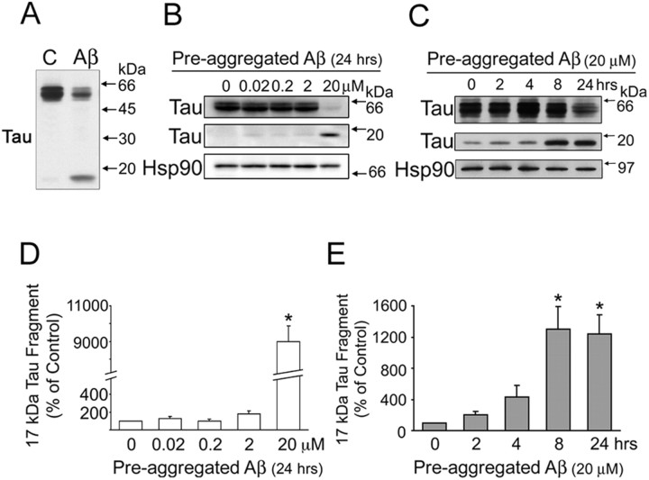
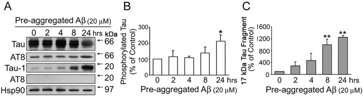
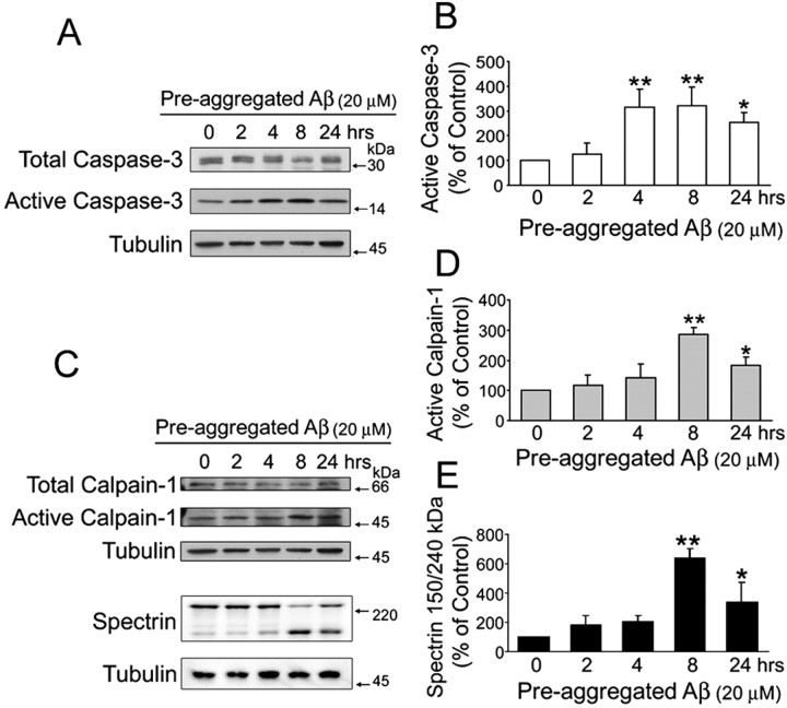
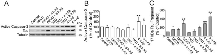
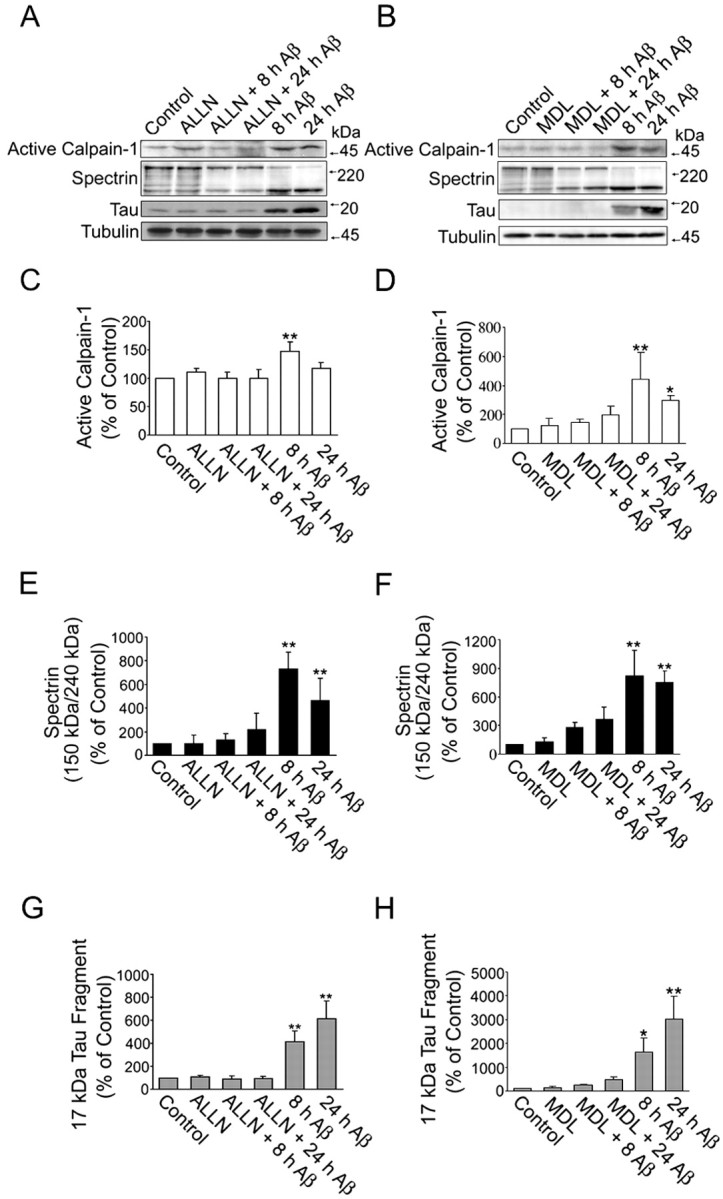
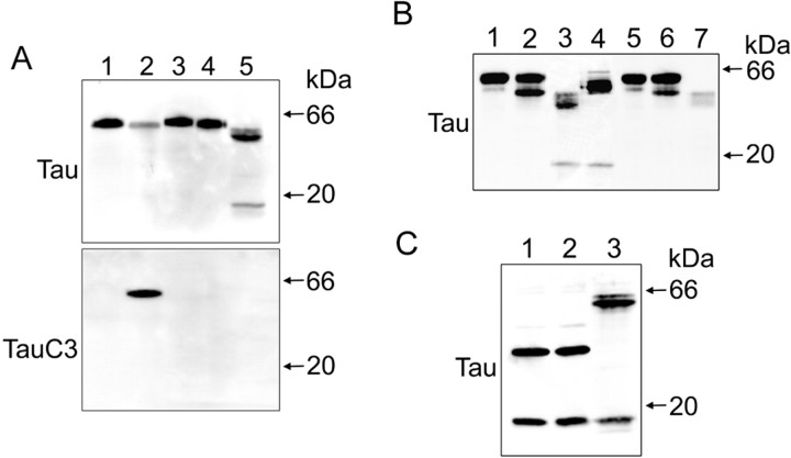
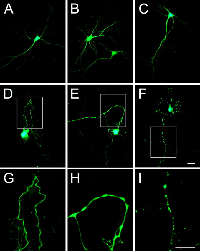
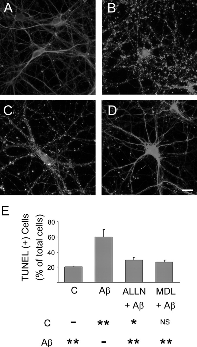
Recently, we have shown that the microtubule-associated protein tau is essential for beta-amyloid (Abeta)-induced neurotoxicity in hippocampal neurons. However, the mechanisms by which tau mediates Abeta-induced neurite degeneration remain poorly understood. In the present study, we analyzed whether tau cleavage played a role in these events. Our results showed that pre-aggregated Abeta induced the generation of a 17 kDa tau fragment in cultured hippocampal neurons. The generation of this fragment was preceded by the activation of calpain-1. Conversely, inhibitors of this protease, but not of caspases, completely prevented tau proteolysis leading to the generation of the 17 kDa fragment and significantly reduced Abeta-induced neuronal death. Furthermore, the expression of this fragment in cultured hippocampal neurons induced the formation of numerous varicosity-bearing tortuous processes, as well as the complete degeneration of some of those neurite processes. These results suggest that Abeta-induced neurotoxicity may be mediated, at least in part, through the calpain-mediated generation of a toxic 17 kDa tau fragment. Collectively, these results provide insight into a novel mechanism by which tau could mediate Abeta-induced neurotoxicity.


Figures

Figure 1.
Figure 1.
Pre-aggregated Aβ induced tau cleavage in mature hippocampal neurons in a dose- and time-dependent manner. A, Western blot analysis of tau content in heat-stable fractions prepared from 21 d in vitro hippocampal neurons cultured in the absence (C) or in the presence of pre-aggregated Aβ (20 μm) for 24 h using a phosphorylation-independent tau antibody (clone tau-5). Note the decrease in full-length tau and the appearance of a tau-immunoreactive band of ∼17 kDa in Aβ-treated neurons. B, Western blot analysis of tau content in heat-stable fractions prepared from 21 d in vitro hippocampal neurons cultured for 24 h in the presence of pre-aggregated Aβ at final concentrations ranging from 0.02 to 20 μm using a tau antibody (clone tau-5). C, Western blot analysis of heat-stable fractions prepared from 21 d in vitro hippocampal neurons cultured in the presence of pre-aggregated Aβ (20 μm) for up to 24 h using a tau antibody (clone tau-5). Equal amounts of protein were loaded in each lane. The Hsp90 was also used as a loading control. D, E, Graphs showing the levels of the 17 kDa tau fragment in the dose-response (D) and time course (E) experiments performed as described above. Data were obtained using Molecular Analyst software and normalized using full-length tau and Hsp90 as internal controls. Values are expressed as a percentage of untreated controls, considering the values obtained in these neurons as 100%. Each number represents the mean ± SEM from three different experiments. *p < 0.01, different from control.
Figure 2.
Figure 2.
Pre-aggregated Aβ-induced tau cleavage preceded the increase in tau phosphorylation. A, Western blot analysis of tau content in heat-stable fractions prepared from 21-d-in-culture hippocampal neurons treated with pre-aggregated Aβ (20 μm) for up to 24 h using nonphosphorylation-dependent tau (clone tau-5), phosphorylated tau (clone AT8), and dephosphorylated tau (clone tau-1) antibodies. Note the absence of AT8 immunoreactivity of the 17 kDa tau fragment. Equal amounts of total protein were loaded in each lane. B, C, Graphs showing the time course of tau phosphorylation and tau cleavage in the presence of pre-aggregated Aβ. Densitometric values were normalized using full-length tau (tau-5) and the Hsp90 as internal controls. Values are expressed as a percentage of untreated controls, considering the values obtained in these neurons as 100%. Each number represents the mean ± SEM from three different experiments. *p < 0.05, **p < 0.01, different from control.
Figure 3.
Figure 3.
Pre-aggregated Aβ induced the activation of caspase-3 and calpain-1 in cultured hippocampal neurons. A, C, Western blot analysis of active caspase-3 (A) and active calpain-1 (C) content in whole-cell extracts prepared from 21 d in vitro hippocampal neurons cultured in the presence of pre-aggregated Aβ (20 μm) for up to 24 h using specific antibodies. Tubulin immunoblots were used as loading controls (B, D, E). The bar graphs show the levels of active caspase-3 (B), active calpain-1 (D), and calpain-specific cleavage products of spectrin (E) in the samples obtained as described above. Note the increase in active calpain-1 levels in samples obtained 8 h after the addition of Aβ and the concomitant decrease in full-length spectrin and increase in the 150 kDa specific spectrin cleaved product (C-E). Densitometric values from active caspase-3, active calpain-1, and cleaved spectrin were normalized using total caspase-3, calpain-1, and full-length spectrin as internal controls. Values are expressed as a percentage of untreated controls, considering the values obtained in these neurons as 100%. Each number represents the mean ± SEM from three different experiments. *p < 0.05, **p < 0.01, different from control.
Figure 4.
Figure 4.
Caspase inhibitors partially blocked tau cleavage induced by pre-aggregated Aβ. A, Western blot analysis of active caspase-3 and 17 kDa tau fragment content in whole-cell extracts prepared from 21-d-in-culture hippocampal neurons treated with either DEVD (50 μm; specific caspase-3 inhibitor) or VAD (50 μm; general caspase inhibitor) 1 h before the treatment with pre-aggregated Aβ (20 μm) for 4 and 8 h using specific antibodies. B, C, Graphs showing changes in active caspase-3 and 17 kDa tau fragment content in samples obtained as described above. Data were obtained using Molecular Analyst software and normalized using total caspase-3 and tubulin as internal controls. Values are expressed as a percentage of untreated controls, considering the values obtained in these neurons as 100%. Each number represents the mean ± SEM from three different experiments. *p < 0.05, **p < 0.01, different from control.
Figure 5.
Figure 5.
Calpain inhibitors completely blocked the generation of the 17 kDa tau fragment induced by pre-aggregated Aβ in cultured hippocampal neurons. A, B, Western blot analysis of active calpain-1, cleaved spectrin, and 17 kDa tau fragment content in whole-cell extracts prepared from 21-d-in-culture hippocampal neurons treated with either ALLN (A) or MDL 28,170 (B), two calpain inhibitors, 1 h before the treatment with pre-aggregated Aβ (20 μm) for 4 and 8 h using specific antibodies. C-H, Graphs showing changes in the content of active calpain-1 (C, D), calpain-specific cleavage product of spectrin (E, F), and 17 kDa tau fragment (G, H) in hippocampal neurons treated with ALLN (C, E, G) or MDL 28, 170 (D, F, H) before the addition of Aβ. Data were obtained using Molecular Analyst software and normalized using total calpain-1, spectrin, tau, and tubulin as internal controls. Values are expressed as a percentage of untreated controls, considering the values obtained in these neurons as 100%. Each number represents the mean ± SEM from three different experiments. *p < 0.05, **p < 0.01, different from control.
Figure 6.
Figure 6.
Calpain-1 cleaved tau in vitro, generating a 17 kDa fragment. A, CHO cells were transfected with tau-pRC/CMV or D421E-tau-pRC/CMV using LipofectAMINE. After 48 h of transfection, CHO cells were scraped in lysis buffer. Whole-cell lysates were incubated with buffer control or recombinant caspase-3 at 37°C for 1 h. Reaction mixtures were analyzed by immunoblot with antibodies directed against phosphorylation-independent tau (clone tau-5) or tau truncated at Asp421 by caspase-3 (clone tau-C3) antibodies. Hippocampal neurons treated with pre-aggregated Aβ (20 μm) for 24 h were used as positive controls. Lane 1, Full-length tau with buffer control; lane 2, full-length tau with caspase-3; lane 3, D421E-tau with buffer control; lane 4, D421E-tau with caspase-3; lane 5, hippocampal neurons treated with pre-aggregated Aβ (20 μm) for 24 h. B, Full-length tau or L43A-V229A-tau-pRC/CMV-transfected CHO cells were harvested in lysis buffer. Whole-cell lysates were incubated with either buffer control or calpain-1 at 30°C for 5 or 60 min. There action mixtures were analyzed by Western blot using an antibody directed against tau (clone tau-5). Hippocampal neurons treated with pre-aggregated Aβ (20 μm) for 24 h were used as positive controls. Lane 1, Full-length tau with buffer control; lane 2, full-length tau with calpain-1 for 5 min; lane 3, full-length tau with calpain-1 for 60 min; lane 4, hippocampal neurons treated with pre-aggregated Aβ (20 μm) for 24 h; lane 5, L43A-V229A-tau with buffer control; lane 6, L43A-V229A-tau with calpain-1 for 5 min; lane 7, L43A-V229A-tau with calpain-1 for 60 min. C, Whole-cell lysates of tau45-230-pcDNA3.1(-)-transfected CHO cells were analyzed by immunoblot reacted with an antibody against tau (clone tau-5). Tau45-230, corresponding to the 17 kDa fragment on tau, was incubated with either buffer control (lane 1) or calpain-1 at 30°C for 1 h (lane 2). Hippocampal neurons treated with pre-aggregated Aβ (20 μm) for 24 h were used as positive controls (lane 3).
Figure 7.
Figure 7.
The 17 kDa tau fragment induced apoptosis in CHO cells. A-D, Detection of TUNEL+ cells in CHO cells transfected with GFP vector alone (A, B) or 17Tau-GFP (C, D). GFP-transfected (A) and 17Tau-GFP-transfected (C) cells are shown in green, and TUNEL+ cells are shown in red. B and D are phase-contrast pictures of A and C, respectively. Scale bar, 50 μm.
Figure 8.
Figure 8.
The 17 kDa tau fragment induced neurodegeneration in cultured hippocampal neurons. Fourteen-day-in-culture hippocampal neurons were transfected with GFP (A), full-length Tau-GFP (B), and 17Tau-GFP (C-I). GFP-transfected (A) and full-length Tau-GFP-transfected (B) neurons did not show any sign of degeneration even 48 h after transfection. Normal morphological characteristics were detected also in 17Tau-GFP-transfected cells 24 h after transfection (C). In contrast, hippocampal neurons transfected with 17Tau-GFP for 48 h showed numerous signs of degeneration, including the formation of tortuous processes (D), varicosity along the neurites (E), and the retraction of neurites (F). G-I, High-power magnification of the boxed areas in D-F. Scale bars, 20 μm.
Figure 9.
Figure 9.
Calpain inhibitors prevented Aβ-induced neurotoxicity in culture hippocampal neurons. Hippocampal neurons cultured for 21 d were treated with calpain inhibitors, ALLN (C) or MDL 28, 170 (D), 1 h before the incubation with pre-aggregated Aβ for 24 h. No signs of neurite degeneration were detected in untreated controls (A). In contrast, severe neurite degeneration was observed in cultures incubated with pre-aggregated Aβ (B). Both calpain inhibitors significantly reduced the appearance of dystrophic neurites induced by Aβ. E, Detection of TUNEL+ cells in hippocampal neurons cultured in the presence or absence of calpain (C) inhibitors and Aβ as described above. Results were expressed as a percentage of the total number of neurons. Each number represents the mean ± SEM from three different experiments. More than 150 neurons were counted for each experimental condition. *p < 0.05, **p < 0.01, different from control. Scale bar, 20 μm. NS, Not statistically different.

Similar articles

Cited by

References

    1. Adamec E, Mohan P, Vonsattel JP, Nixon RA (2002a) Calpain activation in neurodegenerative diseases: confocal immunofluorescence study with antibodies specifically recognizing the active form of calpain 2. Acta Neuropathol (Berl) 104: 92-104. - PubMed
    1. Adamec E, Murrell JR, Takao M, Hobbs W, Nixon RA, Ghetti B, Vonsattel JP (2002b) P301L tauopathy: confocal immunofluorescence study of perinuclear aggregation of the mutated protein. J Neurol Sci 200: 85-93. - PubMed
    1. Allen JW, Eldadah BA, Huang X, Knoblach SM, Faden AI (2001) Multiple caspases are involved in beta-amyloid-induced neuronal apoptosis. J Neurosci Res 65: 45-53. - PubMed
    1. Alvarez A, Toro R, Caceres A, Maccioni RB (1999) Inhibition of tau phosphorylating protein kinase cdk5 prevents beta-amyloid-induced neuronal death. FEBS Lett 459: 421-426. - PubMed
    1. Bensadoun A, Weinstein D (1976) Assay of proteins in the presence of interfering materials. Anal Biochem 70: 241-250. - PubMed

Publication types

MeSH terms