

Review
doi: 10.1172/JCI24761.
The genetic epidemiology of neurodegenerative disease
Affiliations
- PMID: 15931380
- PMCID: PMC1137006
- DOI: 10.1172/JCI24761
Item in Clipboard
Review
The genetic epidemiology of neurodegenerative disease
J Clin Invest.
2005 Jun.
Abstract
Gene defects play a major role in the pathogenesis of degenerative disorders of the nervous system. In fact, it has been the very knowledge gained from genetic studies that has allowed the elucidation of the molecular mechanisms underlying the etiology and pathogenesis of many neurodegenerative disorders. In this review, we discuss the current status of genetic epidemiology of the most common neurodegenerative diseases: Alzheimer disease, Parkinson disease, Lewy body dementia, frontotemporal dementia, amyotrophic lateral sclerosis, Huntington disease, and prion diseases, with a particular focus on similarities and differences among these syndromes.
Figures
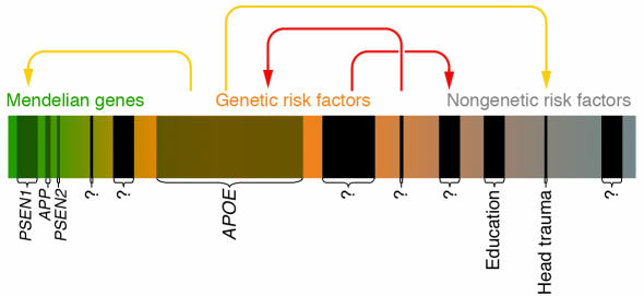
This scheme depicts the risk spectrum predisposing to common diseases as one continuum, using AD as an example. The continuum extends from the most extreme genetic form (“Mendelian genes”; green) to cases influenced by genetic susceptibility factors (“Genetic risk factors”; orange), until reaching into a less well-defined area of cases that may be caused by genes of lesser penetrance/lower effect size and/or altogether nongenetic factors (“Nongenetic risk factors”; gray). Established Mendelian genes (APP, PSEN1, and PSEN2) or genetic risk factors (APOE-ε4) are represented by shaded boxes and represent the most obvious candidates of AD genetics; the width of these boxes approximately represents the relative contribution to the overall risk for disease. Black boxes indicate still-elusive disease genes/risk factors (“?”). Colored arrows indicate possible gene-gene and gene-environment interaction patterns: yellow arrows represent previously suggested interactions (e.g., between PSEN1 and APOE-ε4). Note that some interactions (red arrows) as well as the number of elusive genes are entirely hypothetical and are depicted for didactic purposes only.
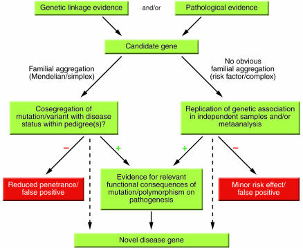
Flow chart of current strategies used to identify novel disease genes. This scheme outlines strategies for identifying mutations and/or polymorphisms causing or predisposing to disease. Candidate genes are chosen based on genetic linkage data and/or known or hypothesized pathobiological relevance to disease mechanisms. This procedure is referred to as the “candidate gene approach.” An alternative and inherently similar strategy is based on the detection of formerly unknown genes/proteins according to genetic linkage data and is referred to as “positional cloning.” Dashed lines indicate “shortcuts” allowing the definition of a novel disease gene based on the genetic evidence alone, e.g., APOE-ε4 in AD, of which the precise functional consequences remain unknown despite an established genetic role. Note that there are examples of genes/mutations with reduced penetrance or minor risk effects (red boxes) within bona fide disease genes (e.g., certain mutations in PSEN1 in AD).
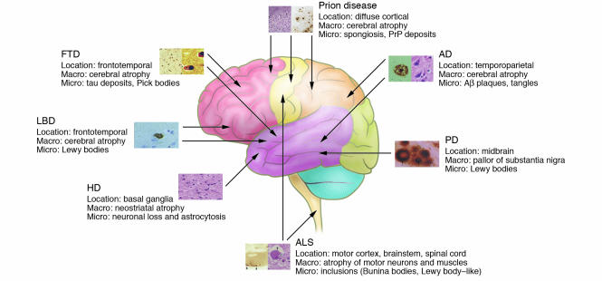
Overview of the anatomical location of and macroscopic and microscopic changes characteristic of the neurodegenerative disorders discussed in this review. Note that the full neuropathological spectrum of these disorders is much more complex than depicted here. When there is more than one characteristic histopathological feature, these are depicted from left to right, as indicated in the labels listing microscopic changes (e.g., the 2 panels for AD depict an Aβ plaque [left] and neurofibrillary tangles [right]). All histopathological images are reprinted with permission from ISN Neuropath Press (ref. 99).
References
-
- Lander ES. The new genomics: global views of biology. Science. 1996;274:536–539. - PubMed
-
- Risch NJ. Searching for genetic determinants in the new millennium. Nature. 2000;405:847–856. - PubMed
-
- Hardy J, Myers A, Wavrant-De Vriese F. Problems and solutions in the genetic analysis of late-onset Alzheimer’s disease. Neurodegenerative Diseases. 2004;1:213–217. - PubMed
-
- Munafo MR, Clark TG, Flint J. Assessing publication bias in genetic association studies: evidence from a recent meta-analysis. Psychiatry Res. 2004;129:39–44. - PubMed
-
- Colhoun HM, McKeigue PM, Davey Smith G. Problems of reporting genetic associations with complex outcomes. Lancet. 2003;361:865–872. - PubMed
Publication types
MeSH terms
LinkOut - more resources
Full Text Sources
Other Literature Sources
Medical
