. 2005 Jun 2;6(1):50.
doi: 10.1186/1465-9921-6-50.

Multiple exposures to swine barn air induce lung inflammation and airway hyper-responsiveness

Affiliations

Multiple exposures to swine barn air induce lung inflammation and airway hyper-responsiveness

Chandrashekhar Charavaryamath et al. Respir Res. .

Abstract

Background: Swine farmers repeatedly exposed to the barn air suffer from respiratory diseases. However the mechanisms of lung dysfunction following repeated exposures to the barn air are still largely unknown. Therefore, we tested a hypothesis in a rat model that multiple interrupted exposures to the barn air will cause chronic lung inflammation and decline in lung function.

Methods: Rats were exposed either to swine barn (8 hours/day for either one or five or 20 days) or ambient air. After the exposure periods, airway hyper-responsiveness (AHR) to methacholine (Mch) was measured and rats were euthanized to collect bronchoalveolar lavage fluid (BALF), blood and lung tissues. Barn air was sampled to determine endotoxin levels and microbial load.

Results: The air in the barn used in this study had a very high concentration of endotoxin (15361.75 +/- 7712.16 EU/m3). Rats exposed to barn air for one and five days showed increase in AHR compared to the 20-day exposed and controls. Lungs from the exposed groups were inflamed as indicated by recruitment of neutrophils in all three exposed groups and eosinophils and an increase in numbers of airway epithelial goblet cells in 5- and 20-day exposure groups. Rats exposed to the barn air for one day or 20 days had more total leukocytes in the BALF and 20-day exposed rats had more airway epithelial goblet cells compared to the controls and those subjected to 1 and 5 exposures (P < 0.05). Bronchus-associated lymphoid tissue (BALT) in the lungs of rats exposed for 20 days contained germinal centers and mitotic cells suggesting activation. There were no differences in the airway smooth muscle cell volume or septal macrophage recruitment among the groups.

Conclusion: We conclude that multiple exposures to endotoxin-containing swine barn air induce AHR, increase in mucus-containing airway epithelial cells and lung inflammation. The data also show that prolonged multiple exposures may also induce adaptation in AHR response in the exposed subjects.


Figures

Figure 1
Figure 1
Airway hyper-responsiveness. Airway hyperresponsiveness to methacholine challenge in rats was measured using a whole-body head-out plathysmograph. Compared to controls, both 1-day and 5-day (P < 0.001) exposed rats showed increased airway hyperresponsiveness. Compared to 20-day exposed rats, 5-day (P = 0.001) and 1-day (P = 0.014) exposed rats showed increased airway hyper-responsiveness There was no difference between control and 20-day exposed (P = 0.207) and 1-day and 5-day exposed (P = 0.249) rats. *: Significantly different from other groups as indicated by line/s.
Figure 2
Figure 2
Total and differential leukocytes in the bronchoalveolar lavage fluid. Bronchoalveolar lavage was performed on the whole lung using 3 ml of cold HBSS. Cells were counted using a hemocytometer. Cytospins were prepared from BAL fluid and cells were differentiated with Wright's staining. 2A. BALF total leukocyte counts. BALF total leukocytes were different among the four groups (P < 0.001). Compared to controls, 5-day and 20-day exposed, 1-day exposed rats showed increased numbers of BALF total leukocytes (P < 0.001). Rats exposed for 20 days showed increased numbers of BALF total leukocytes when compared to controls (P = 0.01) and 5-day (P = 0.008) exposed rats. 5-day exposed rats did not differ from controls in their BALF total leukocyte numbers (P = 0.932). ** Significantly different from control, 5-day and 20-day exposed rats and * significantly different from control, 1-day and 5-day exposed rats. 2B. BALF absolute neutrophil counts. BALF absolute neutrophil counts were different among the groups (P < 0.001). 1-day exposed rats showed higher BALF absolute neutrophils when compared to control, 5-day and 20-day exposed rats (P < 0.001). 20-day exposed rats showed higher BALF absolute neutrophil count when compared to control rats (P = 0.022). There was no difference between control and 5-day exposed (P = 0.538) and 20-day and 5-day exposed (P = 0.119) rats. ** Significantly different from control, 5-day and 20-day exposed rats and * significantly different from control. 2C. BALF absolute macrophage counts. BALF absolute macrophage count was different among the four groups (P < 0.001). BALF absolute macrophage count was higher in 1-day exposed when compared to control, 5-day and 20-day exposed rats (P < 0.001). 20-day exposed rats showed higher BALF absolute macrophage count when compared to control and 5-day (P < 0.001) exposed rats. There was no difference between control and 5-day exposed rats (P = 0.789). ** Significantly different from control, 5-day and 20-day exposed rats and * indicates significantly different from control and 1-day and 5-day exposed rats. D. BALF absolute lymphocyte count (Figure 2D). BALF absolute lymphocyte count was different among the four groups (P < 0.001). BALF absolute lymphocyte count was higher in 1-day exposed when compared to control, 5-day and 20-day exposed rats (P < 0.001). * Significantly different from other three groups.
Figure 3
Figure 3
Total and differential leukocyte count in blood. Blood total leukocytes were counted using hemocytometer and smears were differentiated with Wright's stain. 3A. Blood total leukocyte count did not differ among the groups (Figure 3A; P = 0.090). 3B. Blood absolute neutrophils count was different among the four groups (Figure 3B; P < 0.001). 20-day exposed rats showed higher blood absolute neutrophils count when compared to control, 1-day and 5-day exposed rats (P < 0.001). 1-day exposed rats showed higher blood absolute neutrophil count when compared to 5-day exposed rats (P < 0.038). Both 1-day (P = 0.073) and 5-day exposed rats (P = 0.678) did not differ from controls. ** Indicate significantly different from control, 1-day and 5-day exposed rats and * indicate significantly different from 20-day and 5-day exposed rats. 3C. Blood absolute monocyte count did not differ among the four groups (Figure 3C; P = 0.122). 3D. Blood absolute lymphocyte count was different among the four groups (Figure 4D; P < 0.001). Compared to 20-day exposed, control (P = 0.003), 1-day (P < 0.001) and 5-day (P = 0.011) exposed rats showed increased numbers of blood absolute lymphocytes. * indicates significantly different from other three groups.
Figure 4
Figure 4
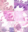
Histopahtological evaluation of lung sections. Histopathological changes in the lungs of swine barn air exposed and control rats were evaluated using hematoxylin and eosin stained sections. Control rat lungs (A) showed no inflammatory cell infiltration. Among the exposed groups, 1-day (B), 5-day (C) and 20-day exposed rats (not shown) showed peribronchiolar neutrophilic (C; arrows) and 5-day (D) and 20-day exposed (not shown) showed eosinophilic (D; arrows and inset) infiltration. Bronchus-associated lymphoid tissue (BALT) in control (E), 1-day and 5-day exposed (both not shown) appeared normal and had no germinal centers, whereas 20-day exposed rat lungs had activated BALT with germinal centers (F; outlined in black line) containing several mitotic cells (F; inset). Original magnification A-C: ×400; D-F: ×100; Insets: ×1000
Figure 5
Figure 5
Quantification of mucus producing cells in the airways. Mucus producing goblet cells in the airways were quantified using PAS staining. Control rats showed no mucus producing cells in the bronchioles (A). 5-day exposed and 20-day exposed rats showed large number of mucus producing cells (B&C; arrows). Quantification of PAS-positive cells showed a significantly higher number of cells in 5-day and 20-day exposed rat lungs compared to the controls (5-day: P = 0.040; 20-day: P < 0.001) and one-day (5-day: P = 0.007; 20-day: P < 0.001) exposed rats (Figure D). Also, the increase in mucus producing cells was higher in 20-day exposed compared to 5-day exposed rat lungs (P < 0.001). Number of mucus producing cells did not differ between control and 1-day exposed rats (P = 0.435). *: Significantly different from control, 1-day and 20-day exposure. **: Significantly different from control, 1-day and 5-day exposure. The bars represent mean ± SD. Original magnification A-C; ×400
Figure 6
Figure 6
Quantification of septal macrophages in the lung. Macrophages were stained using ED-1 antibody. Lungs from control (A), 1-day (not shown in picture), 5-day exposed (B) and 20-day exposed (C) rats appeared to have similar numbers of septal macrophages. To confirm this we quantified ED-1 positive cells in the septum. D: Is a scatter plot showing number of ED-1 cells in the septum, in different groups. The horizontal bars in each group represent the mean for that particular group. There was no difference between the groups (P = 0.350). Original magnification A-C; ×400
Figure 7
Figure 7
Airway smooth muscle quantification. The staining pattern for smooth muscle in controls (A), 1-day (not shown in picture), 5-day exposed (B) and 20-day exposed (C) rat lungs appeared similar. D. The stained area of smooth muscle around the bronchioles was measured using image analyses software. The area of smooth muscle was not significantly different between the groups (P = 0.681). Original magnification A-C; ×400

References

    1. Schenker MB, Christiani D, Cormier Y, Dimich-Ward H, Doekes G, Dosman JA. In: Respiratory health hazards in agriculture. Schenker MB, editor. Vol. 158. American Thoracic Society; 1998. pp. S1–S76.
    1. Asmar S, Pickrell JA, Oehme FW. Pulmonary diseases caused by airborne contaminants in swine confinement buildings. Vet Hum Toxicol. 2001;43:48–53. - PubMed
    1. Zejda JE, Barber EM, Dosman JA, Olenchock SA, McDuffie HH, Rhodes CS, Hurst TS. Respiratory health status in swine producers relates to endotoxin exposure in the presence of low dust levels. J Occup Med. 1994;36:49–56. - PubMed
    1. Zejda JE, Hurst TS, Rhodes CS, Barber EM, McDuffie HH, Dosman JA. Respiratory health of swine producers: Focus on young workers. Chest. 1993;103:702–709. - PubMed
    1. Frevert CW, Warner AE. Respiratory distress resulting from acute lung injury in the veterinary patient. J Vet Int Med. 1999;6:154–165. - PubMed

Publication types