

Immunohistochemical analysis of MCT1, MCT2 and MCT4 expression in rat plantaris muscle
- PMID: 15932892
- PMCID: PMC1474173
- DOI: 10.1113/jphysiol.2005.087411
Immunohistochemical analysis of MCT1, MCT2 and MCT4 expression in rat plantaris muscle
Abstract
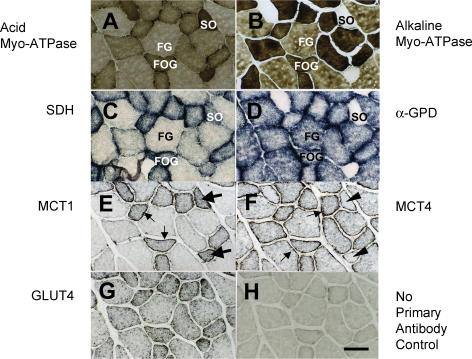
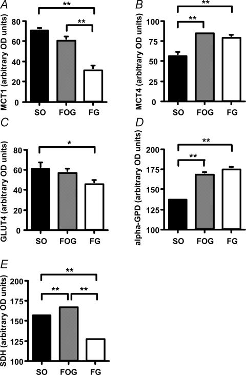
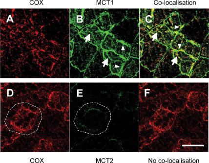
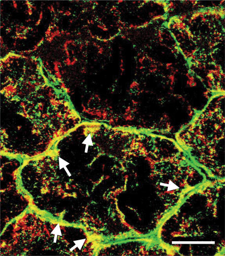
We addressed the need for histological assessment of myocellular domains occupied by monocarboxylate transporters (MCT1, MCT2 and MCT4). From the perspective of lactate shuttle hypotheses we posited that MCT1 would be highly expressed in oxidative fibres, whereas MCT4 would be found in highly glycolytic fibres. Furthermore, we hypothesized that MCT1 would be detected at interfibrillar as well as at subsarcolemmal and sarcolemmal cell domains, whereas MCT2 and MCT4 abundances would be most prominent at the sarcolemma. To test these hypotheses, we examined cellular locations of MCT1, MCT2 and MCT4 transporter proteins in different fibre types (slow oxidative, SO; fast oxidative glycolytic, FOG; fast glycolytic, FG) in rat plantaris muscles by the avidin-biotin complex (ABC) as well as other methods. The plantaris was used as it is a mixed fibre skeletal muscle. MCTs, glucose transporter (GLUT4) protein, and mitochondrial constituent cytochrome oxidase (COX) abundances were assessed by immunohistochemistry and Western blotting using affinity-purified antibodies. The staining method was specific and stable, which allowed for semiquantitative assessment of MCT expression. As well, confocal laser scanning microscopy assessed MCT isoform localizations. Findings of the present study were: (1) MCT1 is located at the sarcolemma and throughout the cell interior in SO and FOG fibres where the mitochondrial reticulum was present; (2) in contrast, MCT4 was highly expressed in the sarcolemmal domain of FG and FOG fibres but poorly expressed in SO fibres; and (3) confocal laser-scanning microscopy demonstrated that MCT1 and COX are co-localised at both interfibrillar and subsarcolemmal cell domains, whereas MCT2 is only faintly detected at the sarcolemma of oxidative fibres. MCTs and associated proteins are positioned to facilitate the function of the lactate shuttles.
Figures
Similar articles
-
Testosterone increases lactate transport, monocarboxylate transporter (MCT) 1 and MCT4 in rat skeletal muscle.J Physiol. 2006 Nov 15;577(Pt 1):433-43. doi: 10.1113/jphysiol.2006.115436. Epub 2006 Sep 7. J Physiol. 2006. PMID: 16959859 Free PMC article.
-
Possible involvement of AMPK in acute exercise-induced expression of monocarboxylate transporters MCT1 and MCT4 mRNA in fast-twitch skeletal muscle.Metabolism. 2013 Nov;62(11):1633-40. doi: 10.1016/j.metabol.2013.06.010. Epub 2013 Jul 23. Metabolism. 2013. PMID: 23886299
-
PGC-1alpha increases skeletal muscle lactate uptake by increasing the expression of MCT1 but not MCT2 or MCT4.Physiol Genomics. 2008 Sep 17;35(1):45-54. doi: 10.1152/physiolgenomics.90217.2008. Epub 2008 Jun 3. Physiol Genomics. 2008. PMID: 18523157
-
The expression of lactate transporters (MCT1 and MCT4) in heart and muscle.Eur J Appl Physiol. 2001 Nov;86(1):6-11. doi: 10.1007/s004210100516. Eur J Appl Physiol. 2001. PMID: 11820324 Review.
-
Effects of acute and chronic exercise on sarcolemmal MCT1 and MCT4 contents in human skeletal muscles: current status.Am J Physiol Regul Integr Comp Physiol. 2012 Jan 1;302(1):R1-14. doi: 10.1152/ajpregu.00250.2011. Epub 2011 Oct 19. Am J Physiol Regul Integr Comp Physiol. 2012. PMID: 22012699 Review.
Cited by
-
Endogenous Nutritive Support after Traumatic Brain Injury: Peripheral Lactate Production for Glucose Supply via Gluconeogenesis.J Neurotrauma. 2015 Jun 1;32(11):811-9. doi: 10.1089/neu.2014.3482. Epub 2015 Mar 11. J Neurotrauma. 2015. PMID: 25279664 Free PMC article.
-
Overview of the proton-coupled MCT (SLC16A) family of transporters: characterization, function and role in the transport of the drug of abuse gamma-hydroxybutyric acid.AAPS J. 2008 Jun;10(2):311-21. doi: 10.1208/s12248-008-9035-6. Epub 2008 Jun 4. AAPS J. 2008. PMID: 18523892 Free PMC article. Review.
-
The mitochondrial lactate oxidation complex: endpoint for carbohydrate carbon disposal.Am J Physiol Endocrinol Metab. 2025 Jan 1;328(1):E126-E136. doi: 10.1152/ajpendo.00306.2024. Epub 2024 Dec 23. Am J Physiol Endocrinol Metab. 2025. PMID: 39714986 Free PMC article.
-
Role of Human Monocarboxylate Transporter 1 (hMCT1) and 4 (hMCT4) in Tumor Cells and the Tumor Microenvironment.Cancer Manag Res. 2023 Sep 4;15:957-975. doi: 10.2147/CMAR.S421771. eCollection 2023. Cancer Manag Res. 2023. PMID: 37693221 Free PMC article. Review.
-
Tumor metabolism of lactate: the influence and therapeutic potential for MCT and CD147 regulation.Future Oncol. 2010 Jan;6(1):127-48. doi: 10.2217/fon.09.145. Future Oncol. 2010. PMID: 20021214 Free PMC article. Review.
References
-
- Benton CR, Campbell SE, Tonouchi M, Hatta H, Bonen A. Monocarboxylate transporters in subsarcolemmal and intermyofibrillar mitochondria. Biochem Biophys Res Commun. 2004;323:249–253. - PubMed
-
- Bergman BC, Wolfel EE, Butterfield GE, Lopaschuk GD, Casazza GA, Horning MA, Brooks GA. Active muscle and whole body lactate kinetics after endurance training in men. J Appl Physiol. 1999;87:1684–1696. - PubMed
-
- Bonen A. The expression of lactate transporters (MCT1 and MCT4) in heart and muscle. Eur J Appl Physiol. 2001;86:6–11. - PubMed
-
- Brooks GA. Mammalian fuel utilization during sustained exercise. Comp Biochem Physiol B Biochem Mol Biol. 1998;120:89–107. - PubMed
Publication types
MeSH terms
Substances
Grants and funding
LinkOut - more resources
Full Text Sources
Research Materials