Comparative Study
. 2005 Jun 14;102(24):8489-94.
doi: 10.1073/pnas.0503525102. Epub 2005 Jun 2.

Structural basis of the Sir1-origin recognition complex interaction in transcriptional silencing

Affiliations
Comparative Study

Structural basis of the Sir1-origin recognition complex interaction in transcriptional silencing

Zhonggang Hou et al. Proc Natl Acad Sci U S A. .

Abstract

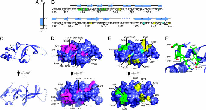
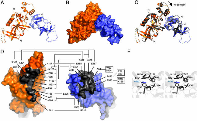
The Sir1 protein plays a key role in establishing a silent chromatin structure at the cryptic mating-type loci HMR and HML in Saccharomyces cerevisiae by interacting with the bromo-adjacent homology (BAH) domain of the Orc1p subunit of the origin recognition complex (ORC). Here, we present the high-resolution crystal structures of the ORC interaction region (OIR) of Sir1p and that of the complex formed between the OIR and BAH domains. Amino acids within the OIR previously shown to be required for a Sir1p/ORC interaction are presented on a conserved, convex surface that forms a complementary interface with a concave region of the Orc1 BAH domain that is critical for transcriptional silencing. The OIR/BAH interaction surface comprises a network of hydrophobic and polar/ionic interactions between discrete structural modules in each protein and involves several residues that were not implicated in previous studies. These data provide important structural insights into a protein-protein interaction critical for the formation of a specialized chromatin domain within eukaryotic chromosomes.


Figures

Fig. 1.
Fig. 1.
Structure of the Sir1p-OIR. (A) Schematic diagram of S. cerevisiae Sir1p indicating the position of the Sir1p-OIR (residues 473–611) in blue. (B) Sequence, conservation, genetically determined roles, and secondary structure of Sir1p-OIR*. The Sir1p-OIR* sequence is colored to identify residues that are important for Orc1p binding (green), Sir4p binding (yellow), or binding for Orc1p and Sir4p (light blue). Sir1p residues that are invariant among the four yeast Sir1p homolog sequences are underlined. Residue 593, labeled in bold, is alanine in the described structure but is a cysteine in the natural S. cerevisiae Sir1p sequence. Secondary structural elements in the Sir1p-OIR* domain are shown above the sequence; the dotted line indicates a region where electron density was not observed. (C) Orthogonal views of a ribbon diagram of the crystal structure of Sir1p-OIR*. Secondary structure elements from B are labeled on the lower diagram. Ribbon diagrams and surface representations were rendered with pymol (26). (D) Orthogonal views of the surface conservation of Sir1p-OIR* with invariant residues (underlined in B) colored magenta and labeled. Views correspond to those in C. (E) Surface representation of Sir1p-OIR* colored as in B to indicate residues important for interaction with Orc1p, Sir4p, or both proteins (11). (F) Close-up view of previously identified Orc1p-interacting residues (ball-and-stick form) within the SRD module. Residues are labeled, and selected distances are indicated.
Fig. 2.
Fig. 2.
Structure of the Sir1p-OIR*/Orc1p-BAH complex. (A) Ribbon diagram of the crystal structure of the Sir1p-OIR*/Orc1p-BAH complex, with Orc1p-BAH in orange and OIR* in blue. (B) Surface representation of structure shown in A. (C) Overlay of the complex structure from A, with the isolated Orc1p-BAH and Sir1 OIR structures shown in gray. The apparent movement of the H-domain of Orc1 is indicated by an arrow. (D) The two domains from the OIR*/BAH complex are rotated orthogonally and shown independently to expose the interacting surfaces on each domain. Residues that form the interface are colored gray and labeled. Specific residue contacts are indicated with connecting lines. (E) Stereoview P492 of OIR* in a binding pocket formed by several amino acids in the Orc1p-BAH domain.

References

    1. Grewal, S. I. & Moazed, D. (2003) Science 301, 798-802. - PubMed
    1. Weinreich, M., Palacios DeBeer, M. A. & Fox, C. A. (2004) Biochim. Biophys. Acta 1677, 142-157. - PubMed
    1. Fischle, W., Wang, Y. & Allis, C. D. (2003) Curr. Opin. Cell Biol. 15, 172-183. - PubMed
    1. Rusche, L. N., Kirchmaier, A. L. & Rine, J. (2003) Annu. Rev. Biochem. 72, 481-516. - PubMed
    1. Bell, S. P. (2002) Genes Dev. 16, 659-672. - PubMed

Publication types

MeSH terms

Substances