. 2005 Jun;71(6):2862-9.
doi: 10.1128/AEM.71.6.2862-2869.2005.

Characterization of a nucleus-encoded chitinase from the yeast Kluyveromyces lactis

Affiliations

Characterization of a nucleus-encoded chitinase from the yeast Kluyveromyces lactis

Paul A Colussi et al. Appl Environ Microbiol. 2005 Jun.

Abstract

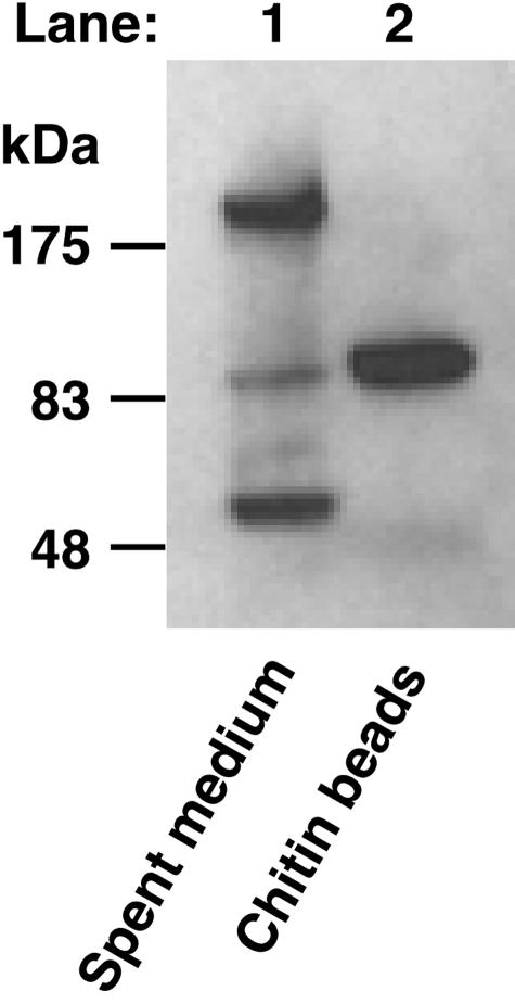
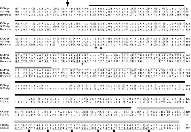
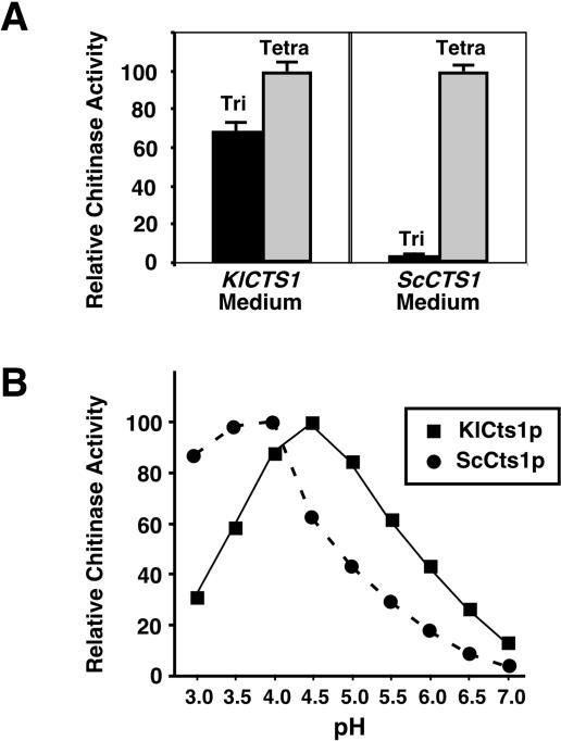
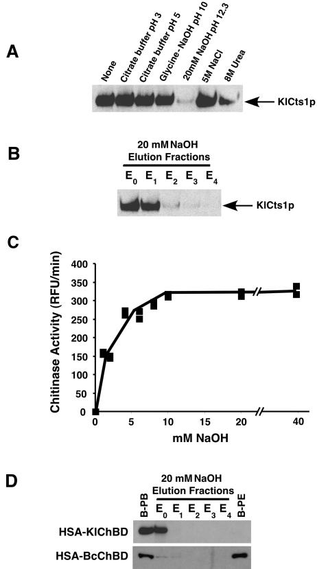
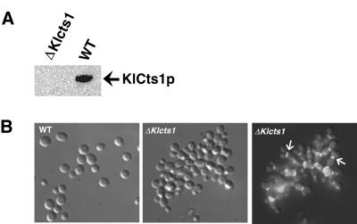
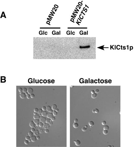
Endogenous proteins secreted from Kluyveromyces lactis were screened for their ability to bind to or to hydrolyze chitin. This analysis resulted in identification of a nucleus-encoded extracellular chitinase (KlCts1p) with a chitinolytic activity distinct from that of the plasmid-encoded killer toxin alpha-subunit. Sequence analysis of cloned KlCTS1 indicated that it encodes a 551-amino-acid chitinase having a secretion signal peptide, an amino-terminal family 18 chitinase catalytic domain, a serine-threonine-rich domain, and a carboxy-terminal type 2 chitin-binding domain. The association of purified KlCts1p with chitin is stable in the presence of high salt concentrations and pH 3 to 10 buffers; however, complete dissociation and release of fully active KlCts1p occur in 20 mM NaOH. Similarly, secreted human serum albumin harboring a carboxy-terminal fusion with the chitin-binding domain derived from KlCts1p also dissociates from chitin in 20 mM NaOH, demonstrating the domain's potential utility as an affinity tag for reversible chitin immobilization or purification of alkaliphilic or alkali-tolerant recombinant fusion proteins. Finally, haploid K. lactis cells harboring a cts1 null mutation are viable but exhibit a cell separation defect, suggesting that KlCts1p is required for normal cytokinesis, probably by facilitating the degradation of septum-localized chitin.


Figures

FIG. 1.
FIG. 1.
Identification of a secreted 85-kDa K. lactis chitin-binding protein. Secreted proteins in 10 μl of K. lactis GG799 spent culture medium (after 96 h of growth) were separated by SDS-PAGE and screened for the presence of a chitin-binding domain by Western blotting with α-ChBD antibodies (lane 1). Secreted proteins were incubated with chitin beads, after which chitin-bound proteins were eluted by boiling, separated by SDS-PAGE, and detected by α-ChBD Western blotting (lane 2).
FIG. 2.
FIG. 2.
Domain organization and alignment of KlCts1p with family 18 chitinases. The KlCts1p sequence was aligned with family 18 chitinases from S. cerevisiae (Cts1p; GenBank accession no. A41035) and the rubber tree, Hevea brasiliensis (hevamine; GenBank accession no. AJ007701). The amino acids enclosed in boxes are identical in at least two of the three sequences. KlCts1p consists of a signal peptide that is cleaved after A19 (arrow), a family 18 chitinase catalytic domain (single line), a serine-threonine-rich domain (double line), and a type 2 chitin-binding domain (gray box). Conserved catalytic amino acids predicted from the hevamine crystal structure (4) are indicated by asterisks. Six cysteines conserved in type 2 ChBDs are indicated by solid dots. Hevamine (311 amino acids) does not contain a Ser-Thr-rich domain or a ChBD. Therefore, seven amino acids were truncated from its carboxy terminus to allow alignment of the Ser-Thr-rich domains of KlCts1p and ScCts1p.
FIG. 3.
FIG. 3.
Enzymatic properties of secreted KlCts1p. (A) Substrate preferences of KlCts1p and ScCts1p. Spent culture media from cultures of K. lactis GG799 and S. cerevisiae BY4741 cells were incubated with 50 μM 4MU-GlcNAc3 (Tri) (black bars) and with 50 μM 4MU-GlcNAc4 (Tetra) (gray bars) at pH 4.5 and 37°C. The relative rates of 4-MU release for each reaction were measured. (B) pH optima of KlCts1p and ScCts1p. The relative rates of 4-MU release were measured at 37°C for KlCts1p using 50 μM 4MU-GlcNAc3 and for ScCts1p using 50 μM 4MU-GlcNAc4 in McIlvaine's buffers with pHs ranging from 3.0 to 7.0.
FIG. 4.
FIG. 4.
KlCts1p dissociates from chitin at high pH. (A) KlCts1p was bound to chitin beads in minicolumns as described in Materials and Methods. The solutions were passed over separate minicolumns, after which KlCts1p that was still chitin bound was eluted by boiling, separated by SDS-PAGE, and detected by α-ChBD Western blotting. (B) KlCts1p elution from chitin with 20 mM NaOH. Chitin-bound KlCts1p was eluted in five successive 1-ml fractions of 20 mM NaOH (E0 to E4, where E0 represents the column void volume), separated by SDS-PAGE, and detected by α-ChBD Western blotting. (C) Base-eluted KlCts1p retains chitinolytic activity. Chitin-bound KlCts1p was eluted from chitin with various concentrations of NaOH, and the eluates were assayed for chitinase activity with 4MU-GlcNAc3 at pH 4.5 and 37°C. RFU, relative fluorescence units. (D) KlCts1p-ChBD can function as an elutable affinity tag. HSA fusions to ChBDs derived from KlCts1p or B. circulans ChiA1 were secreted from K. lactis, bound to chitin beads, and eluted with five 1-ml 20 mM NaOH fractions (E0 to E4). Proteins that were bound to chitin beads prior to elution (B-PB) or that were still bound after elution with 20 mM NaOH (B-PE) were eluted by boiling. All samples were separated by SDS-PAGE and examined by α-ChBD Western blotting.
FIG. 5.
FIG. 5.
(A) K. lactis Δcts1 cells do not secrete KlCts1p. Proteins in spent culture medium from wild-type GG799 (WT) and Δcts1 K. lactis cells were incubated with chitin beads. Bound proteins were eluted by boiling and were detected by α-ChBD Western blotting. (B) KlCTS1 is required for efficient cell separation. Wild-type and Δcts1 K. lactis cells were grown in YPD medium and fixed in 2.5% glutaraldehyde. Septum-localized chitin was stained with calcofluor white and detected by fluorescence microscopy using a DAPI filter. The middle and right panels show the same cells visualized by phase-contrast and fluorescence microscopy, respectively. The arrows in the right panel indicate the locations of septa in certain cells.
FIG. 6.
FIG. 6.
(A) S. cerevisiae Δcts1 cells expressing KlCTS1 secrete KlCts1p. S. cerevisiae Δcts1 cells harboring either an empty vector (pMW20) or a vector containing KlCTS1 under the control of a galactose-inducible glucose-repressible promoter (pMW20-KlCTS1) were grown in medium containing glucose or galactose overnight at 30°C. Spent culture medium was tested for the presence of KlCts1p by passage over chitin beads, elution of chitin-bound proteins by boiling, separation by SDS-PAGE, and α-ChBD Western blotting. (B) Expression of KlCTS1 corrects the aggregation phenotype of S. cerevisiae Δcts1 cells. S. cerevisiae Δcts1 cells harboring pMW20-KlCTS1 were grown in medium containing galactose or glucose. Cells were fixed in 2.5% glutaraldehyde and visualized by phase-contrast microscopy. Cells harboring an empty vector maintained an aggregation phenotype regardless of the carbon source (data not shown).

References

    1. Arakane, Y., Q. Zhu, M. Matsumiya, S. Muthukrishnan, and K. J. Kramer. 2003. Properties of catalytic, linker and chitin-binding domains of insect chitinase. Insect Biochem. Mol. Biol. 33:631-648. - PubMed
    1. Asensio, J. L., F. J.Canada, H. C. Siebert, J. Laynez, A. Poveda, P. M. Nieto, U. M. Soedjanaamadja, H. J. Gabius, and J. Jimenez-Barbero. 2000. Structural basis for chitin recognition by defense proteins: GlcNAc residues are bound in a multivalent fashion by extended binding sites in hevein domains. Chem. Biol. 7:529-543. - PubMed
    1. Bernard, M. P., D. Cao, R. V. Myers, and W. R. Moyle. 2004. Tight attachment of chitin-binding-domain-tagged proteins to surfaces coated with acetylated chitosan. Anal. Biochem. 327:278-283. - PubMed
    1. Bokma, E., H. J. Rozeboom, M. Sibbald, B. W. Dijkstra, and J. J. Beintema. 2002. Expression and characterization of active site mutants of hevamine, a chitinase from the rubber tree Hevea brasiliensis. Eur. J. Biochem. 269:893-901. - PubMed
    1. Bulik, D. A., M. Olczak, H. A. Lucero, B. C. Osmond, P. W. Robbins, and C. A. Specht. 2003. Chitin synthesis in Saccharomyces cerevisiae in response to supplementation of growth medium with glucosamine and cell wall stress. Eukaryot. Cell 2:886-900. - PMC - PubMed

MeSH terms