. 2005 Jun 15;19(12):1455-65.
doi: 10.1101/gad.1318405. Epub 2005 Jun 2.

Structural and functional analysis of SET8, a histone H4 Lys-20 methyltransferase

Affiliations

Structural and functional analysis of SET8, a histone H4 Lys-20 methyltransferase

Jean-François Couture et al. Genes Dev. .

Abstract

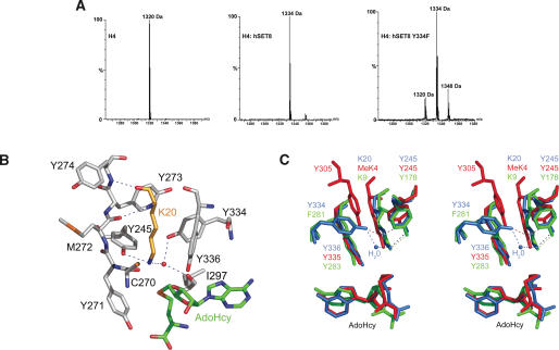
SET8 (also known as PR-SET7) is a histone H4-Lys-20-specific methyltransferase that is implicated in cell-cycle-dependent transcriptional silencing and mitotic regulation in metazoans. Herein we report the crystal structure of human SET8 (hSET8) bound to a histone H4 peptide bearing Lys-20 and the product cofactor S-adenosylhomocysteine. Histone H4 intercalates in the substrate-binding cleft as an extended parallel beta-strand. Residues preceding Lys-20 in H4 engage in an extensive array of salt bridge, hydrogen bond, and van der Waals interactions with hSET8, while the C-terminal residues bind through predominantly hydrophobic interactions. Mutational analysis of both the substrate-binding cleft and histone H4 reveals that interactions with residues in the N and C termini of the H4 peptide are critical for conferring substrate specificity. Finally, analysis of the product specificity indicates that hSET8 is a monomethylase, consistent with its role in the maintenance of Lys-20 monomethylation during cell division.


Figures

Figure 1.
Figure 1.
Crystal structure of hSET8. (A) Experimental Se-MAD phased electron density map at 2.0 Å resolution illustrating the active site of hSET8 bound to Lys-20 in histone H4. The map is contoured at 1.0 σ. The carbon atoms of hSET8 and the histone H4 residues are displayed and labeled in green and gold, respectively. (B) Ribbon diagram of the secondary structure of hSET8 with the β-strands and α- and 310-helices denoted. The SET, nSET, iSET, and cSET regions are depicted in green, yellow, blue, and red, respectively, while AdoHcy and the H4 peptide are represented in magenta and orange, respectively.
Figure 2.
Figure 2.
Cofactor-binding pocket. (A) Stereoview of the cofactor-binding cavity in complex with AdoHcy. The carbon atoms in hSET8, the cofactor, and the H4 peptide are depicted in gray, green, and gold, respectively. Hydrogen bonds are illustrated as dashed blue lines. (B) ITC titration experiment with the wild-type histone H4 peptide and hSET8 (upper trace) and the fitted binding curve (lower trace). (C) Michaelis-Menten plot of the initial velocity versus substrate concentration for the methylation of the H4 peptide by hSET8.
Figure 3.
Figure 3.
Histone substrate-binding cleft. (A) Two orthogonal views of the molecular surface of the histone-binding cleft of hSET8 in which the β6-strand is denoted through the transparent surface. The H4 peptide and cofactor are rendered in orange and magenta, respectively, while the secondary structure of the enzyme is depicted as in Figure 1B. (B) Substrate-binding cleft of hSET8 in which the carbon atoms of the protein, the H4 peptide, and the cofactor are illustrated in gray, gold, and green, respectively. Hydrogen bonds are displayed as dashed blue lines. Protein and ligands atoms are labeled according to the color of their carbon atoms, except for mutations in hSET8 that are labeled in red.
Figure 4.
Figure 4.
Product specificity of hSET8. (A) MALDI mass spectrometry of the 10-residue histone H4 peptide (m/z = 1320) and its methylation by wild-type hSET8 and the Y334F mutant. (B) Close-up view of hSET8 lysine-binding channel. Residues and the water molecule that interact with the side chain of Lys-20 are illustrated. Hydrogen bonds are rendered as dashed blue lines. The carbon atoms of Lys-20, AdoHcy, and the enzyme are depicted in gold, green, and gray, respectively. (C) Stereoview of a superimposition of the lysine-binding channels of hSET8:H4 Lys-20 (blue), hSET7/9:H3 MeLys4 (1O9S.pdb, red), and DIM-5:H3 Lys-9 (1PEG.pdb, green). Hydrogen bonds are rendered with dashed lines corresponding to the color of each enzyme.

References

    1. Aasland R., Abrams, C., Ampe, C., Ball, L.J., Bedford, M.T., Cesareni, G., Gimona, M., Hurley, J.H., Jarchau, T., Lehto, V.P., et al. 2002. Normalization of nomenclature for peptide motifs as ligands of modular protein domains. FEBS Lett. 513: 141-144. - PubMed
    1. Abrahams J.P. and Leslie, A.G.W. 1996. Methods used in the structure determination of bovine mitochondrial F1 ATPase. Acta Crystallogr. D Biol. Crystallogr. 52: 30-42. - PubMed
    1. Beisel C., Imhof, A., Greene, J., Kremmer, E., and Sauer, F. 2002. Histone methylation by the Drosophila epigenetic transcriptional regulator Ash1. Nature 419: 857-862. - PubMed
    1. Brunzelle J.S., Shafaee, P., Yang, X., Weigand, S., Ren, Z., and Anderson, W.F. 2003. Automated crystallographic system for high-throughput protein structure determination. Acta Crystallogr. D Biol. Crystallogr. 59: 1138-1144. - PubMed
    1. Chirpich T.P., Zappia, V., Costilow, R.N., and Barker, H.A. 1970. Lysine 2,3-aminomutase. Purification and properties of a pyridoxal phosphate and S-adenosylmethionine-activated enzyme. J. Biol. Chem. 245: 1778-1789. - PubMed

Publication types

MeSH terms