. 2005 Jul 6;24(13):2425-35.
doi: 10.1038/sj.emboj.7600712. Epub 2005 Jun 2.

Activating transcription factor 3, a stress sensor, activates p53 by blocking its ubiquitination

Affiliations

Activating transcription factor 3, a stress sensor, activates p53 by blocking its ubiquitination

Chunhong Yan et al. EMBO J. .

Abstract

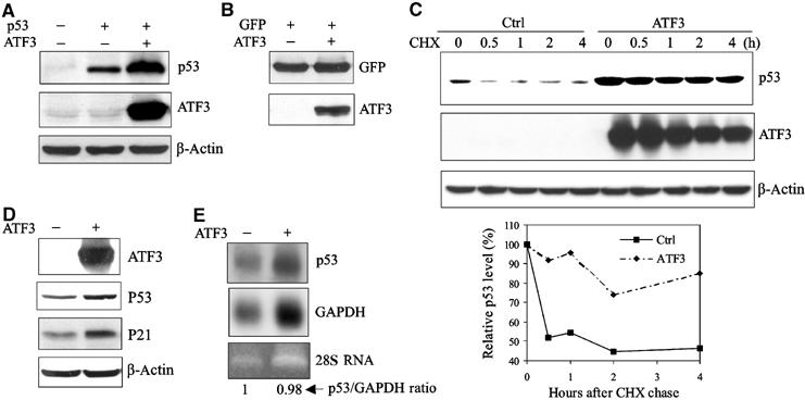
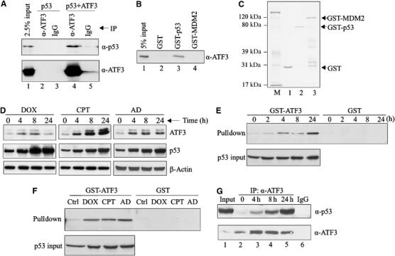
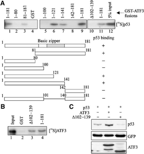
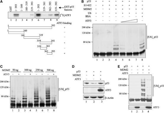
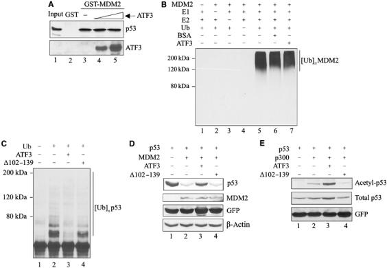
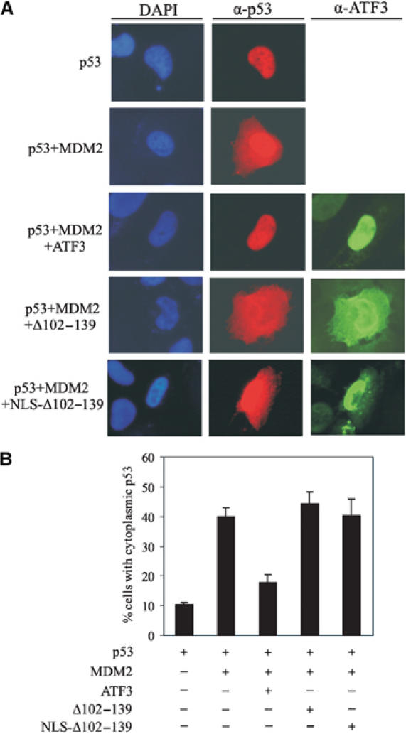
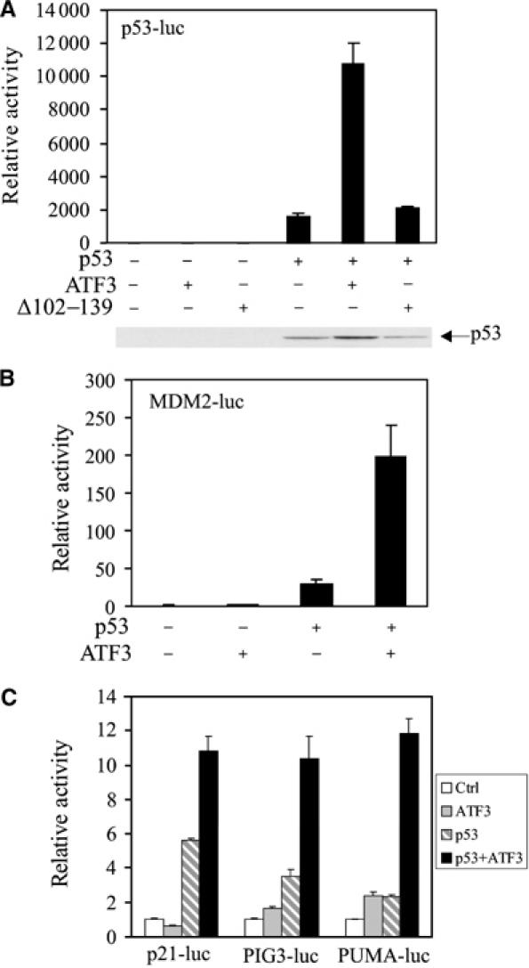
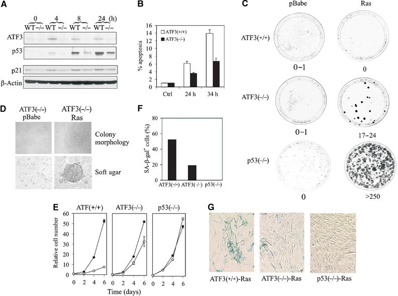
Activating transcription factor 3 (ATF3) is rapidly induced by diverse environmental insults including genotoxic stress. We report herein that its interaction with p53, enhanced by genotoxic stress, stabilizes the tumor suppressor thereby augmenting functions of the latter. Overexpression of ATF3 (but not a mutated ATF3 protein (Delta102-139) devoid of its p53-binding region) prevents p53 from MDM2-mediated degradation and leads to increased transcription from p53-regulated promoters. ATF3, but not the Delta102-139 protein, binds the p53 carboxy-terminus and diminishes its ubiquitination and nuclear export. Genotoxic-stressed ATF3-null mouse embryonic fibroblasts, or cells in which ATF3 was reduced by small interference RNA, show inefficient p53 induction and impaired apoptosis compared with wild-type cells. ATF3-null cells (but not wild-type cells), which poorly accumulate p53, are transformed by oncogenic Ras. Thus, ATF3 is a novel stress-activated regulator of p53 protein stability/function providing the cell with a means of responding to a wide range of environmental insult, thus maintaining DNA integrity and protecting against cell transformation.


Figures

Figure 1
Figure 1
ATF3 increases p53 protein stability. (A) H1299 cells were transfected with, as indicated, 0.05 μg p53, 0.8 μg ATF3 or its vector pCG, and 0.1 μg RSV-luc. Cell lysates normalized according to luciferase activity were subjected to Western blotting. (B) H1299 cells were transfected, as indicated, with 0.05 μg pEGFP-N1, 0.8 μg ATF3 or pCG (vector), and 0.1 μg RSV-luc. Cell lysates normalized as described in (A) were subjected to Western blotting. (C) H1299 cells were transfected with a p53 expression construct and, where indicated, an expression plasmid bearing ATF3 and then treated with cycloheximide. Cells were harvested at the indicated time points, and cell lysates with an equal protein amount were subjected to Western blotting. Relative p53 levels were quantified by densitometry (D, E) HCT116 cells were transfected with pEGFP-N1 and, where indicated, with ATF3 or its vector. GFP-positive cells were collected by FACS, and subjected to Western blotting (D) or Northern blotting (E).
Figure 2
Figure 2
Genotoxic stress increases the direct interaction of the ATF3 and p53 proteins. (A) H1299 cells were transfected with p53 and, where indicated, with pCG/ATF3. Cell lysates (1 mg protein) were incubated with 1 μg of anti-ATF3 antibody or IgG at 4°C overnight, immunocomplexes captured with protein A-agarose and subjected to Western blotting for p53 and ATF3. (B) The indicated GST fusion proteins from bacteria culture were immobilized on glutathione-agarose and incubated with a purified recombinant ATF3 protein 4 h at 4°C. The agarose was extensively washed and eluates subjected to Western blotting for ATF3. (C) The indicated GST fusion proteins were resolved by SDS–PAGE and stained with Coomassie blue. (D) HCT116 cells were treated with 0.4 μg/ml DOX, 2 μM CPT or 5 nM AD as indicated. After the indicated times, cells were lysed and subjected to Western blotting for ATF3 and p53. (E, F) HCT116 cell lysates, adjusted to equal p53 amounts (p53 input), from unstressed and stressed cells (treated as in (D); for (E), the cells were treated with DOX) were incubated with glutathione-agarose beads bound with GST-ATF3 fusion protein or GST protein. After extensive washes, bound material was eluted and immunoblotted for p53. (G) HCT116 cells were treated with 0.4 μg/ml DOX and harvested at the indicated time points. Cell lysates (2 mg protein) were incubated with 0.5 μg ATF3 antibody overnight. Immunoprecipitates were captured and subjected to Western blotting for p53 and ATF3. Lysate from 24-h-treated cells was also immunoprecipitated with 0.5 μg normal IgG as a control (lane 6).
Figure 3
Figure 3
Interaction of the ATF3 and p53 proteins is required for stabilization of the tumor suppressor. (A) GST-ATF3 fusion proteins were adsorbed to glutathione-agarose, and incubated with in vitro-translated [35S]methionine-labeled p53 protein. After extensive washes, the bound p53 was eluted, subjected to SDS–PAGE and visualized by fluorography. (B) GST or GST fused to the mutant (Δ102–139) or full-length ATF3 protein (1–181) was adsorbed to glutathione-agarose, incubated with in vitro-translated [35S]methionine-labeled ATF3 protein (full length) and subjected to pulldown assays as in (A). (C) Plasmids encoding p53, the full-length ATF3 protein or mutant ATF3 protein (Δ102–139) were transfected where indicated with 0.05 μg pEGFP-N1 into H1299 cells and Western blotting performed as described in Figure 1A with the exception that the HA-tagged ATF3 proteins were detected with an anti-HA antibody.
Figure 4
Figure 4
ATF3 prevents MDM2-mediated p53 degradation by blocking its ubiquitination. (A) Truncated GST-p53 proteins were immobilized onto glutathione-agarose and incubated with 35S-labeled ATF3 protein. After extensive washes, the bound ATF3 was eluted, subjected to SDS–PAGE and visualized by fluorography. (B) A 0.5 μl portion of p53 protein prepared with rabbit reticulocyte lysate was preincubated for 1 h with different amounts of purified ATF3 protein (200, 250 and 400 ng) or 400 ng BSA and then mixed with 50 ng MDM2 and other URCs at 37°C for 1 h. The reactions were then subjected to Western blotting for p53. (C) A 0.5 μl portion of p53 protein was preincubated with 250 ng ATF3 protein and subjected to ubiquitination reactions with the indicated amounts of MDM2. (D) H1299 cells were transfected, where indicated, with 0.05 μg p53, 0.4 μg MDM2, 1.5 μg ATF3 or pCG, and 0.1 μg RSV-luc plasmids, and harvested for Western blotting. (E) H1299 cells were transfected with 0.4 μg p53, 0.9 μg MDM2, 0.9 μg HA-ub, and 3.8 μg ATF3 or pCG, as indicated, and treated with 25 μM MG125 and 100 μM MG101 for 3 h before harvest. Cell lysates were adjusted to equal p53 level and subjected to Western blotting with DO-1.
Figure 5
Figure 5
ATF3 does not interfere with either the binding of MDM2 to p53 or the ubiquitinating activity of MDM2. (A) p53 protein prepared from rabbit reticulocyte lysates (2 μl) was incubated with (1 and 1.6 μg) purified ATF3 protein at 37°C and then incubated with immobilized GST-MDM2. After extensive washes, the bound p53 was eluted and subjected to Western blotting for p53 and then reprobed with an anti-ATF3 antibody. (B) Purified ATF3 protein (250 ng) was mixed with 250 ng MDM2, URCs and BSA (250 ng) for ubiquitination reactions as indicated. The reaction mixtures were subjected to Western blotting for ubiquitinated MDM2 using anti-polyubiquitin antibody (FK-1). (C) p53 protein was preincubated with 250 ng of the full-length or the mutant ATF3 protein and then subjected to ubiquitination reactions as in Figure 4C. (D) Transfections of H1299 cells were as in Figure 4E with the exception that a plasmid encoding ATF3 deleted of the p53-binding domain (Δ102–139) and pEGFP-N1 were included. (E) H1299 cells were transfected with the following expression vectors: 0.1 μg p53, 0.1 μg RSV-luc and 0.05 μg pEGFP-N1 and, where indicated, 2 μg p300, 1.75 μg pCG, the mutant or full-length ATF3. The cells were lysed in a buffer containing 5 μM TSA and subjected to Western blotting with antibodies against Lys382 acetylated p53, total p53 (DO-1) or GFP.
Figure 6
Figure 6
ATF3 prevents MDM2-mediated p53 nuclear export. H1299 cells were cotransfected, where indicated, with 0.1 μg p53, 0.2 μg MDM2 and 0.7 μg ATF3 expression vectors, fixed and stained using anti-p53 and anti-ATF3 antibodies for visualization (A) and counting (B). At least 300 transfected cells were scored for p53 subcellular distribution.
Figure 7
Figure 7
ATF3 augments trans-activation of p53 responsive promoters. H1299 cells were transfected, where indicated, with vectors bearing p53 (0.02 μg), ATF3 (0.4 μg) or Δ102–139 (0.4 μg), pRL-TK (0.001 μg), and a reporter regulated by tandem repeated p53 response elements (0.01 μg) (A) or 0.1 μg of the following promoters: MDM2 (B), p21, PIG3 or PUMA (C). Cells were lysed 30 h later and subjected to luciferase assays using the Dual-luciferase Assay System. p53 protein level was determined by Western blotting and shown for (A).
Figure 8
Figure 8
Loss of ATF3 impairs the p53-dependent cellular response to DNA damage and allows cellular transformation by an oncogenic Ras. (A) ATF3-null (−/−) or wild-type (WT) MEFs were treated with 2 μM CPT. Cells were lysed and subjected to Western blotting for ATF3, p53 or p21. (B) MEFs were infected with a retrovirus bearing E1A 12S, treated with 2 μM CPT and harvested and fixed at the indicated times. Apoptotic cells were defined as sub-G0/G1 cell as determined by FACS analysis. (C) MEFs were infected with a retrovirus bearing H-rasV12 or empty vector (pBabe). After selecting in puromycin for 4 days, the cells were plated in triplicate for colony formation assays. (D) Colony morphology of Ras-transformed ATF3−/− cells in monolayer and in soft agar. (E) Infected MEFs were plated in 12-well plates, harvested at the indicated times and cell numbers determined by crystal violet staining. Open circle indicates Ras and closed circle pBabe. (F, G) Ras-infected MEFs were stained with X-gal (pH 6.0) overnight. X-gal staining-positive cells were counted (F) under a microscope.

Similar articles

Cited by

References

    1. An WG, Kanekal M, Aaronswon SA, Simon MC, Maltepe E, Blagosklonny MV, Neckers LM (1998) Stabilization of wild-type p53 by hypoxia-inducible factor 1α. Nature 392: 405–408 - PubMed
    1. Buschmann T, Lin Y, Aithmitti N, Fuchs SY, Lu H, Resnick-Silverman L, Manfredi JJ, Ronai Z, Wu X (2001) Stabilization and activation of p53 by the coactivator protein TAFII31. J Biol Chem 276: 13852–13857 - PubMed
    1. Chen BP, Wolfgang CD, Hai T (1996) Analysis of ATF3, a transcription factor induced by physiological stresses and modulated by gadd153/Chop10. Mol Cell Biol 16: 1157–1168 - PMC - PubMed
    1. Chen D, Li M, Luo J, Gu W (2003) Direct interactions between HIF-1α and Mdm2 modulate p53 function. J Biol Chem 278: 13595–13598 - PubMed
    1. Cheng T, Liu D, Griffin JH, Fernandez JA, Castellino F, Rosen ED, Fukudome K, Zlokovic BV (2003) Activated protein C blocks p53-mediated apoptosis in ischemic human brain endothelium and is neuroprotective. Nat Med 9: 338–342 - PubMed

Publication types

MeSH terms