. 2005 Jul 6;24(13):2414-24.
doi: 10.1038/sj.emboj.7600710. Epub 2005 Jun 2.

The Rsp5 ubiquitin ligase is coupled to and antagonized by the Ubp2 deubiquitinating enzyme

Affiliations

The Rsp5 ubiquitin ligase is coupled to and antagonized by the Ubp2 deubiquitinating enzyme

Younghoon Kee et al. EMBO J. .

Abstract

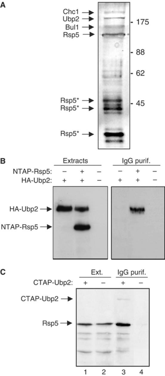
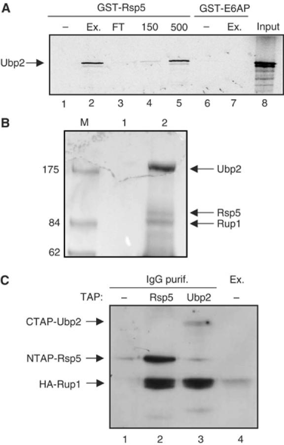
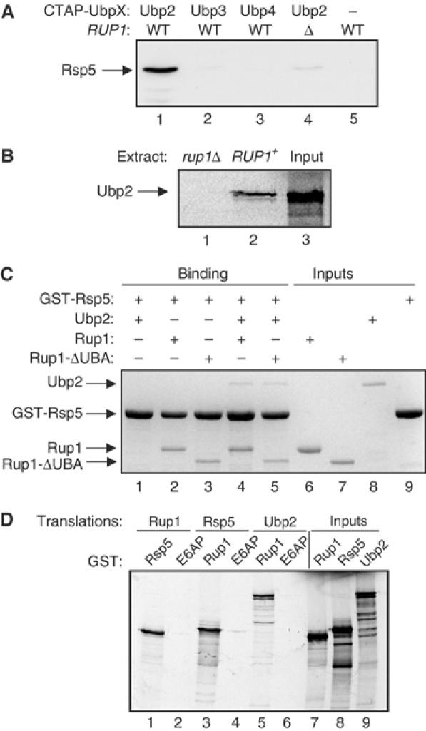
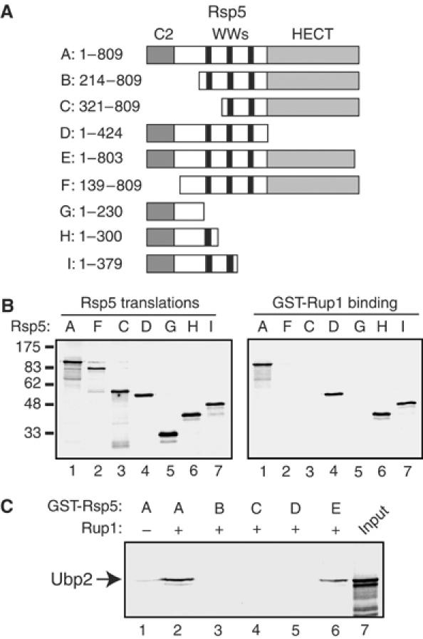
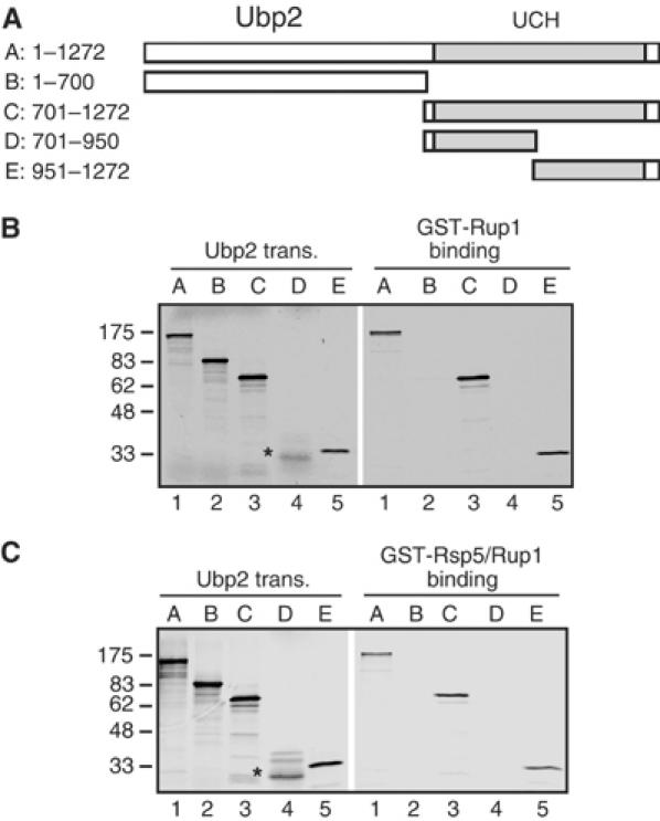
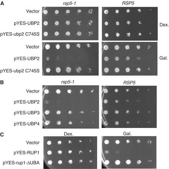
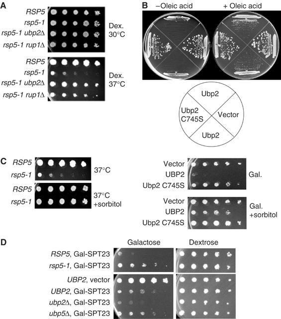
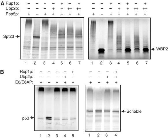
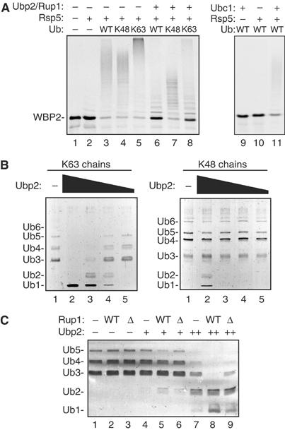
Saccharomyces cerevisiae Rsp5 is an essential HECT ubiquitin ligase involved in several biological processes. To gain further insight into regulation of this enzyme, we identified proteins that copurified with epitope-tagged Rsp5. Ubp2, a deubiquitinating enzyme, was a prominent copurifying protein. Rup1, a previously uncharacterized UBA domain protein, was required for binding of Rsp5 to Ubp2 both in vitro and in vivo. Overexpression of Ubp2 or Rup1 in the rsp5-1 mutant elicited a strong growth defect, while overexpression of a catalytically inactive Ubp2 mutant or Rup1 deleted of the UBA domain did not, suggesting an antagonistic relationship between Rsp5 and the Ubp2/Rup1 complex. Consistent with this model, rsp5-1 temperature sensitivity was suppressed by either ubp2Delta or rup1Delta mutations. Ubp2 reversed Rsp5-catalyzed substrate ubiquitination in vitro, and Rsp5 and Ubp2 preferentially assembled and disassembled, respectively, K63-linked polyubiquitin chains. Together, these results indicate that Rsp5 activity is modulated by being physically coupled to the Rup1/Ubp2 deubiquitinating enzyme complex, representing a novel mode of regulation for an HECT ubiquitin ligase.


Figures

Figure 1
Figure 1
(A) Purification of NTAP-Rsp5 and associated proteins from extract of an 8 l culture (YK001). The final eluate was concentrated and separated by SDS–PAGE and silver stained. The positions and identities of bands identified by LC/MS are indicated; Rsp5* indicates breakdown products of NTAP-Rsp5. Molecular weight markers (kDa) are indicated. (B) Confirmation of the Rsp5–Ubp2 interaction. Extracts were prepared from YK002 (NTAP-RSP5, HA-UBP2), YK008 (RSP5, HA-UBP2), and BY4741 (RSP5, UBP2). Proteins were affinity selected on IgG Sepharose and bound proteins were released with SDS–PAGE loading buffer. Total extracts (lanes 1–3) and eluates (lanes 4–6) were analyzed by SDS–PAGE and immunoblotted with anti-HA antibody. (C) Extracts were prepared from YK005 (TAP-UBP2) and BY4741 (RSP5, UBP2) and proteins were affinity selected on IgG Sepharose. Total extracts (lanes 1 and 2) and eluates (lanes 3 and 4) were analyzed by SDS–PAGE and immunoblotting with anti-Rsp5 antibody.
Figure 2
Figure 2
A cellular factor mediates the association of Rsp5 and Ubp2. (A) In vitro-translated 35S-labelled Ubp2 was incubated with GST-Rsp5 or GST-E6AP immobilized on glutathione Sepharose, in the absence (−lanes) or presence of cell extracts (Ex.; lanes 2 and 7) or fractionated extracts (lanes 3–5) from the ubp2Δ strain. Bound Ubp2 was detected by SDS–PAGE and autoradiography. DEAE high-salt fractions were either 150 or 500 mM NaCl eluates; FT represents the flow-through fraction. Input (lane 8) shows 50% of translation mixture used in the binding assays. (B) TAP purification of Ubp2-associating proteins. Proteins from YK005 (CTAP-UBP2) were affinity selected on IgG Sepharose followed by elution and cleavage with TEV protease. The eluate (lane 2) was separated on 4–15% gradient gel and stained with Coomassie blue. A parallel purification was performed using the control BY4741 strain (lane 1). Arrows indicate the position of Ubp2, Rsp5, and Rup1 proteins, as identified by LC/MS. Molecular weight markers (kDa) are indicated. (C) HA-Rup1 binds to both NTAP-Rsp5 and CTAP-Ubp2. HA-Rup1 was expressed from a galactose-inducible promoter plasmid in strains YK001 (NTAP-RSP5), YK005 (CTAP-UBP2), and BY4741 (control, lane 1, not expressing any TAP-tagged protein). Extracts were prepared and TAP proteins purified on IgG Sepharose. Eluates (lanes 1–3) were analyzed by SDS–PAGE and immunoblotting with anti-HA antibody. Lane 4 shows HA-Rup1 in total lysate from BY4741. Detection of NTAP-Rsp5 and CTAP-Ubp2 (lanes 2 and 3, respectively) was a result of anti-mouse IgG secondary antibody recognizing the TAP epitope.
Figure 3
Figure 3
Rup1 mediates the association of Rsp5 and Ubp2. (A) Cell extracts were prepared from strains expressing the indicated CTAP-tagged Ubp proteins in either an RUP1 or rup1Δ background. Proteins were affinity selected on IgG Sepharose and eluates were analyzed by SDS–PAGE and immunblotting with anti-Rsp5 antibody. (B) 35S-labeled Ubp2 was assayed for binding to GST-Rsp5 (as in Figure 2A) in the presence of cell extract from either the rup1Δ strain (lane 1) or an RUP1 wild-type strain (lane 2). Lane 3 shows 50% of the input amount of Ubp2 used in the binding reactions. (C) Purified Ubp2, Rup1, and Rup1ΔUBA proteins were used in GST-Rsp5 pull-down assays. A 100% of input amounts of each protein is shown in lanes 6–9. Bound proteins were analyzed by SDS–PAGE and Coomassie blue staining. (D) Rup1, Rsp5, and Ubp2 were 35S-labeled by in vitro translation and assayed for binding to purified GST-Rsp5, E6AP, or Rup1. Inputs (lanes 7–9) show 50% of the translation mixture used in the binding assays.
Figure 4
Figure 4
Domains of Rsp5 required for binding to Rup1 and Ubp2. (A) Schematic of Rsp5 truncation mutants used in binding assays. (B) Binding of GST-Rup1 to Rsp5 proteins. 35S-labeled in vitro-translated Rsp5 proteins were incubated with GST-Rup1 and bound proteins were resolved by SDS–PAGE and visualized by autoradiography. Input amounts (left panel) represent 50% of the translations used in the binding reaction (right panel). (C) Binding of GST-Rsp5 proteins to Ubp2. Purified GST fusion Rsp5 proteins, on glutathione Sepharose, were incubated with 35S-labeled in vitro-translated Ubp2 in the presence (lanes 2–6) or absence (lane 1) of purified Rup1, and bound proteins were resolved by SDS–PAGE and visualized by autoradiography. Input (lane 7) indicates 50% of translation used in the binding reaction.
Figure 5
Figure 5
Domains of Ubp2 required for binding to Rsp5 and Rup1. (A) Schematic Ubp2 mutants used in binding assays. (B) Binding of GST-Rup1 to Ubp2 proteins. 35S-labeled in vitro-translated Ubp2 proteins were incubated with GST-Rup1 and bound proteins were resolved by SDS–PAGE and visualized by autoradiography. Input amounts (left panel) represent 50% of the translations used in the binding reaction (right panel). * indicates predicted size of primary translation product for Ubp2-D. (C) Binding of GST-Rsp5 to Ubp2, in the presence of Rup1. 35S-labeled in vitro-translated Ubp2 proteins were incubated with GST-Rsp5 in the presence of added Rup1 protein, and bound proteins were resolved by SDS–PAGE and visualized by autoradiography. Input amounts (left panel) represent 50% of the translations used in the binding reaction (right panel).
Figure 6
Figure 6
Overexpression of Ubp2 or Rup1 inhibits growth of the rsp5-1 mutant. (A) FY56 (RSP5) and FW1808 (rsp5-1) were transformed with pYES2 (vector), pYES-UBP2, or pYES-ubp2 C745S plasmids. The transformants were serially diluted (10-fold at each step) and plated onto either dextrose (Dex.) or galactose (Gal.) media and grown for 2 and 3 days, respectively, at 30°C. (B) FY56 (RSP5) and FW1808 (rsp5-1) were transformed with empty vector, or pYES-UBP2, UBP3, or UBP4 plasmids and serially diluted on galactose media and grown for 3 days at 30°C. (C) FW1808 (rsp5-1) was transformed with empty vector, pYES-RUP1, and pYes-rup1ΔUBA plasmids and serial dilutions were plated onto either dextrose or galactose media and grown for 3 and 4 days, respectively, at 34°C.
Figure 7
Figure 7
Genetic interactions between RSP5, RUP1, and UBP2. (A) ubp2Δ or rup1Δ mutations partially rescue the temperature sensitivity phenotype of the rsp5-1 mutant. RSP5 (FY56), rsp5-1 (FW1808), rsp5-1, ubp2Δ (YK003), and rsp5-1, rup1Δ (YK004) strains were serially diluted and grown on dextrose-containing media at 30 or 37°C for 2 and 4 days, respectively. (B) Exogenous oleic acid partially suppresses the growth inhibition due to Ubp2 overexpression. FW1808 (rsp5-1) transformed with pYES2 (vector), pYES-UBP2 (in duplicate), or pYES-ubp2 C745S was streaked onto galactose-containing media, in the absence or presence of added oleic acid, and grown at 30°C for 4 days. (C) The Ubp2 overexpression phenotype in the rsp5-1 mutant is rescued by 1 M sorbitol. Left panel: RSP5 (FY56) and rsp5-1 (FW1808) strains were plated on dextrose-containing media at 37°C, with or without 1 M sorbitol. Right panel: The indicated FW1808 transformants were serially diluted and plated onto galactose-containing media at 30°C, with or without the addition of 1 M sorbitol to the media. The – sorbitol plate was grown for 3 days, and the + sorbitol plate was grown for 4 days. (D) Toxicity due to Spt23 overexpression is suppressed by rsp5-1 and enhanced by ubp2Δ. Upper panel: The pYES-SPT23 plasmid was transformed into FY56 (RSP5) or FW1808 (rsp5-1) and plated on galactose- or dextrose-containing media. Lower panel: The empty pYES vector or pYES-SPT23 plasmid was transformed into either UBP2, ubp2Δ, or ubp5Δ strains and plated onto galactose- or dextrose-containing media and grown for 4 days at 30°C.
Figure 8
Figure 8
Ubp2 antagonizes Rsp5-catalyzed ubiquitination in vitro. (A) Rsp5 ubiquitination assays utilized 35S-labeled in vitro-translated Spt23 and WBP2. Each reaction contained added E1, E2 (Ubc1), Ub, and ATP in the absence or presence of Rsp5. At 30 min after initiating the ubiquitination reactions, Rup1 and/or Ubp2 (two different amounts) were added, followed by an additional 30 min incubation. Total reaction products were analyzed by SDS–PAGE and autoradiography. (B) E6AP ubiquitination of p53 and Scribble was performed in the presence of added HPV39 E6 protein, E1, and E2 (UbcH7), followed by the addition of Rup1 and/or Ubp2 as described above, and total reaction products were analyzed by SDS–PAGE and autoradiography.
Figure 9
Figure 9
Rsp5 and Ubp2 preferentially assemble and disassemble K63-linked polyubiquitin chains. (A) Rsp5-catalyzed ubiquitination reactions were carried out with WBP2 as a substrate, as in Figure 8A, except that the endogenous ubiquitin present in the translation reaction was first removed by anion exchange chromatography. The reactions were then performed in the absence of added ubiquitin (left panel, lane 2) or the presence of wild-type ubiquitin, K48-only ubiquitin, or K63-only ubiquitin (left panel, lanes 3–5). Deubiquitination (lanes 6–8) was initiated after 30 min by the addition of Ubp2 and Rup1. Control reactions (lanes 9–11) show that the ubiquitination reaction was dependent on added E2 (Ubc1) and Rsp5. (B) Purified K63 and K48 polyubiquitin chains (Ub3–Ub7; Boston Biochem) were assayed as substrates of purified Ubp2 protein over a 100-fold range of Ubp2 concentration. The deubiquitination reactions were performed for 30 min at room temperature. Products were analyzed by 12% SDS–PAGE and Coomassie blue staining. (C) Rup1 stimulates Ubp2-catalyzed disassembly of K63 chains, dependent on the UBA domain. Disassembly of free K63 chains was assayed at the two lowest concentrations of Ubp2 shown in panel B, in the absence (−) or presence of purified Rup1 (WT) or Rup1ΔUBA (Δ) proteins. Products were analyzed by SDS–PAGE and Coomassie blue staining.

Similar articles

Cited by

References

    1. Amerik AY, Hochstrasser M (2004) Mechanism and function of deubiquitinating enzymes. Biochim Biophys Acta 1695: 189–207 - PubMed
    1. Beaudenon SL, Huacani MR, Wang G, McDonnell DP, Huibregtse JM (1999) Rsp5 ubiquitin-protein ligase mediates DNA damage-induced degradation of the large subunit of RNA polymerase II in Saccharomyces cerevisiae. Mol Cell Biol 19: 6972–6979 - PMC - PubMed
    1. Brummelkamp TR, Nijman SM, Dirac AM, Bernards R (2003) Loss of the cylindromatosis tumour suppressor inhibits apoptosis by activating NF-kappaB. Nature 424: 797–801 - PubMed
    1. Canning M, Boutell C, Parkinson J, Everett RD (2004) A RING finger ubiquitin ligase is protected from autocatalyzed ubiquitination and degradation by binding to ubiquitin-specific protease USP7. J Biol Chem 279: 38160–38168 - PubMed
    1. Chen L, Madura K (2002) Rad23 promotes the targeting of proteolytic substrates to the proteasome. Mol Cell Biol 22: 4902–4913 - PMC - PubMed

Publication types

MeSH terms

Substances