. 2005 Jun 15;24(12):2172-82.
doi: 10.1038/sj.emboj.7600700. Epub 2005 Jun 2.

PIASy mediates SUMO-2 conjugation of Topoisomerase-II on mitotic chromosomes

Affiliations

PIASy mediates SUMO-2 conjugation of Topoisomerase-II on mitotic chromosomes

Yoshiaki Azuma et al. EMBO J. .

Abstract

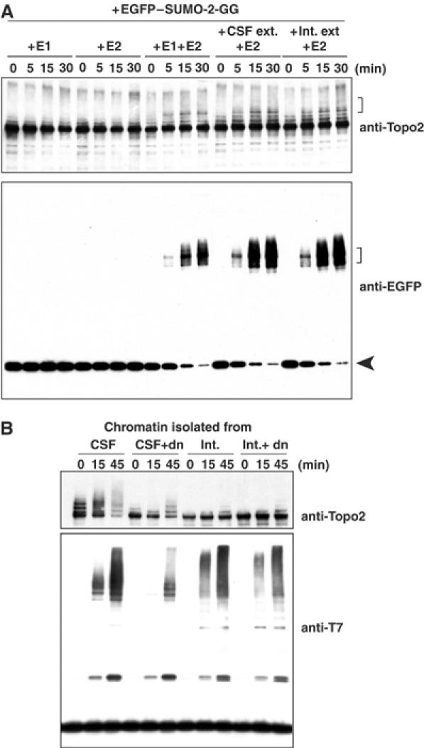
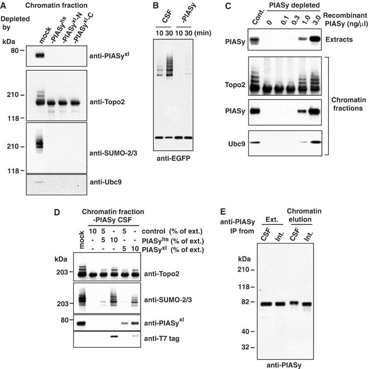
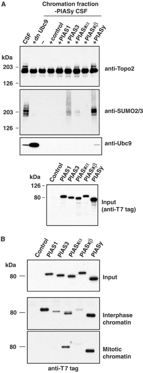
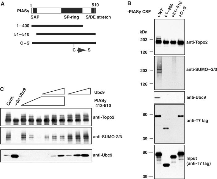
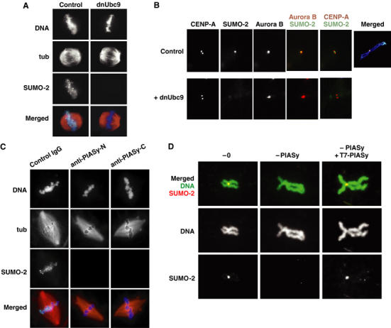
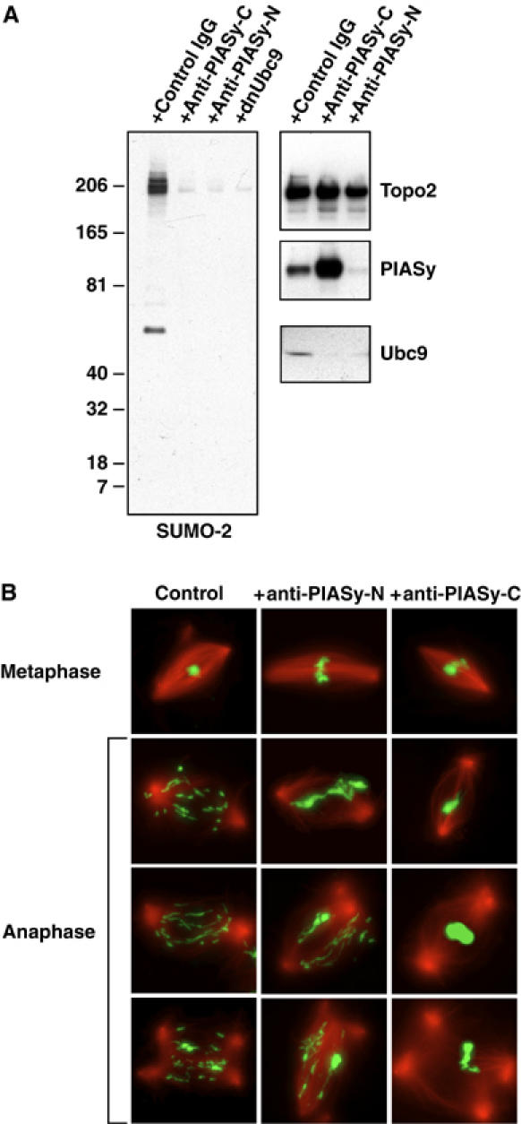
Here we show that the PIASy protein is specifically required for mitotic modification of Topoisomerase-II by SUMO-2 conjugation in Xenopus egg extracts. PIASy was unique among the PIAS family members in its capacity to bind mitotic chromosomes and recruit Ubc9 onto chromatin. These properties were essential, since PIASy mutants that did not bind chromatin or failed to recruit Ubc9 were functionally inactive. We observed that PIASy depletion eliminated essentially all chromosomal accumulation of EGFP-SUMO-2-conjugated species, suggesting that it is the primary E3-like factor for mitotic chromosomal substrates of SUMO-2. PIASy-dependent SUMO-2-conjugated species were highly concentrated on the inner centromere, and inhibition of PIASy blocked anaphase sister chromatid segregation in egg extracts. Taken together, our observations suggest that PIASy is a critical regulator of mitotic SUMO-2 conjugation for Topoisomerase-II and other chromosomal substrates, and that its activity may have particular relevance for centromeric functions required for proper chromosome segregation.


Figures

Figure 1
Figure 1
Mitotic chromatin is competent to promote SUMO-2 modification of Topoisomerase-II in vitro. (A) Mitotic chromatin promotes Topoisomerase-II conjugation. Mitotic chromatin was assembled in CSF extract containing dnUbc9, and isolated as described in Materials and methods. The chromatin was incubated with purified Uba2/Aos1 (20 nM/reaction; ‘+E1'), purified Ubc9 (300 nM/reaction; ‘+E2') or both (‘+E1 +E2'), as indicated. Alternatively, the chromatin was incubated with M-phase egg extract (1/100 volume of reaction mixture; ‘+CSF ext. +E2') or interphase egg extract (1/100 volume of reaction mixture; ‘+Int. ext. +E2') plus purified Ubc9. Aliquots were taken from the reaction mixtures at the indicated times (in minutes), and conjugation of EGFP–SUMO-2 to Topoisomerase-II was analyzed by Western blotting with anti-EGFP and anti-TopoII antibodies. Brackets indicate EGFP–SUMO-2-conjugated Topoisomerase-II, and arrowhead indicates unconjugated EGFP–SUMO-2. (B) Interphase chromatin does not support Topoisomerase-II conjugation. Chromatin was prepared from CSF or interphase extracts in the presence or absence of dnUbc9. After isolation, the chromatin was incubated in a reaction mixture containing purified Uba2/Aos1, purified Ubc9 and T7–SUMO-2-GG. At the indicated times (min), aliquots were taken and incorporation of T7–SUMO-2 to Topoisomerase-II was analyzed by Western blotting with anti-Topo2 and anti-T7 antibodies.
Figure 2
Figure 2
PIASy protein is essential for mitotic SUMO-2 conjugation to Topoisomerase-II. (A) PIASy is essential for SUMO-2 conjugation of Topoisomerase-II and for Ubc9 recruitment to chromatin. CSF extracts were immunodepleted, using antibodies against human PIASy (-PIASyhs), an N-terminal fragment of Xenopus PIASy (-PIASyxl-N) or a C-terminal fragment of Xenopus PIASy (-PIASyxl-C). Depleted extracts were incubated with 5000 sperm nuclei/μl. After 45 min, chromatin from each reaction was isolated and analyzed by Western blotting with the antibodies indicated to the right of each panel. (B) Mitotic chromatin lacking PIASy does not support Topoisomerase-II conjugation in vitro. CSF egg extracts were depleted of PIASy using affinity-purified antibodies, or mock-treated with nonspecific IgG. Sperm chromatin was added to each of the extracts, and allowed to assemble in the presence of dnUbc9. The assembled chromatin was isolated and incubated in reaction mixtures containing E1, E2 and EGFP–SUMO-2, as in Figure 1. Incorporation of EGFP–SUMO-2 was analyzed by Western blotting with anti-EGFP antibody. (C) Recombinant Xenopus PIASy protein restores Topoisomerase-II modification in PIASy-depleted extracts. His6-tagged Xenopus PIASy protein was expressed in bacteria and purified affinity chromatography and gel filtration. The indicated concentrations of purified protein were added to CSF extract that had been previously depleted using anti-PIASyxl-N antibodies. Samples from each reaction were analyzed by Western blotting with anti-PIASy antibody to compare the amount of endogenous protein and recombinant protein (top panel). After 45 min, chromatin from each reaction was isolated and analyzed by Western blotting with the antibodies indicated to the left of each panel (lower three panels). (D) Rescue of Topoisomerase-II modification with human and Xenopus PIASy proteins. Human (PIASyhs) and Xenopus (PIASyxl) PIASy proteins were translated in reticulocyte lysates. The translation reactions were added to CSF extract that had been previously depleted using anti-PIASyxl-N antibodies. The amount of reticulocyte lysate added was adjusted to 10% of the final reaction volume by mixing PIASy-expressed reticulocyte lysate with unprogrammed reticulocyte lysate (control), as indicated. After 45 min, chromatin from each reaction was isolated and analyzed by Western blotting with the antibodies indicated to the right of each panel. Note that both the human and Xenopus proteins were T7-tagged on their N-termini. (E) Reduced electrophoretic mobility of PIASy associated with mitotic chromatin. PIASy was immunoprecipitated from complete extracts (Ext.) or from fractions obtained by salt elution from chromatin (chromatin elution). The precipitated proteins were analyzed by Western blotting with anti-PIASy antibody. Precipitates from both M-phase (CSF) and interphase (Int.) extracts are shown.
Figure 3
Figure 3
Topoisomerase-II conjugation specifically requires PIASy. (A) Other PIAS proteins do not efficiently promote Topoisomerase-II modification. Human, PIAS3, PIASxα, PIASxβ PIASy and mouse PIAS1 were translated in reticulocyte lysates. The translation reactions or unprogrammed reticulocyte lysates (control) were added to CSF extracts with 5000 sperm nuclei/μl (10% final reaction volume). All reactions were incubated for 45 min to allow chromatin remodeling and chromosome condensation. Chromatin from the reactions was isolated and analyzed by Western blotting with the antibodies indicated to the right of the upper three panels. The bottom panel shows a Western blot of the reticulocyte translation assays. Since all of the human PIAS proteins had T7 tags on their N-terminus, this provides quantitation of the relative amounts of each protein added to the CSF extracts. (B) Regulated association of PIAS proteins with mitotic and interphase chromatin. T7-tagged mammalian PIAS proteins were expressed in reticulocyte lysates and mixed with either interphase or CSF extracts in the presence of 5000 sperm nuclei/μl at room temperature. After 45 min, chromatin fractions were isolated, and the association of PIAS proteins was analyzed by Western blotting with anti-T7 antibodies.
Figure 4
Figure 4
Three conserved domains of PIASy are required for SUMO-2 modification of Topoisomerase-II. (A) Representation of conserved domains within PIASy and mutants analyzed. (B) PIASy domains required for chromatin binding and Ubc9 recruitment. Reticulocyte lysates expressing wild-type or mutant human PIASy proteins were added into PIASy-depleted extracts and chromatin fractions were analyzed by Western blotting with the indicated antibodies. (C) Exogenous C-terminal fragment of PIASy blocks Ubc9 recruitment to chromatin. Increasing amounts of human PIASy C-terminal fragment (PIASyhs413–510) and/or Ubc9 were added to CSF extracts containing 5000 sperm nuclei/μl (PIASyhs413–510=0.5, 1, 2 μM; Ubc9=0.2, 0.6, 2 μM). After isolation of the chromatin fraction from these reactions, SUMO-2 conjugation to Topoisomerase-II and Ubc9 recruitment to the chromatin fraction were analyzed by Western blotting with the indicated antibodies.
Figure 5
Figure 5
PIASy mediates SUMO-2 incorporation on the IC of mitotic chromosomes. (A) EGFP–SUMO-2 foci are conjugation-dependent. CSF extracts containing rhodamine-labeled tubulin (red) and EGFP–SUMO-2 (green) were released into interphase by CaCl2 addition. In all, 200 sperm/μl were added 5 min after CaCl2 and the extract was incubated at 23°C for 55 min. Re-entry into mitosis was induced by addition of fresh CSF extract (50% of reaction volume) with or without dnUbc9 (final concentration at 150 ng/μl). After 30 min, samples were removed and analyzed for microtubule and EGFP–SUMO-2 distribution, as well as by staining with Hoechst 33342 DNA dye (blue). (B) EGFP–SUMO-2 foci coincide with ICs. Reactions containing EGFP–SUMO-2 (green) were treated as in (A), except that nocodazole was added coincident with induction of M-phase re-entry with fresh CSF extracts. Samples from each reaction were spun onto coverslips and analyzed by immunofluorescence with antibodies against CENP-A and Aurora B (shown red in merged panels). For comparison, EGFP–SUMO-2 signal from the control extract is also shown in a merged image with Hoechst 33342 staining (far right panel). (C) EGFP–SUMO-2 foci require mitotic PIASy activity. Mitotic chromatin and spindles were assembled and analyzed as in (A). Anti-PIASy antibodies were added coincident with induction of M-phase re-entry with fresh CSF extract. (D) IC accumulation of EGFP–SUMO-2 is PIASy-dependent. EGFP–SUMO-2 (shown in red) was added to untreated CSF extracts, PIASy-depleted extracts or PIASy-depleted extracts supplemented with reticulocyte-translated human PIASy. The extracts were released into interphase by CaCl2 addition. Sperm nuclei addition, re-entry into mitosis and preparation of samples were as described in (B). Samples from each reaction were simultaneously stained with Hoechst 33342 (shown in green).
Figure 6
Figure 6
Perturbation of PIASy function in mitosis by anti-PIASy antibodies causes chromosome segregation defects. (A) Anti-PIASyxl-N and anti-PIASyxl-C block SUMO-2 conjugation on mitotic chromatin. CSF extract was released into interphase by CaCl2 addition. In all, 2000 sperm/μl were added 5 min after CaCl2, and the extract was incubated at 23°C for 55 min. Re-entry into mitosis was induced with fresh CSF extract (50% of reaction volume) containing control IgG, antibodies against the C-terminus of Xenopus PIASy (anti-PIASyxl-C), antibodies against the N-terminus of Xenopus PIASy (anti-PIASyxl-N) (final antibody concentration in each case=0.3 μg/μl), or dnUbc9 (final concentration at 150 ng/μl). Mitotic chromatin was isolated from the control reaction and each of the antibody-containing reactions, as indicated. The chromatin was subjected to Western blotting with antibodies indicated to the right of each panel. (B) Anti-PIASyxl-N and anti-PIASyxl-C block sister chromatid segregation in anaphase. Reactions as in (A) were supplemented with rhodamine-labeled tubulin. 30 min after the induction of metaphase by CSF extracts, anaphase was induced with an addition of CaCl2. Aliquots were removed prior to CaCl2 addition (upper row) or 13 min after CaCl2 addition (bottom three rows). The samples were analyzed for DNA morphology using Hoechst 33342 DNA dye (green) and for microtubule structures (red) after fixation.

References

    1. Aravind L, Koonin EV (2000) SAP—a putative DNA-binding motif involved in chromosomal organization. Trends Biochem Sci 25: 112–114 - PubMed
    1. Arnaoutov A, Dasso M (2003) The Ran GTPase regulates kinetochore function. Dev Cell 5: 99–111 - PubMed
    1. Ayaydin F, Dasso M (2004) Distinct in vivo dynamics of vertebrate SUMO paralogues. Mol Biol Cell 15: 5208–5218 - PMC - PubMed
    1. Azuma Y, Arnaoutov A, Dasso M (2003) SUMO-2/3 regulates topoisomerase II in mitosis. J Cell Biol 163: 477–487 - PMC - PubMed
    1. Azuma Y, Tan SH, Cavenagh MM, Ainsztein AM, Saitoh H, Dasso M (2001) Expression and regulation of the mammalian SUMO-1 E1 enzyme. FASEB J 15: 1825–1827 - PubMed

Publication types

MeSH terms

Substances