. 2005 Apr 20;24(8):1584-95.
doi: 10.1038/sj.emboj.7600636. Epub 2005 Mar 31.

Translation of aberrant mRNAs lacking a termination codon or with a shortened 3'-UTR is repressed after initiation in yeast

Affiliations

Translation of aberrant mRNAs lacking a termination codon or with a shortened 3'-UTR is repressed after initiation in yeast

Toshifumi Inada et al. EMBO J. .

Abstract

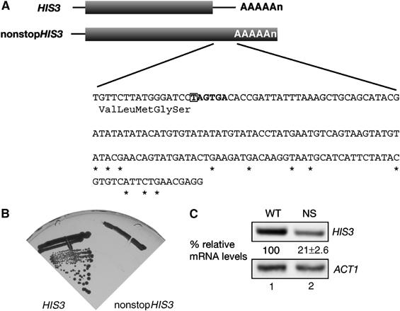
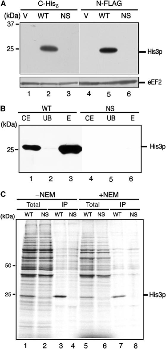
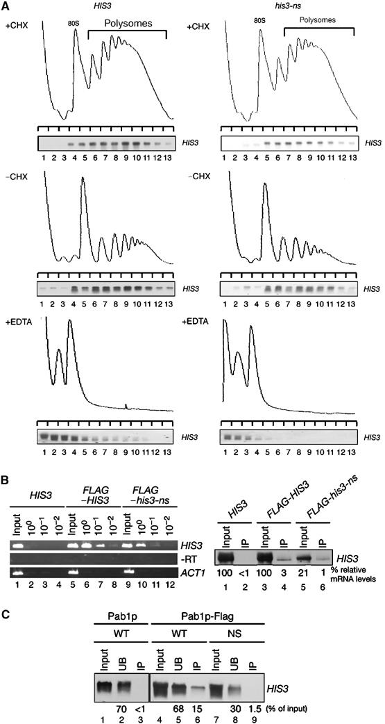
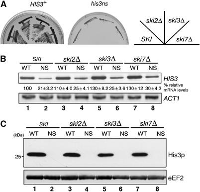
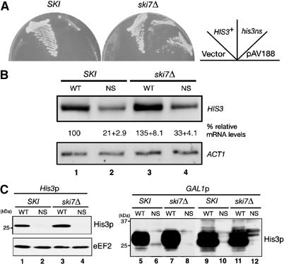
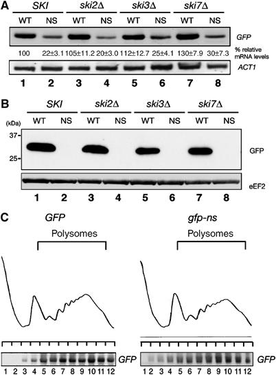
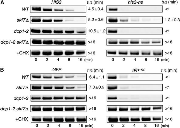
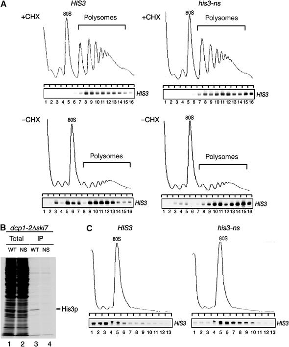
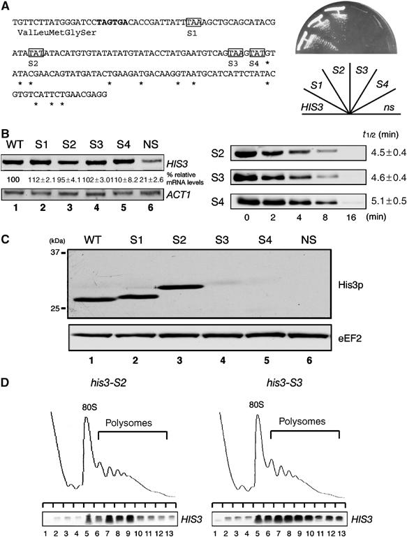
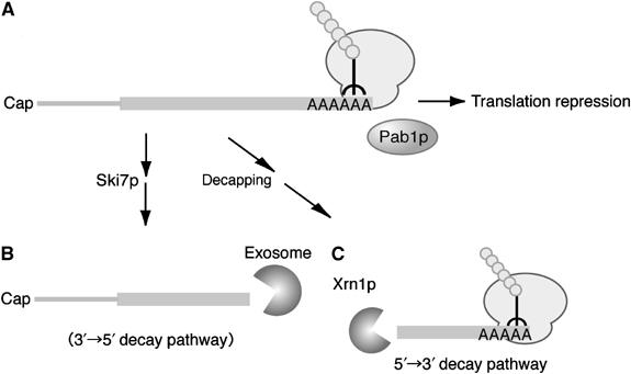
A novel mRNA surveillance for mRNA lacking a termination codon (nonstop mRNA) has been proposed in which Ski7p is thought to recognize stalled ribosomes at the 3' end of mRNA. Here we report our analysis of translation and decay of nonstop mRNAs in Saccharomyces cerevisiae. Although the reduction of nonstop mRNAs was only 4.5-fold, a level that is sufficient for residual protein synthesis, translation products of nonstop mRNAs were hardly detectable. We show that nonstop mRNAs were associated with polysomes, but not with Pab1p. We also show that ribosomes translating nonstop mRNA formed stable and heavy polysome complexes with mRNA. These data suggest that ribosome stalling at the 3' end of nonstop mRNA may block further rounds of translation, hence repressing protein synthesis. Furthermore, it was found that the 5' --> 3' decay pathway was accelerated for nonstop mRNA decay in the absence of Ski7p. We also found that translation of aberrant mRNAs with a shortened 3'-UTR was repressed, suggesting that an improper spatial distance between the termination codon and the 3' end of mRNA results in translation repression.


Figures

Figure 1
Figure 1
A nonstopHIS3 reporter gene cannot complement his3 mutant. (A) Schematic drawing of HIS3 reporter genes used in this study. The shaded boxes indicate open reading frames (ORFs). DNA sequences of the 3′-UTR region are shown and asterisks represent the poly(A) addition sites determined previously (Mahadevan et al, 1997). Translation termination codon of the HIS3 gene is indicated by bold letters, and the first nucleotide that was deleted to construct nonstopHIS3 reporter gene is boxed. (B) W303 cells were transformed with pIT709 (pHIS3-His6) or pIT711 (phis3-His6-ns), and transformants were streaked on SC-His plate and incubated for 3 days at 30°C. (C) W303 cells harboring pIT709 (pHIS3-His6) or pIT711 (phis3-His6-ns) were grown on SC-Leu medium and total RNAs were prepared. HIS3 or ACT1 mRNAs in strains were detected with Northern blot analysis with DIG-labeled probe. The values under Northern blot show the relative intensity to the amount of wild-type mRNA normalized by control ACT1 mRNA and are shown as the mean values±standard deviations (s.d.), obtained from at least three independent experiments.
Figure 2
Figure 2
Translation product of nonstopHIS3 mRNA is hardly detectable. (A) W303 cells were transformed with the following plasmids. Lanes 1 and 4: yCplac111 (V); lane 2: pIT709 (pHIS3-His6, WT); lane 3: pIT711 (phis3-His6-ns, NS); lane 5: pIT798 (pFLAG-HIS3, WT); lane 6: pIT799 (pFLAG-his3-ns, NS). Cells were grown on SC-Leu medium, and proteins from cell extracts resolved by SDS–PAGE were blotted to detect His3p or eEF2 protein with anti-His6 antibodies (lanes 1–3), anti-FLAG antibodies (lanes 4–6) or anti-eEF2 antibodies (bottom panel). (B) W303 cells harboring pIT765 (pGAL1p-HIS3-His6, WT) or pIT766 (pGAL1p-his3-His6-ns, NS) were grown on SG-Ura medium. Cell extracts equivalent to 10 OD600 were used for affinity purification with Ni2+-NTA agarose (QIAGEN). Total cell extracts (CE), unbound fractions (UB) and purified samples (E) were resolved by 12% SDS–PAGE and visualized by immunoblot analysis with anti-His6 antibodies. (C) Pulse labeling and immunoprecipitation. W303 cells harboring pIT826 (pGAL1p-FLAG-HIS3, WT) or pIT827 (pGAL1p-FLAG-his3-ns, NS) were grown on SG-UraMet, pulse labeled with [35S]methionine and immunoprecipitated. To inhibit proteasome activity, 0.1 M NEM was added after the pulse label. Total cell extracts (lanes 1, 2, 5 and 6) and immunoprecipitated samples (lanes 3, 4, 7 and 8) were subjected to SDS–PAGE and visualized by autoradiography.
Figure 3
Figure 3
Nonstop mRNAs are associated with ribosomes. (A) W303 cells harboring pIT709 (pHIS3-His6, left panels) or pIT711 (phis3-His6-ns, right panels) were grown on SC-Leu medium. Cell extracts were prepared in the presence (top panels) or absence of (middle panels) CHX. Cell extracts were resolved by velocity sedimentation on 10–50% sucrose gradients. RNA samples prepared from the indicated fractions were analyzed by Northern blotting. Polysome analysis performed in the presence of 30 mM EDTA to separate ribosome subunits is shown in bottom panels. (B) Left: W303 cells harboring the indicated plasmids were grown on SG-Ura medium and cell extracts were prepared in the presence of CHX for affinity purification. RNA samples prepared from purified samples were subjected to RT reaction and cDNA was amplified by PCR with the series of dilutions indicated. Lane 1–4: pIT765 (pGAL1p-HIS3-His6, HIS3); lanes 5–8: pIT826 (pGAL1p-FLAG-HIS3, FLAG-HIS3); lanes 9–12: pIT827 (pGAL1p-FLAG-his3-ns, FLAG-his3-ns). Right: HIS3 mRNAs in samples prepared from total extract or purified samples were detected with Northern blot analysis. (C) W303 cells harboring pIT765 (pGAL1p-HIS3-His6, WT), YIT874 (pab1-FLAGHA TRP1) cells containing pIT765 (pGAL1p-HIS3-His6, WT) or pIT766 (pGAL1p-his3-His6-ns, NS) were grown on SG-Ura medium. An amount of cell extracts equivalent to 40 A260 was used for affinity purification. RNA samples were prepared from cell extracts (lanes 1, 4 and 7), unbound fractions (lanes 2, 5 and 8) and purified fractions (lanes 3, 6 and 9), and analyzed by Northern blotting.
Figure 4
Figure 4
The ski mutations have minimal effect on nonstopHIS3 expression. (A) Yeast strains (WT, W303; ski2Δ, YIT888; ski3Δ, YIT890; ski7Δ, YIT929) were transformed with pIT709 (pHIS3-His6, left panel) or pIT711 (phis3-His6-ns, right panel). Transformants were streaked on SC-His plate and incubated for 3 days at 30°C. (B) Sample preparation and hybridization were performed as described for Figure 1C. Lanes 1 and 2: W303; lanes 3 and 4: YIT888 (ski2Δ); lanes 5 and 6: YIT890 (ski3Δ); lanes 7 and 8: YIT929 (ski7Δ). Cells were transformed with pIT709 (pHIS3-His6, odd lanes) or pIT711 (phis3-His6-ns, even lanes). (C) Yeast cells described in (B) were grown on SC-Leu medium. Protein separation and Western analysis were performed as described for Figure 2A.
Figure 5
Figure 5
NonstopHIS3 expression in BY4741ski7 mutant. (A) Yeast strains (SKI, BY4741; ski7Δ, KO1852) were transformed with pIT798 (pFLAG-HIS3), pIT799 (pFLAG-his3-ns) or pAV188 (his3-ns). Transformants were streaked on SC-His plates and incubated for 3 days at 30°C. (B) Northern analysis. RNA samples (1 μg) were analyzed as described for Figure 1C. Yeast cells BY4741 (lanes 1 and 2) or KO1852 (ski7Δ; lanes 3 and 4) were transformed with pIT826 (pGAL1p-FLAG-HIS3, odd lanes) or pIT827 (pGAL1p-FLAG-his3-ns, even lanes). (C) Western analysis was performed with anti-FLAG antibodies. Sample preparation was performed as described for Figure 2. Left: Yeast strains (SKI, BY4741; ski7Δ, KO1852) were transformed with pIT798 (pFLAG-HIS3, odd lanes) or pIT799 (pFLAG-his3-ns, even lanes). Right: Yeast cells W303 (SKI, lanes 5–6) or YIT929 (ski7Δ, lanes 7–8), BY4741 (SKI, lanes 9–10) or KO1852 (ski7Δ, lanes 11–12) were transformed with pIT826 (pGAL1p-FLAG-HIS3, odd lanes) or pIT827 (pGAL1p-FLAG-his3-ns, even lanes).
Figure 6
Figure 6
Translation of nonstop mRNA is generally repressed. (A) Yeast cells were grown on SC-Leu medium, and HIS3 mRNAs were detected by Northern blotting with DIG-labeled GFP probe. Lanes 1 and 2: W303; lanes 3 and 4: YIT888 (ski2Δ); lanes 5 and 6: YIT890 (ski3Δ); lanes 7 and 8: YIT929 (ski7Δ). Cells were transformed with pIT810 (pGFP, odd lanes) or pIT811 (pgfp-ns, even lanes). (B) Yeast cells as described in (A) were grown on SC-Leu medium, and proteins from cell extracts equivalent to 0.1 OD600 were subjected to 12% SDS–PAGE and analyzed by immunoblot analysis with anti-GFP antibodies. (C) W303 cells harboring pIT810 (pGFP) or pIT811 (pgfp-ns) were grown on SC-Leu medium and cell extracts were prepared after the addition of 0.1 mg/ml CHX. Cell extracts equivalent to 40 A260 were resolved by velocity sedimentation on 10–50% sucrose gradients. RNA samples prepared from the indicated fractions were analyzed by Northern blotting with DIG-labeled GFP probe.
Figure 7
Figure 7
The 5′ → 3′ decay pathway is accelerated for NSD in the absence of Ski7p. (A) Yeast strains (WT, W303; ski7Δ, YS002; dcp1-2, YS088; dcp1-2 ski7Δ, YS091) were transformed with pIT765 (pGAL1p-HIS3-His6, HIS3) or pIT766 (pGAL1p-his3-His6-ns, nonstopHIS3). Cells were grown in SG-Ura. At the beginning of the experiment, glucose was added to inhibit transcription from the GAL1 promoter and samples were harvested at the indicated times. For analysis of mRNA decay in the YS088 (dcp1-2) or YS091 (dcp1-2 ski7Δ) mutant, transcription from the GAL1 promoter was inhibited after incubation at 37°C for 90 min. RNA samples subjected to agarose gel electrophoresis were analyzed by Northern blotting with DIG-labeled HIS3 probe. Where indicated, CHX was added to inhibit translation elongation. The half-lives (t1/2; min) are shown as the mean values±standard deviations (s.d.), which are obtained from at least three independent experiments. (B) Yeast strains (WT, W303; ski7Δ, YS002; dcp1-2, YS088; dcp1-2 ski7Δ, YS091) were transformed with pIT859 (pGAL1p-GFP, GFP) or pIT860 (pGAL1p-gfp-ns, nonstopGFP). Transcriptional repression and hybridization were performed as described for (A), but DIG-labeled GFP probe was used.
Figure 8
Figure 8
Ribosomes form stable complexes with nonstop mRNA. (A) YS091 (dcp1-2 ski7Δ) cells harboring pIT765 (pGAL1p-HIS3-His6, HIS3, left panels) or pIT766 (pGAL1p-his3-His6-ns, nonstopHIS3, right panels) were grown on SG-UraMet at 25°C. After incubation at 37°C for 90 min, cells were harvested in the presence (top panels) or absence (bottom panels) of CHX. Cell extracts were resolved by velocity sedimentation on 10–50% sucrose gradients. RNA samples prepared from the indicated fractions were analyzed as shown in Figure 3A. (B) YS091 (dcp1-2 ski7Δ) cells harboring pIT826 (pGAL1p-FLAG-HIS3, WT) or pIT827 (pGAL1p-FLAG-his3-ns, NS) were grown on SG-Ura medium at 25°C. After incubation at 37°C for 90 min, cells were labeled with [35S]methionine for 10 min followed by immunoprecipitation. Total cell extracts (lanes 1 and 2) and immunoprecipitated samples (lanes 3 and 4) were subjected to SDS–PAGE and visualized by autoradiography. (C) W303 cells harboring pIT922 (pGPDp-FLAG-HIS3, left panel) or pIT923 (pGPDp-FLAG-his3-ns, right panel) were grown on SC-Ura medium at 30°C. After incubation in medium without glucose for 10 min, cells were harvested. Sample preparation and hybridization were performed as described in Figure 3A.
Figure 9
Figure 9
Translation of aberrant mRNA with a shortened 3′-UTR is also repressed. (A) Left: Schematic drawing of HIS3 reporter genes with a termination codon in the 3′-UTR. DNA sequences of 3′-UTR region are shown and asterisks represent the poly(A) addition sites (Mahadevan et al, 1997). An authentic translation termination codon of HIS3 gene is indicated in bold letters, and the shaded boxes indicate termination codons for each HIS3 mutant. Right: The complementation test. W303 cells were transformed with mutant plasmids or pIT798 (WT) or pIT799 (NS), and transformants were streaked on SC-His plate and incubated for 2 days at 30°C. (B) Left: W303 cells harboring pIT938 (pFLAG-his3-S1), pIT939 (pFLAG-his3-S2), pIT940 (pFLAG-his3-S3) or pIT941 (pFLAG-his3-S4) were grown on SC-Leu medium. Sample preparation and hybridization were performed as described for Figure 1C. Right: W303 cells were transformed with pIT927 (pGAL1p-FLAG-his3-S2, S2), pIT928 (pGAL1p-FLAG-his3-S3, S3) or pIT929 (pGAL1p-FLAG-his3-S4, S4). Cells were grown in SG-Ura. Sample preparation and hybridization were performed as described for Figure 7A. (C) Yeast cells as indicated in (B) were grown on SC-Leu medium. Sample preparation and Western blot analysis were performed as described for Figure 5C. (D) W303 cells harboring pIT927 (pGAL1p-FLAG-his3-S2, left panel) or pIT928 (pGAL1p-FLAG-his3-S3, right panel) were grown on SG-Ura medium. Cell extracts were prepared in the absence of CHX. Sample preparation and hybridization were performed as described for Figure 3A.
Figure 10
Figure 10
A model for translation repression and degradation pathways of nonstop mRNA in yeast. The first ribosome translating nonstop mRNA dislodges Pab1p from the poly(A) tail and the stalled ribosome at the 3′ end of mRNA represses multiround translation by blocking the completion of translation by subsequent ribosomes (A). The stable complex formed by ribosome and nonstop mRNA might be recognized by Ski7p and degraded by the Ski complex–exosome-dependent 3′ → 5′ degradation pathway (B). The removal of Pab1p from the 3′ end of mRNA results in accelerated 5′ → 3′ decay (C).

Similar articles

Cited by

References

    1. Amrani N, Ganesan R, Kervestin S, Mangus DA, Ghosh S, Jacobson A (2004) A faux 3′-UTR promotes aberrant termination and triggers nonsense-mediated mRNA decay. Nature 432: 112–118 - PubMed
    1. Ashe MP, De Long SK, Sachs AB (2000) Glucose depletion rapidly inhibits translation initiation in yeast. Mol Biol Cell 11: 833–848 - PMC - PubMed
    1. Baudin A, Ozier-Kalogeropoulos O, Denouel A, Lacroute F, Cullin C (1993) A simple and efficient method for direct gene deletion in Saccharomyces cerevisiae. Nucleic Acids Res 21: 3329–3330 - PMC - PubMed
    1. Caponigro G, Parker R (1995) Multiple functions for the poly(A)-binding protein in mRNA decapping and deadenylation in yeast. Genes Dev 9: 2421–2432 - PubMed
    1. Coller J, Parker R (2004) Eukaryotic mRNA decapping. Annu Rev Biochem 73: 861–890 - PubMed

Publication types

MeSH terms

Substances