. 2005 Apr 20;24(8):1608-20.
doi: 10.1038/sj.emboj.7600630. Epub 2005 Mar 31.

Nova autoregulation reveals dual functions in neuronal splicing

Affiliations

Nova autoregulation reveals dual functions in neuronal splicing

B Kate Dredge et al. EMBO J. .

Abstract

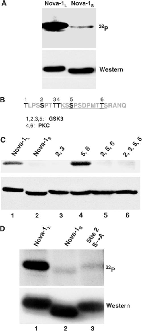
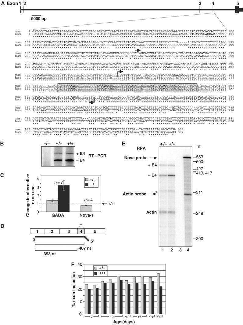
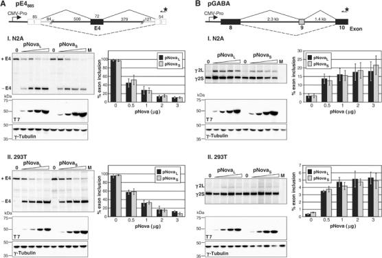
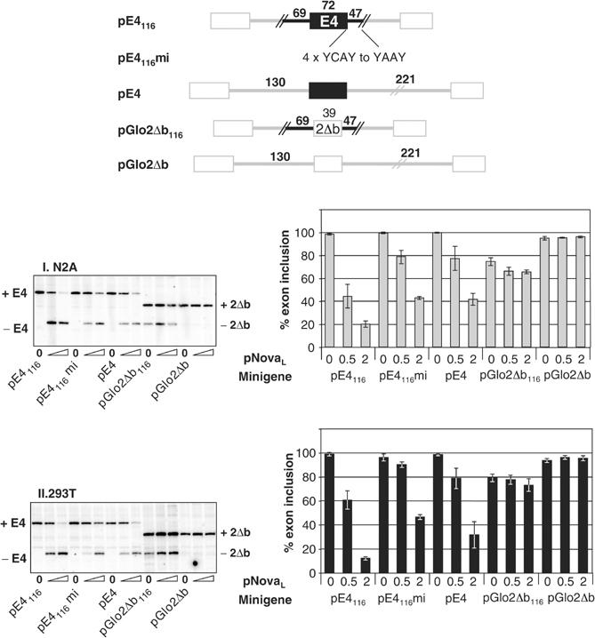
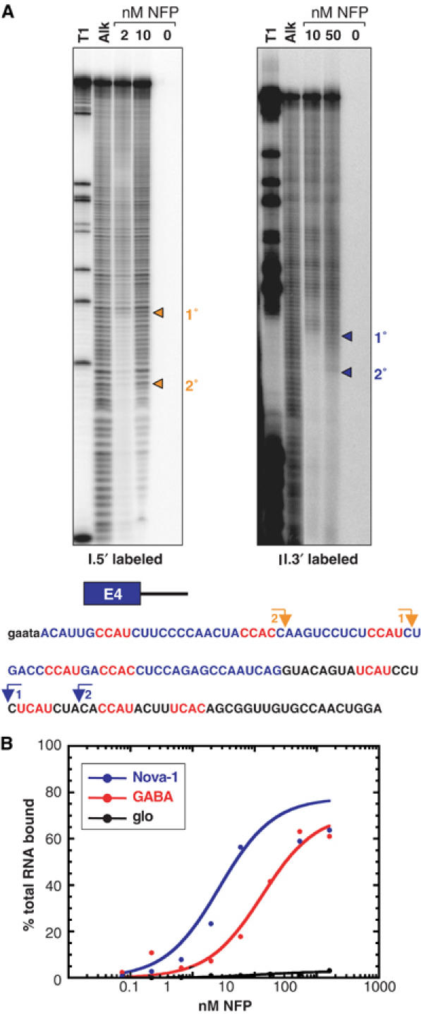
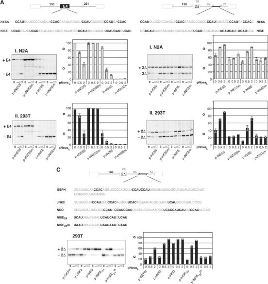
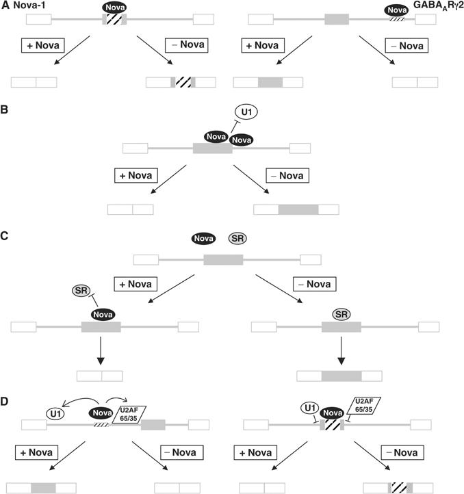
The Nova family of neuron-specific RNA-binding proteins were originally identified as targets in an autoimmune neurologic disease characterized by failure of motor inhibition. Nova-1 regulates alternative splicing of pre-mRNAs encoding the inhibitory neurotransmitter receptor subunits GABA(A)Rgamma2 and GlyRalpha2 by directly binding intronic elements, resulting in enhancement of exon inclusion. Here we identify exon E4 in the Nova-1 pre-mRNA itself, encoding a phosphorylated protein domain, as an additional target of Nova-dependent splicing regulation in the mouse spinal cord. Nova binding to E4 is necessary and sufficient for Nova-dependent exon exclusion. E4 harbors five repeats of the known Nova-binding tetranucleotide YCAY and mutation of these elements destroys Nova-dependent regulation. Furthermore, swapping of the sites from Nova-1 and GABA(A)Rgamma2 indicates that the ability of Nova to enhance or repress alternative exon inclusion is dependent on the position of the Nova-binding element within the pre-mRNA. These studies demonstrate that in addition to its previously described role as a splicing activator, Nova autoregulates its own expression by acting as a splicing repressor.


Figures

Figure 1
Figure 1
Nova-1 domain 4 is phosphorylated in vivo. (A) N2A cells were transiently transfected with T7-tagged Nova constructs with (Nova-1L) or without (Nova-1S) inclusion of domain 4, and 32P-orthophosphate was added to the medium. After 5 h incubation, cells were harvested, lysed and immunoprecipitated with anti-T7-tag antibody. The pellet was treated with RNase prior to separation by SDS–PAGE, transferred to nitrocellulose and exposed to autoradiography film (upper panel). Subsequently, the membrane was probed by Western blot with anti-T7-tag antibody (lower panel). (B) Amino-acid sequence encoded by E4. The residues in bold and numbered 1–6 are phosphorylation sites predicted by Prosite (http://us.expasy.org/prosite/) and Scansite (http://scansite.mit.edu). The underlined segment is a PEST region predicted by PESTfind (http://emb1.bcc.univie.ac.at/embnet/tools/bio/PESTfind/). (C) Identification of the phosphorylation site by site-directed mutagenesis. Metabolic phosphorylation assays were performed as in (A). Expression constructs encoding Nova-1L (lane 1) or Nova-1S (lane 2), and point mutants in which the indicated residue numbers indicated in (B) were mutated in Nova-1L to alanines (lanes 3–6) were utilized. (D) Expression constructs encoding Nova-1L (lane 1), Nova-1S (lane 2) or Nova-1L with a serine to alanine mutation at site 2 were utilized for the transfection/metabolic labeling assay as in (A).
Figure 2
Figure 2
Change in alternative splicing of Nova-1 E4 in Nova-1 mouse spinal cord. (A) Exon structure of the mouse Nova-1 gene, adapted from http://genome.ucsc.edu/. Scale bar represents intronic lengths, and exons are to approximate scale only. Below is a comparison of the mouse (mus) and human (hum) sequence surrounding alternatively spliced E4 (gray box) by ClustalW alignment (MacVector). YCAY motifs are in boldface type, and arrows indicate regions included in minigene constructs pE4307 and pE4116 (see Figure 4). (B) RT–PCR. Spinal cord RNA isolated from P17 Nova-1 null, heterozygous and wild-type mouse littermates was analyzed by semiquantitative RT–PCR using primers to exons 3 and 5. The reverse primer was 32P-labeled. (C) Comparison of the magnitude of splicing defects in heterozygous mice analyzed by RT–PCR. The RT–PCR data from panel B together with additional samples were plotted as the change in alternative exon use (with the ratio of alternative exon included:excluded form from the wild type set to 1) for comparison with previously published RT–PCR data; Jensen et al, 2000a). Error bars indicate the standard deviation, n=number of litters assayed. (D) Schematic representation of the 32P-labeled antisense RNA probe used for RPA and the resulting product sizes used for quantitation. An antisense RNA probe specific for β-actin mRNA was also made. (E) RPA. Spinal cord RNA isolated from P16 Nova-1 heterozygous mouse and wild-type littermate was analyzed by RPA. The specific activities of the probes were adjusted to give protected fragments of approximately equal intensities for each message. Lane 3: yeast RNA control; lane 4: undigested probes. The asterisk marks an unidentified product that was protected by the Nova-1 probe. (F) Quantitation of the data presented in (E) together with additional RNA samples. Products were quantitated by phosphorimager and corrected for the expected number of 32P-labeled uridines per protected RNA fragment.
Figure 3
Figure 3
Nova-1 regulates alternative splicing of Nova-1 and GABAARγ2 in cell lines independent of domain E4 inclusion. (A) Nova-1 minigene construct used for transient transfection assays harboring 957 of Nova-1 mouse genomic sequence (black lines, black filled box; sequence is shown in Figure 2) flanked by donor and acceptor sites derived from the human β-globin gene (gray lines, empty boxes). The sizes (in base pairs) are shown above each element. The starred arrow depicts the 32P-labeled primer used for primer extension analysis. Distances are not to scale. RNA was isolated from N2A (I) or 293T (II) cells transiently transfected with pE4885 and increasing amounts of pNova-1L or pNova-1S (0, 0.5, 1, 2 and 3 μg) and analyzed by primer extension (upper panels). Mock transfections were performed using no plasmid DNA (lanes M). Below are Western blots using anti-T7 antibody (middle panels) showing the titration of T7-tagged Nova-1 protein levels. The blots were stripped and reprobed with anti-γ-tubulin antibody (lower panels) as a loading control. The graphs depict quantitation of three independent transfection experiments, displayed as the average percent central exon inclusion [Phosphorimager counts in the exon included product/(counts in the exon included product+counts in the exon excluded product) × 100], ±standard deviation. (B) Transient transfection assays were performed and analyzed as in (A) using a GABAARγ2 (pGABA) minigene containing the full mouse intronic regions surrounding and including alternatively spliced exon 9 plus shortened exons 8 and 10.
Figure 4
Figure 4
Exon E4 is necessary and sufficient for regulation of alternative splicing by Nova-1. (A) The minigenes depicted were cotransfected into N2A (I) or 293T (II) cells with increasing amounts of pNova-1L (0, 0.5 and 2.0 μg); spliced products were measured by primer extension and phosphorimage analysis as in Figure 3 and titration of transfected pNova-1L expression was monitored by Western blotting (not shown). The asterisk denotes an uncharacterized product consistently seen with construct pE4307. (B) Mutagenesis of the YCAY repeats in E4 abrogates Nova-dependent regulation of alternative splicing. YAAY mutations were made in the first two (m1, 2), last three (m3, 4, 5) or all five (m1–5) YCAY motifs of E4 in construct pE4. Plasmid pGlo2Δb was used as a Nova-independent control. The minigenes were transfected and analyzed as in (A).
Figure 5
Figure 5
Downstream intronic sequences enhance Nova- dependent splicing of E4, but are neither necessary nor sufficient for the effect. Four YCAY repeats within the Nova-1 intronic sequence downstream of E4 in construct pE4116 were mutated to YAAY to generate construct pE4116mi. Intronic sequences in pGlo2Δb were replaced by the Nova-1 intronic sequences present in pE4116 to generate construct pGlo2Δb116. Constructs pE4 and pGlo2Δb were used as Nova-dependent and -independent controls, respectively. Minigenes were cotransfected into N2A (I) or 293T (II) cells with increasing amounts of pNova-1L (0, 0.5 and 2.0 μg); spliced products were measured by primer extension and phosphorimage analysis as in Figure 3 and titration of transfected pNova-1L expression was monitored by Western blotting (not shown).
Figure 6
Figure 6
Nova-1 binds with high affinity to its own RNA in vitro. (A) Mapping the boundaries of Nova-1 protein binding to Nova-1 E4 plus downstream intronic RNA. RNA was labeled with 32P at either the 5′ or 3′ end and subjected to mild alkaline hydrolysis (Alk). The RNA was then incubated with Nova-1 fusion protein (NFP) at a final concentration of 2, 10 or 50 nM and protein:RNA complexes were captured by filtration through nitrocellulose. Bound RNAs were eluted and analyzed by denaturing PAGE. RNase T1 digestion (T1) was used to generate size standards. Boundaries are highlighted by arrows and indicated on the transcribed RNA sequence shown at the bottom. A schematic representation of the RNA transcribed is shown above the sequence; exonic sequence is shown in blue, intronic sequence in black and YCAY motifs are highlighted in red. Small letters represent sequences derived from primers. (B) Nitrocellulose filter-binding assays were performed using Nova-1 RNA as in (A) or control RNAs. GABA: 159 nt RNA corresponding to the GABAARγ2 NISE element (Dredge and Darnell, 2003); glo: 175 nt RNA derived from human β-globin, which spans regions of exon 1 and intron 1 and contains no YCAY tetranucleotides.
Figure 7
Figure 7
Nova-dependent regulation is dependent on the position of the Nova-binding site within the pre-mRNA. (A) A 50 nt stretch of Nova-1 E4 (NESS) containing all five YCAY repeats was replaced in pE4 (renamed p-eNESS) by a 54 nt stretch of the GABAARγ2 intron 9 NISE region (NISE) encompassing six UCAY repeats to generate plasmid p-eNISE. Corresponding constructs with YCAY to YAAY mutations in the introduced segments (p-eNESSm and p-eNISEm) were also tested. These constructs were cotransfected into N2A (I) or 293T (II) cells with increasing amounts (0, 0.5 and 2.0 μg) of pNova-1L; spliced products were measured by primer extension and phosphorimage analysis as in Figure 3 and titration of transfected pNova-1L expression was monitored by Western blotting (not shown). (B) A total of 51 nt from Nova-1 E4 or GABAARγ2 NISE, each harboring five YCAY repeats, were cloned into pGlo2Δ 72 nt upstream of the downstream splice acceptor site (plasmids p-iNESS and p-iNISE). Corresponding constructs with YCAY to YAAY mutations in the introduced segments (p-iNESSm and p-iNISEm) were also tested. These constructs were transfected and analyzed as in (A). (C) Three additional putative Nova target sequences in gephyrin, JNK2 and neogenin identified by CLIP (Ule et al, 2003) were cloned into pGlo2Δ (plasmids p-iGEPH, p-iJNK2 and p-iNEO). Construct p-iNISE24 harboring a 24 nt stretch of the GABAARγ2 NISE and a corresponding construct with YCAY to YAAY mutations (p-iNISE24m) were used as Nova-dependent and -independent controls, respectively (Dredge and Darnell, 2003). These minigenes were transfected and analyzed as in (A).
Figure 8
Figure 8
Models for Nova's action on alternative splicing. (A) Position-dependent action of Nova on alternative splicing. Nova binding to a high-affinity site within an alternative exon represses exon inclusion. In contrast, Nova binding within the intron downstream of an alternative exon enhances exon inclusion. (B) Nova-1 binding to exon E4 may inhibit binding and/or assembly of constitutive splicing factors such as U1 as shown, or sterically inhibit bridging interactions across the exon (not shown). (C) Nova-dependent inhibition may occur through competitive inhibition of nonessential splicing factors such as SR protein(s). (D) Nova may inhibit exon inclusion via incorrect recruitment of splicing factors. Empty boxes represent constitutively included exons, and filled boxes represent alternative exons. Hatching represents high-affinity Nova-binding sites. Note that while Nova protein is represented as a single oval, a number of Nova molecules are likely to be bound at each binding site.

Similar articles

Cited by

References

    1. Bernardi G (1995) The human genome: organization and evolutionary history. Annu Rev Genet 29: 445–476 - PubMed
    1. Biondi RM, Nebreda AR (2003) Signalling specificity of Ser/Thr protein kinases through docking-site-mediated interactions. Biochem J 372: 1–13 - PMC - PubMed
    1. Black DL (2003) Mechanisms of alternative pre-messenger RNA splicing. Annu Rev Biochem 72: 291–336 - PubMed
    1. Blanchette M, Chabot B (1999) Modulation of exon skipping by high-affinity hnRNP A1-binding sites and by intron elements that repress splice site utilization. EMBO J 18: 1939–1952 - PMC - PubMed
    1. Buckanovich RJ, Darnell RB (1997) The neuronal RNA binding protein Nova-1 recognizes specific RNA targets in vitro and in vivo. Mol Cell Biol 17: 3194–3201 - PMC - PubMed

Publication types

MeSH terms