

Damage and threshold shift resulting from cochlear exposure to paraquat-generated superoxide
- PMID: 15935579
- PMCID: PMC1987394
- DOI: 10.1016/j.heares.2005.03.025
Damage and threshold shift resulting from cochlear exposure to paraquat-generated superoxide
Abstract
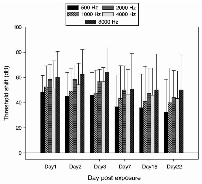
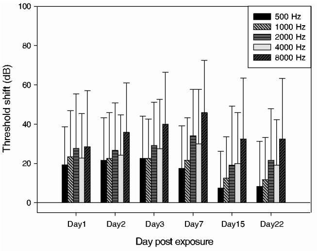
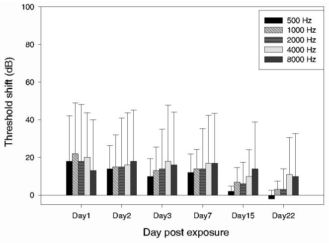
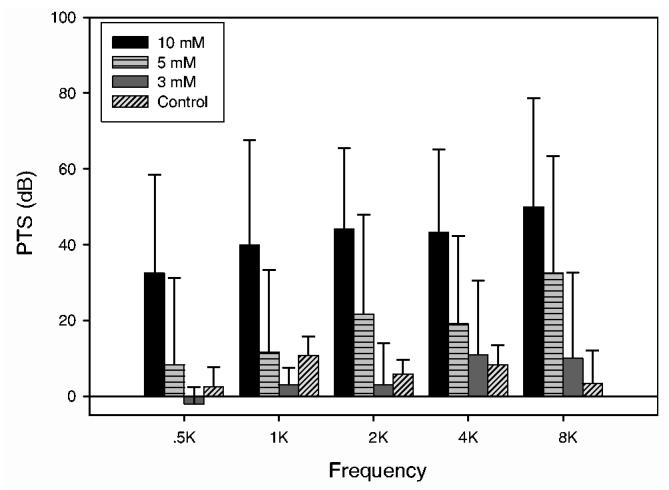
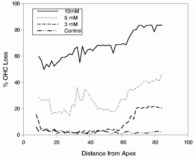
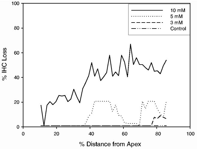
Superoxide has been implicated as a contributing factor to cochlear pathology from a number of sources, including noise and ototoxic drugs. The effects of NADPH oxidase-dependent superoxide on the cochlea were investigated in the current study using paraquat (PQ). PQ is a toxic herbicide that causes tissue damage by generating superoxide through reduction of molecular oxygen in a reaction catalyzed by NADPH oxidase. The current study examined the effects of round window PQ administration on inferior colliculus (IC) evoked potential thresholds (EVP) and hair cell damage. Using implanted IC electrodes, chinchillas were tested for IC EVP thresholds before and after PQ exposure. Ears were exposed to PQ at one of four concentrations: 10, 5, 3 mM, and vehicle control. Thresholds were increased in a dose-dependent manner, and peaked between one and seven days post-exposure. Thresholds then showed a small amount of recovery before reaching PTS by Day 22. Outer and inner hair cell losses were consistent with PTS. The similarities between PQ ototoxicity and noise-induced hearing loss suggest the possibility of similar biochemical pathways involving superoxide.
Figures
References
-
- Böheim K, Bichler E. Cisplatin-induced ototoxicity: Audiometric findings and experimental cochlear pathology. Arch. Otorhinolaryngol. 1985;242:1–6. - PubMed
-
- Campbell KC, Rybak LP, Meech RP, Hughes L. d-methionine provides excellent protection from cisplatin ototoxicity in the rat. Hear. Res. 1996;102:90–98. - PubMed
-
- Clerici WJ, Yang L. Direct effects of intraperilymphatic reactive oxygen species generation on cochlear function. Hear. Res. 1996;101:14–22. - PubMed
-
- Clerici WJ, Hensley K, DiMartino DL, Butterfield DA. Direct detection of ototoxicant-induced reactive oxygen species generation in cochlear explants. Hear. Res. 1996;98:116–124. - PubMed
Publication types
MeSH terms
Substances
Grants and funding
LinkOut - more resources
Full Text Sources
